|
|
||||
|
Published by : PROFESSIONAL MEDICAL PUBLICATIONS |
||||
|
ISSN 1681-715X |
||||
|
||||
|
- |
||||
|
ORIGINAL ARTICLE |
||||
|
- |
||||
|
Volume 22 |
April - June 2006 |
Number 2 |
||
|
|
||||
|
|
||||
|
|
||||
|
Published by : PROFESSIONAL MEDICAL PUBLICATIONS |
||||
|
ISSN 1681-715X |
||||
|
||||
|
- |
||||
|
ORIGINAL ARTICLE |
||||
|
- |
||||
|
Volume 22 |
April - June 2006 |
Number 2 |
||
|
|
||||
|
|
||||
Evaluation of the Enzyme-linked Immuno-electro
Transfer Blot (EITB) technique using hydatid cyst
antigens B/5 and total IgG antibodies in laboratory
diagnosis of human hydatidosis
MB Rokni1, B Aminian2
Abstract
Objective: To evaluate the validity of the Enzyme-linked Immuno-electro Transfer Blot (EITB) technique to diagnose human hydatidosis using sheep hydatid fluid antigens and human sera infected with hydatidosis.
Design: After preparing parasite antigen from sheep hydatid cyst fluid, all collected human sera infected with hydatidosis and other parasitic diseases as well as normal individuals, were analyzed by EITB test to evaluate its validity in diagnosing of hydatidosis.
Setting: Department of Medical Parasitology and Mycology, School of Public Health, Tehran University of Medical Sciences, Iran.
Subjects: Seventy patients infected with hydatidosis confirmed by surgery, 15 with fasciolosis, 10 with toxocariasis, 15 with strongyloidiasis, 5 with amoebiasis, 10 with trichostrongylosis and 30 normal controls.
Main Outcome Measures: The sensitivity, specificity, positive and negative predictive values of EITB test.
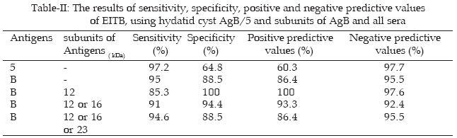
Result: Using total IgG antibody isotype, the sensitivity, specificity, positive & negative predictive values were 95%, 88.5%, 86.4%, 95.5 % 97.3%, 64.8%, 60.3% & 97.7% for antigens B&5 respectively. The total IgG antibodies in hydatidosis patients documented the parasite AgB subunits i.e 12, 16, 23 kDa, also larger subunit of Ag5, namely 39 kDa.
Conclusion: The study showed that although, EITB method was a time consuming test, but due to high validity could be considered as an authentic technique, especially when the diagnosis is vague and time is not imperative.
Source of funding: Vic- Chancellery for Research, Tehran University of Medical Sciences, Iran.
Conflicts of interest: No Conflicts of interest exists.
Key words: Hydatidosis, SDS-PAGE, EITB, Antigen B, Antigen 5
Pak J Med Sci April - June 2006 Vol. 22 No. 2 127 - 131
1. Dr. MB Rokni
Assistant Professor of Medical Parasitology
2. Dr. B Aminian MSPH
Medical Parasitology
1-2: Department of Medical Parasitology & Mycology,
School of Public Health,
Tehran University of Medical Sciences, Iran.
Correspondence:
Dr. MB Rokni
P.O. Box: 14155-6446,
Tehran, IRAN.
E-Mail: roknimoh@tums.ac.ir
* Received for publication September 16, 2005
Accepted February 3, 2006
Introduction
Hydatidosis is an important zoonotic infection of humans caused by larva of Echinococcus granulosos.1 It has a world wide distribution as well as in Iran reported throughout the country.
In spite of much progress in various lab facilities and imaging techniques in the world for diagnosis of this disease, immunologic tests are now extensively used to supplement its diagnosis of this disease.2 The search for increasing assay validity has involved research on cyst fluid fractions and individual cyst proteins as possible diagnostic molecules.3 Discovery of two major parasite antigens, characterized as antigen 5 and B, resulted in a set of immunological tests to be evaluated as potential approaches to diagnose human hydatidosis.4 Serological reactivity of antigen B has also been documented, with recent interest focusing on its lowest 12 kDa subunit as putative diagnostic molecule using immuno blot analysis.5
The main purpose of this study was to evaluate the sensitivity and specificity of the Enzyme-linked Immuno-electro Transfer Blot (EITB) technique using cyst antigen B and total IgG antibodies for diagnosis of human hydatidosis.
Materials and Methods
Sera: Blood samples were collected from individuals infected with hydatidosis, confirmed by surgery in the Imam Khomeini Hospital, Tehran, Iran, and patients with other parasitic diseases including 15 with fasciolosis, 10 with toxocariasis, 15 with strongyloidiasis, 5 with amoebiasis and 10 with trichostrongylosis. All these sera were obtained from serum blood bank of the School of Public Health, Tehran University of Medical Sciences, Iran. These patients had been diagnosed based on stool examination, ELISA, IFA as well as at surgery, as appropriate. Control serum samples were obtained from 30 healthy persons. The Human Ethics Committee at the above-mentioned school approved the study. Informed consent was obtained from patients or their legal guardians.
Preparation of antigens: Unilocular hydatid cysts were obtained from the livers of sheep killed at local abattoirs frequently, Ahmad Abad abattoir in Tehran. Cyst fluid was then aseptically aspirated from cysts, pooled, clarified by centrifugation at 1500 g at 4oC for 30 min, and afterwards the supernatant was sterilized by filtration through a Millipore filter (pore diameter 0.45μm), then it was concentrated by membrane analysis in front of fan or putting in glycol powder (2000) at 4oC for 5-7 h and then poured in apendorf tubes and preserved at -20oC until used. The concentration of antigen preparation was measured using Bradford method.6
SDS-PAGE (sodium dodecyle sulfate � polyacrylamide gel electrophoresis): SDS-PAGE was preformed as described previously.7,8 The antigens were separated by SDS-PAGE with 12% gel of 0.75 mm thickness at constant voltage at 110 v in the Bio Rad mini � gel apparatus (Bio-Rad laboratories, Richmond, CA).
The molecular mass of the antigen was estimated by comparing the migration distance of the sample to that of known molecular markers (electrophoresis calibration kit; pharmacia, piscatway. NJ). Separating gel was 12% (Acryleamid-bisacrylamid, 2M Tris [pH 8.8], 10% SDS, 10% Ammonium persulfate, TEMED 10μm) and stacking gel was 6% (Acryleamid-bisacrylamid, 1M Tris-Hcl [pH 6.8], 10% SDS, 10% Ammonium persulfate, TEMED 10�m), the ratio of acrylamide to bisacrylamid was 37.5:1 throughout. Before electrophoresis the dialyzed hydatid fluid was diluted 1:2 with electrophoresis buffer and sample buffer (1M Tris-Hcl [pH 6.8], 10% [W/V] SDS, 50% [V/V] glycerol, 1% [W/V] bromophenol blue and 5% [V/V] 2-mercaptoethanol and boiled in boiling water bath for 5-10 min. the final protein concentration was between 60 and 100 �m/ml. After running antigens, in order to visualize the protein profiles from the antigen preparations was obtained, the gel stained with cumassi-brilliant blue R-250 for overnight at room temperature.
EITB: The antigens were transferred from unstained gel to nitrocellulose membrane using the electrophoresis method and Akhtarian mini-transfer apparatus (Akhtarian co; Tehran) run at 45 mA overnight. Transfer buffer was 15.6 mM Tris-Base, 120 mM glycine, methanol 100% [V/V].Efficiency of transfer proteins was checked by staining nitrocellulose membrane with 0.2% Ponceau S (Sigma), then it was destained by washing the paper with distilled water. The paper was blocked by incubating with blocking solution (3% gelatin in TBS ) for 24h at RT, TBS (Tris buffer saline:2M Tris-Hcl [pH 7.5], 4M NaCl ). Nitrocellulose membrane blotted antigens was cut to strips and put in sera diluted 1:40 with TBS and incubated for 4h in 37o�, the strips were washed 3 times and then incubated in a 1:800 dilution antibody (HRP= horse- radish peroxidas) conjugate rabbit antihuman IgG for 2 h at RT, the strips were washed and stained with DAB (3, 3- diaminobenzidine) in H2O2.
Statistical analysis: The sensitivity, specifity and the predictive values were calculated as previously described.9,10
Results
SDS-PAGE of the hydatid fluid performed in reducing conditions revealed AgB and Ag5 subunits as 12, 16, 23, and 38 kDa, respectively, not mention of some invaluable molecules. Immunoblotting of these proteins showed that antigens B and 5 were immunogenic (Fig-I).
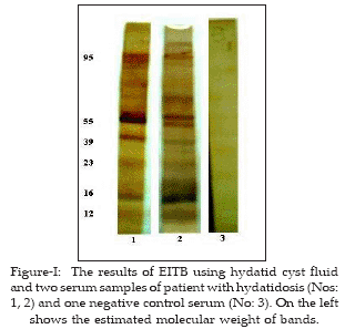
The 38 kDa subunit of Ag5 was immunoreactive with the majority of infected sera with hydatidosis (68/70) and other parasitic diseases as well as with a proportion (12/30) of normal controls. The Table-I is a summary of immunoreactity to the subunits of AgB/5 by IgG from various parasitic infections. Subunit 12kDa band of AgB was highly specific for hydatid disease (100%) while the sensitivity was 85.3%.
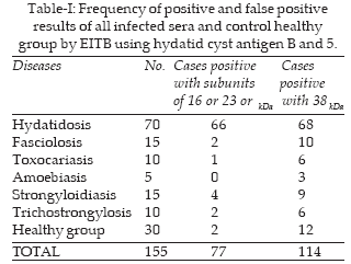
Table-II shows the sensitivity, specifity, positive and negative predictive values for AgB subunits as 12 kDa 12 or 16 kDa and 12 or 16 or 23 as well as Ag5.
Discussion
Hydatidosis is an important zoonotic infection of humans caused by larva of Echinococcus granulosus.1 Immunodiagnosis of this disease is conducted by different serological methods using cyst hydatid fluid. The fluid is generally obtained from infected sheep as this is a readily available source in most countries where hydatid disease is common, despite the qualitative and quantitative antigenic differences in hydatid fluid obtained from different hosts.8 Ag5 migrates in SDS-PAGE in the area of 37 kDa under reduced conditions. Leggate et al (1992) attributed higher sensitivity and specificity to immunodetection of 12 kDa molecule from AgB, when compared with a 38 kDa molecule from Ag5.11 Many researchers reported that in humans infected with Echinococcus, subunits of 12, 16, and 23 kDa of AgB were specific for diagnosis of hydatidosis.11 But in the present study we find that only subunit 8 or 12 is specific. Doiz et al using subunit 39 kDa of hydatid cyst fluid by EITB method, obtained a sensitivity of 92%, with subunit of 20 kDa, a sensitivity of 69% while with Ag5/B the sensitivity and specificity were detected as 97% and 95.7%, respectively.12,13 Gadea et al by EITB technique using AgB of hydatid cyst fluid reported the sensitivity, specificity, positive and negative predictive value as 95.7%, 89%, 82%, and 97.6%, respectively.5 Haniloo using different subunits of AgB/5 obtained the sensitivity, specificity, positive and negative predictive values of 8 kDa band of AgB as 70%, 100%,100% and 89%, correspondingly and with 8 and 16kDa as 81%, 100%, 100% and 92.5% while with 8, 16, 22 kDa as 90.5%, 95.5%, 89%, and 93%, finally with 8, 16, 22 and 38 kDa of AgB/5 as 93.5%, 91%, 81% and 97%, in that order.14 Al-sherbiny et al, using dipstick and EITB method for serological diagnosis of human hydatidosis obtained the sensitivity and specificity as 100% and 91.4%, in that order.15
El-Zayyat et al using AgB/5 of hydatid cyst fluid from camel and EITB method reported that the most diagnostic band was 21 kDa and obtained the sensitivity and specificity of this band as 96% and 98.5%, respectively.16 Kormaz et al for serological diagnosis of alveolar echinococcusis cases, using EITB method and Em70, 90 yielded the sensitivity, specificity as 100% and 95.51%, correspondingly.17
Xu et al using different antibodies and circulating antigens and EITB method for diagnosis of human hydatidosis only reported a low sensitivity.18 While Poretti et al using subunits 8, 29 and 34 kDa of hydatid cyst fluid AgB/5 and EITB method, accounted the sensitivity and specificity as 91% and 97%, in that order.19 Frinder et al reported that AgB was a lipoprotein antigen and at reduced condition produced fractions from 8 to 24 kDa range.20 Using purified AgB by the EITB test it was reported that 8 kDa subunit of AgB was more specific and less cross reactive with other parasitic diseases were seen.21
We conclude from the present study that although, EITB method is a time consuming procdedure but due to its high validity it could be considered as an authentic diagnostic technique, especially when the diagnosis is vague and time is not imperative.
Acknowledgements
The kind cooperation of Miss Neda Mirsepahi, Ali Rahimi, Hoda Bazranbar and Mrs. Shirin Jaafarian from the School of Public Health, Tehran University of Medical Science, Iran, is greatly appreciated. The study was supported financially by the Vice Chancellor for Research, Tehran University of Medical Sciences, Iran.
References
1. Devi CS, Parija SC. A new serum hydatid antigen detection test for diagnosis of cystic echinococcosis. Am J Trop Med Hyg 2003; 69(5):525-8.
2. Sbihi Y, Gil KR, Alvares PA, Orduma A, Rodarigues-Torres A, Osuna A. Development of a dipstick dye immunoassay for diagnosing hydatidosis. J Clin Lab Anal 2003; 17(6):219-22.
3. Maddison SE, Slemenda SB, Schantz PM, Fried JA, Wilson M, Tsang VCW. A specific diagnostic antigen Echinococcus granulosus with an apparent molecular weight of 8 kDa. Am J Trop Med Hyg 1989; 40(4):377- 83.
4. Lihtowlers MW, Gottstien B. Echinococcosis / Hydatidosis: antigens, immuno logical and molecular diagnosis In: Thompson R.C.A and Lymbery AJ (Eds), Echinococcus and Hydatid disease. CAB International Wallingfrod. UK; 1995: 355-410.
5. Siracusano A, Ioppolo S, Notargiacomo S, Ortona E, Rigano R, Teggi A, et al. Dettection of antibodies against Echinococcus granulosus major antigens and their subunits by Immunoblotting. Trans R Soc Trop Med Hyg 1991; 85: 239-43.
6. Bradford MM. A rapid and sensitive method for the quantitation of microgram quantitive protein utilizing the principle of protein-dye binding. Anal Biochem 1976; 72: 248-54.
7. Gadea I, Ayala G, Diago MT, Cunat A, Lomas JG. Immunological diagnosis of human hydatid cyst relapse: Utility of the Enzyme-Linked Immunoelectrotransfer blot and discriminate analysis. Clinic Diag Lab Immunol 1999; 7(4): 549-52.
8. Verastegui M, Moro P, Guevara A, Rodriguez T, Miranda E, Gilman RH. Enzyme-Linked Immunoelectrotransfer blot test for diagnosis of human hydatid disease. J Clin Microbiol 1992; 3 (6): 1557-61.
9. Galen RS. Predictive values and efficiency of laboratory testing. Pediat J Clin North Am 1980; 27: 861-89.
10. Ferragut G, Liungstrom I, Nieto A. Relevance of circulating antigen detection to follow-up experimental and human cystic hydatid infections. Parasit Immuno 1998; 20: 541-9.
11. Leggatt GR, Yang W, McManus DP. Serological evaluation of 12 kDa subunit of antigen B in Echinococcus granulosus cyst fluid by immunoblot analysis. Trans Roy Soc Trop Med Hyg 1992; 86:189-92.
12. Doiz O, Benito R, Sbihi Y, Osuna A, Clavel A, Gomez-Lus R. Western blot applied to the diagnosis and post treatment monitoring of human hydatidosis. Diag Microb Infec Dis 2001; 41: 139-42.
13. Doiz O, Benito R, Gil J, Rojas A, Rubio MC, Osuna A. Pre- and post surgical detection of IgG, IgM and IgA specific to hydatidosis by ELISA purified antigen enriched with the 5/B antigen complex. J Clin Lab Anal 2002; 16(6): 295-8.
14. Haniloo A. Preparation, purification and evaluation of hydatid cyst fluid antigens for immunological diagnosis of hydatidosis. Thesis for PhD degree, School of public Health and Institute of Health Research, Tehran University of Medical Science. 2002.
15. AL-Sherbiny MM, Farrag, Fayad MH, Malked MK, Tawfeek GM, Ali NM. Application and assessment of a dipstick assay in the diagnosis of hydatidosis and trichinosis. Parasitol Res 2004; 93(2): 87-95.
16. El Zayyat EA, Ramzy RM, Abdel-Baki MH, Fahmi IA, Rifaat M, Helmy H, Abdel H, DM. Human cystic echinococcosis: diagnostic Value of different antigenic fractions of hydatid cyst fluid with different specific immunoglobulin G subclasses by enzyme linked immunoclectrotransfer blot. J Egypt Soc Parasitol 1999; 29(3): 817-30.
17. Korkamz M, Inceboz T, Celebi F, Babaoglu A, Uner A. Use of two sensitive and specific immunoblot markers, em 70 and em 90 for diagnosis of alveolar echinococcusis. J Clin Microbiol 2004; 42 (7): 3350-2.
18. Xu MQ, Zhu B, Xue HC, Li Guo XR, Zhang YH, Li H, et al. Reexamination of specitic antibodies in sera of cystic echinococcosis patients with IgG negative seroresponse. Zhongguo Ji Sheng Chong Xue YuJi sheng chong bing Za Zhi 2002; 20(3):148-51.
19. Poretti D, Felleisen E, Grimm F, Pfister M, Teuscher F, Zuercher C, et al. Differential immunodiagnosis between cystic hydatid disease and other cross- reactive pathologies. Am Soc Trop Med Hyg 1999; 60(2): 193-8.
20. Frinder B, Moguilensky J, Salvitti JC, Odriozole M, Cantoni G, Larrieu E. Epidmiological surveillance of human of human hydatidosis: Its contribution to the evaluation of control programs. Acta Tropica 2001; 79: 219-23.
21. Oriol R, Williams JF, Perez-Esnndi MV, Oriol C. Puritication of lipoprotein antigens of Echinococcus granulosus from sheep hydatid fluid. Am J Trop Med Hyg 1971; 20(4): 569-74.
HOME | SEARCH | CURRENT ISSUE | PAST ISSUES
Professional
Medical Publications
Room No. 522, 5th Floor, Panorama Centre
Building No. 2, P.O. Box 8766, Saddar, Karachi - Pakistan.
Phones : 5688791, 5689285 Fax : 5689860
pjms@pjms.com.pk