|
|
||||
|
Published by : PROFESSIONAL MEDICAL PUBLICATIONS |
||||
|
ISSN 1681-715X |
||||
|
||||
|
- |
||||
|
ORIGINAL ARTICLE |
||||
|
- |
||||
|
Volume 23 |
April - June 2007 (Part-I) |
Number 2 |
||
|
|
||||
|
|
||||
|
|
||||
|
Published by : PROFESSIONAL MEDICAL PUBLICATIONS |
||||
|
ISSN 1681-715X |
||||
|
||||
|
- |
||||
|
ORIGINAL ARTICLE |
||||
|
- |
||||
|
Volume 23 |
April - June 2007 (Part-I) |
Number 2 |
||
|
|
||||
|
|
||||
Laparoscopic management of atypical
presentation of ectopic pregnancy
Shamsun Nahar1, Shahida Begum2, M. Saiful Islam3
ABSTRACT
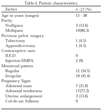
Objective: Analysis of management in 22 ectopic pregnancies clinically presented as an atypical form.
Patients and Methods: Twenty two patients with suspected ectopic pregnancy were successfully managed laparoscopically during three years period at Victory Nursing and infertility management centre, Khulna, Bangladesh.
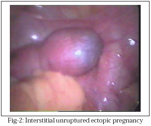
Results: All patients had dull ache pain in lower abdomen or any one of the iliac fossa. All cases were hemodynamically stable and ambulatory. Ultrasonography findings were complex Heterogeneous mass without any free fluid in 8 cases, definitive gestational sac in 6, and sac like structure in 8 cases. Urinary ß–hCG was positive in 40.9% and negative in 59% cases. Pre-operative diagnoses were chronic ectopic pregnancy in 11, ruptured corpus luteum in six and chocolate cyst in five cases. Operating diagnosis were chronic ectopic pregnancy 14, unruptured tubal and cornual pregnancy 5, ovarian ectopic in three cases. Surgical procedures were salpingostomy, salpingectomy, salpingo-ophrectomy, partial ovarian resection and only sac removal with peritoneal toileting. Operating time was 30–120 minutes. The average post- operative stay was 24–48 hours without any complications.
Conclusion: Unruptured early ectopic may present with minimal symptomotology. Hence in all women of child bearing age the provisional diagnosis of ectopic pregnancy may be kept in mind while examining and investigating a patient.
KEY WORDS: Ectopic Pregnancy, Laparoscopic Management.
Pak J Med Sci April 2007 Vol. 23 No. 2 198-201
1. Dr. Shamsun Nahar
Associate Professor
2. Dr. Shahida Begum
Consultant
3. Dr. M. Saiful Islam
Assistant Professor
1-3 Khulna Medical College,
Khulna – Bangladesh.
Correspondence
Dr. Shamsun Nahar
E-mail: sara@khulna.bangla.net
* Received for Publication: June 19, 2006
* Accepted: November 15, 2006
INTRODUCTION
Ectopic pregnancy was first described in the 11th century and until the middle of the 18th century and its outcome was usually fatal. It occurs in approximately 2% of all pregnancies and is a leading cause of first trimester maternal death.1 Historically, the hallmark of ectopic pregnancy has been abdominal pain with spotting usually occurring at six to eight weeks after the last normal menstrual period. This remains the most common presentation of tubal pregnancy in symptomatic patients. Other presentations depend on the location of the ectopic pregnancy. Less commonly, ectopic pregnancy presents with pain radiation to the shoulder, vaginal bleeding, syncope and hypovolaemic shock. Between 40 and 50 percent of ectopic pregnancies are misdiagnosed at the initial visit to an emergency department.2,3 Pregnancy can even occur in both the womb and the tube at the same time (Heterotopic pregnancy), but this is rare (about 1in 10000 pregnancies) many factors are known to increase the risk of having an ectopic pregnancy. Anything that alters the tubal function may affect further pregnancies. Often none of the risk factors or there is no obvious reason are present it was just bad luck.
A proper history and physical examination remain the foundation for initiating an appropriate work-up that will result in the accurate and timely diagnosis of an ectopic pregnancy. Modern advances in ultrasound technology and the determination of serum beta-subunit human chorionic gonadotropin (â-hCG) levels have made it easier to diagnose. Here we discuss doubtful presentation of ectopic pregnancy and their management.PATIENTS AND METHODS
We retrospectively evaluated 22 haemodynamically stable patients with atypical presentation of ectopic pregnancy during the period of January 2002 to December 2004 at Victory Nursing and Infertility Management Centre in Bangladesh. Careful history taking included menstrual cycle pattern, pain criteria and duration, any history of fainting attack and vaginal bleeding. Previous history included any pelvic surgery specially attention to previous surgery for ectopic pregnancy or tubo-ovarian surgery. History of prolonged infertility, tuberculosis and contraceptive in details were taken. Vaginal examination was performed very carefully. Routine investigations were done. Ultrasonography was offered to all patients. Urinary pregnancy test or â-hCG level was done and followed up in all cases.
Operative Techniques: Laparoscopy was performed under general anesthesia. The abdominal cavity was examined thoroughly; separation of adhesions if present, removal blood clot and ectopic mass was detected. Unilateral salpingostomy, salpingectomy, salpingo-ophrectomy, partial ovarian resection, removal of the sac was done by mono and bipolar coagulation. Peritoneal cavity was irrigated thoroughly with Ringer’s lactate solution and proper hemostasis was established.RESULTS
Twenty two women were enrolled and underwent the laparoscopy and their general characteristics are summarized in Table-I.
Table-II shows pre-operative and operative diagnosis and surgical procedure. Chronic ectopic pregnancy presented as a heterogeneous mass in 63.6% cases.
DISCUSSION
The classic presentation of ectopic pregnancy which includes the triad of abdominal pain, vaginal bleeding and amenorrhea are found in 60% cases only. Most of the patients in this study presented with the complaints of abdominal pain of sudden onset, severe in intensity and localized to the ipsilateral lower abdomen (due to sudden rupture). Atypical presentation was seen with gradual onset, vague in character, non-localized and of varying intensity. Vaginal bleeding, scanty and brownish in colour presented in 50-75% of cases.
History of amenorrhea may be absent in 5-25% of patients.4 In our cases the patient presentations were not typical; it was difficult to determine the size and status of the ectopic pregnancy pre-operatively. Careful ultrasound and â-hCG evaluation were done in all cases. Ectopic presentation should be suspected even in women who have undergone Tubal sterilization.4 Majority of study group of patients presented with abdominal pain without vaginal bleeding and no missed cycle or irregular for last few years 54.5% and 45.4% respectively. Clinical and ultrasound findings have lead to diagnosis in 41.1% of cases in a literature review5 and up to 89% of cases in a recent study.6 The diagnosis was made by trans vaginal ultrasound alone in the absence of any clinical abnormality in 36.3% of our cases. Some factors that reduce the sensitivity of ultrasound such as lack of experience by the ultrasonologists, obesity of the patient, uterine myoma or more commonly “inconclusive ultrasound” which then must be repeated by more expert several times.7 The ultrasound prevents from undue surgical intervention as well as undue delay in surgery when it is required.8 The transvaginal approach (TVUS) is more sensitive than the standard abdominal approach in diagnosis. A hemorrhagic corpus luteum cyst may appear complex and may be ultrasonically distinguishable from a tubal ectopic pregnancy cystic structure distinctly separate from the ovary only by its closer proximity to the ovary. There is sudden onset of pelvic or abdominal pain in the second half of the menstrual cycle. The course varies from no symptoms or signs to severe peritoneal signs and ever life threatening shock.9,10 Some gynecologists prefer to perform a primary diagnostic laparoscopy to rule-out an ectopic pregnancy (as the initial procedure of choice) if the clinical picture is suspicious and the patient very symptomatic.
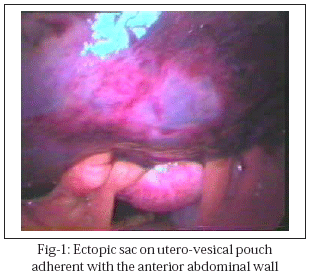
Previously, salpingectomy by laparatomy was the gold standard for the treatment of ectopic pregnancy. Currently laparatomy is the preferred technique when the patient is haemodynamically unstable. We performed linear salpingostomy and the incision was then left to heal by secondary intention in two cases whose opposite tube was sacculated and short. In majority of the cases tubal abortion was in cul-de-sac but in one cases it was anteriorly above the bladder and adherent with the anterior abdominal wall which made a diagnostic confusion.CONCLUSION
Ectopic pregnancy dose not always appear as a typical form. Modern advances in ultrasound technology and the determination of serum â-hCG levels have made it easier to diagnose ectopic pregnancy. But laparoscopy is still an excellent diagnostic and therapeutic modality for doubtful patients. Nonetheless the diagnosis remains a challenge.
REFERENCE
1. Gazvani MR, Baruah DN, Alfirevic Z. Mifepristone in combination with methotrexate for the medical treatment of tubal pregnancy: A randomized controlled trial. Hum Report 1998;13:7.
2. Abbott J, Emmans LS, Lowenstein SR. Ectopic pregnancy ten common Pif falls in diagnosis. Am J Emerg Med 1990;8:515-22.
3. Kaplan BC, Don’t RG, Moskos M, Kuligowska E. Ectopic pregnancy prospective study with improved diagnostic accuracy. Ann Emerg Med 1996;28:10-17.
4. Tenore JL. Ectopic pregnancy. Am Acad Family Physicians 2000;16(4)1-8.
5. Tal J, Haddad S, Gordon N. Heterotopic pregnancy after ovulation induction and assisted reproductive technologies: a literature review from 1971 to 1993. Fertil Steril 1996;66:1-12.
6. Chichia A, Kaubaa A, Tarrask, Makhloaft T. Ultrasonographic diagnosis of ectopic pregnancy. A report of 109 cases. Tunis Med J 2000;78(10)589-94.
7. Gracia CR, Bamhart KT: Diagnosing ectopic pregnancy: decision analysis comparing six strategies. Obstet Gynecol 2001;93:464-70.
8. Engelsbel S, Mol BW, Haienius PJ, Ankum WM, Vander VF, Hemrika DJ. Non-invasive management of patients with suspected of pregnancy. A Survey among Dutch gynaecologists. Eur J Obstet Gynaecol 2001;95(1):81-5.
9. Wang PH, Chao HT, Tseng JY. Laparoscopic surgery for heterotopic pregnancies: A case report and a brief review. Eur J Obestet Gynecol Reprod Biol 1999;80:267-71.
10. Teng S, Tseng J, Chang CK. Comparison of laparoscopy and laparatomy in Managing Hemodynamically Stable Patients with Ruptured Corpus Luteum with Hemoperitoneum. J Am Assoc Gynecol laparosc 2003;10(4):474-7.
HOME | SEARCH | CURRENT ISSUE | PAST ISSUES
Professional
Medical Publications
Room No. 522, 5th Floor, Panorama Centre
Building No. 2, P.O. Box 8766, Saddar, Karachi - Pakistan.
Phones : 5688791, 5689285 Fax : 5689860
pjms@pjms.com.pk