|
|
||||
|
Published by : PROFESSIONAL MEDICAL PUBLICATIONS |
||||
|
ISSN 1681-715X |
||||
|
||||
|
- |
||||
|
ORIGINAL ARTICLE |
||||
|
- |
||||
|
Volume 23 |
April - June (Part-I) 2007 |
Number 2 |
||
|
|
||||
|
|
||||
|
|
||||
|
Published by : PROFESSIONAL MEDICAL PUBLICATIONS |
||||
|
ISSN 1681-715X |
||||
|
||||
|
- |
||||
|
ORIGINAL ARTICLE |
||||
|
- |
||||
|
Volume 23 |
April - June (Part-I) 2007 |
Number 2 |
||
|
|
||||
|
|
||||
Differentiation of common gram negative
pathogens by PCR-ribotyping
Mehwish Jamil1, Saira Bashir2, Mashkoor Mohsin3, Ayesha Tariq4, Aasia Bashir5,
Asma Haque6, Yasra Sarwar7, Aamir Ali8, Abdul Haque9ABSTRACT
Background: Gram negative bacteria especially members of family Enterobacteriaceae are among the most frequently isolated organisms from the clinical specimens. Rapid diagnosis of the pathogen in a clinical sample is always very important. Conventional methods are time-consuming. Among molecular techniques, PCR is very useful but unless very specific primers are used, non-specific amplifications are a problem.
Objectives: PCR-ribotying is a technique that gives very specific multiple bands by use of a single primer set. This study was designed to establish patterns for five common pathogens of Enterobacteriaceae, namely Escherichia coli, Salmonella enterica serovar Typhi (Salmonella Typhi), Proteus vulgaris, Klebsiella aerogenes, and Cirtobacter freundii along with another very common and problematic gram negative pathogen Pseudomonas aeruginosa.
Results: Each species gave a specific ribotyping pattern. Escherichia coli gave four amplification products of 1200, 850, 800, and 700 bps. Four amplification products of different sizes were also observed in Citrobacter freundii (3000, 850, 700, and 580 bps), Proteus vulgaris (900, 800, 750 and 700 bps), and Klebisella aerogenes (3000, 870, 700 and 520 bps). More discrimination with five amplification products was seen in Salmonella Typhi (3000, 1200, 900, 850, and 700 bps). On the other side of spectrum was Pseudomonas aeruginosa only a single amplification product of 750 bps was observed.
Conclusion: PCR-ribotyping can very efficiently and specifically differentiate between opportunistic gram negative human pathogens.
KEY WORDS: Gram negative pathogens, Diagnosis, PCR-ribotyping.Pak J Med Sci April 2007 Vol. 23 No. 2 233-237
1. Mehwish Jamil
2. Saira Bashir
3. Mashkoor Mohsin
4. Ayesha Tariq
5. Aasia Bashir
6. Asma Haque
7. Yasra Sarwar
8. Aamir Ali
9. Abdul Haque:
1-9: Health Biotechnology Division, (NIBGE),
Jhang Road, Faisalabad.
Correspondence:
Dr. Abdul Haque,
Principal Scientific Officer,
Health Biotechnology Division,
National Institute for Biotechnology and
Genetic Engineering (NIBGE),
P.O. Box: 577, Jhang Road,
Faisalabad - Pakistan.
E-mail: ahaq_nibge@yahoo.com
abdulhaq@nibge.org
* Received for Publication: September 7, 2006
* Revision Received: October 13, 2006
* Revision Accepted: October 28, 2006
INTRODUCTION
The bacterial flora of human intestinal tract consists of a large variety of aerobic and anaerobic organisms. Majority are commensals with notable exceptions of Salmonella and Shigella. However, outside the intestine they assume the role of troublesome pathogens which are the most frequent causes of urinary tract infections, wound infections and various other types of infection. Most of these pathogens belong to family Enterobacteriaceae, but some others such as Pseudomonas species are also very important.
It has been reported that in the US, about 50% of all the urinary tract pathogens belong to the enteric group.1 According to data from the US centers for disease control and prevention, about 30% of all identified nosocomial pathogens belong to this group.2
Individualization of pathogenic strains is essential to study the association between clinical cases and possible sources of infection and for this purpose various typing methods have been devised. Conventional methods that include antibiotic resistance patterns, biochemical reactions, bacteriocin typing and phage typing are usually inefficient, time-consuming and expensive. Serotyping has been the reference method for strain characterization of certain organisms3 but it fails to differentiate all isolates originating from different regions or sources.4
More recently, there has been an increasing interest in the application of molecular techniques to type bacterial pathogens. The techniques used include multilocus enzyme electrophoresis, biotyping, restriction endonuclease analysis, ribotyping, PFGE (pulse field gel electrophoresis), nucleotide sequence analysis, protein analysis and plasmid profiling. But all these techniques are technically demanding and expensive. PCR has been the most successful technique that is rapid and sensitive.
During last ten years, a new technique PCR-ribotyping has been widely used for differentiating bacteria up to species and in some cases even strain level. Sequencing of rRNA genes has been a very reliable method of characterizing bacterial species but it has not enough heterogeneity for further classification. PCR-ribotyping is based on the amplification of spacer regions or IVs (intervening sequences) between 16s and 23s RNA genes. The variability in length and number of copies provide means for classification of strains of different bacteria and mycoplasma.5,6 For example, in a study PCR-ribotyping allowed for the identification of seven serovars of Salmonellae.7
This technique has superceded the conventional PCR for confirmation of different species as it later may be complicated by non-specific amplifications and thus need supporting morphological and biochemical data.
The aim of present study was to establish a single step (single-primer set based) PCR-ribotyping reference pattern for molecular confirmation of common gram negative pathogens including members of family Entrobacteriacae and Pseudomonas aeruginosa.MATERIALS AND METHODS
Bacterial strains: Five isolates each of Salmonella Typhi, Proteus vulgaris, Pseudomonas aeruginosa, and Escherichia coli, Klebsiella aerogenes and Citrobacter freundii were taken from stock cultures of NIBGE collection. These isolates from urinary tract and wound infections had been identified and confirmed by conventional methods,8 grown in Trypticase Soy Broth (TSB), and stored in TSB with 10% dimethylsulfoxide at –20°C.
Isolation and Purification of Bacterial Isolates: An aliquot of stock cultures was thawed and 100µL was inoculated in 3mL of Trypticase Soya Broth followed by overnight incubation at 37şC. After 24 hours, a loopful from each tube was streaked on MacConkey agar plates and after overnight incubation at 37şC, a single colony was picked for further studies.
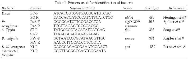
PCR for confirmation of enteric bacteria: For confirmation by PCR different sets of primers targeting specific genes were used9-13 (Table-I). The PCR conditions were as recommended by the respective authors.
PCR Ribotyping of Isolates: Two primers complementary to conserved regions of the 16s and 23s rRNA genes were synthesized on a Pharmacia KLB Gene Assembler Special. The sequences of P1 (5'-TTGTACACA CCGCCCGTCA-3') and P2 (5'-GGTACTTAGA TGTTTCAGTTC-3') have previously been described.14
PCR reaction mixture contained 1.5 mM MgCl2, 50µM of each dNTP, 300 pmol of each primer, 4U of recombinant Taq polymerase (Fermentas) and 0.1µg/µl of DNA template. The thermal cycler conditions were 30 cycles each of 94°C for 1 minute, 50°C for 1 minute, and 72°C for1.5 minute followed by 5 minutes at 72°C.
Detection of PCR Products: A quantity of 40µl of amplified product mixed with 15µl of Bromophenol blue (tracking dye) was fractionated electrophoretically on 2.5% agarose gel for four hours. The voltage was kept constant at 150V. After staining by ethidium bromide (0.5µg per ml), the gel was photographed by using Eagle Eye (Strategene, USA). PCR was performed in duplicate for each sample.RESULTS
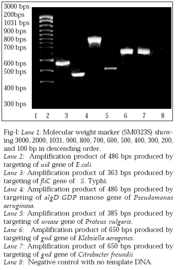
PCR for the confirmation of enteric bacteria: The results are shown in Fig 1. All isolates of E. coli were confirmed through PCR by targeting uid A gene (encoding glucuronidase).
A single amplification product of 486 bps was obtained. Salmonella Typhi isolates were confirmed by targeting fliC gene by specific primers that produce a 363 bps product. For confirmation of Proteus vulgaris strains, PCR was performed to amplify urease gene to get a 385 bps fragment. The algD GDP mannose gene is specific for Pseudomonsa aeruginosa. Its presence was ascertained by PCR targeting a 956 bps fragment. Both Citrobacter freundii and Klebsiella aerogenes have a specific gnd gene. Primers were used to amplify a 650 bps region.
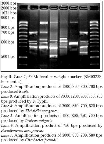
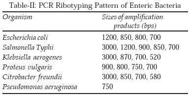
PCR Ribotyping: The results are shown in Fig-II and summarized in (Table-II). PCR- Ribotyping of Escherichia coli gave 4 amplification products of 1200, 850, 800, and 700 bps. As in E.coli, four amplification products were observed in Citrobacter freundii but the pattern was different. The bands were of 3000, 850, 700, and 580 bps.
A different pattern with 4 amplification products was seen in Proteus vulgaris as bands of 900, 800, 750 and 700 bps were observed. Klebisella aerogenes also produced 4 amplification products having a different pattern with bands of 3000, 870, 700 and 520 bps. More discrimination with 5 amplification products was observed with Salmonella Typhi. The sizes were 3000, 1200, 900, 850, and 700 bps. On the other side of spectrum with Pseudomonas aeroginosa only a single amplification product of 750 bps was observed.
DISCUSSION
During last few decades the strength of molecular biology has revolutionized biological sciences. The molecular techniques have opened a new chapter for characterization of different bacteria. These methods can discriminate to a better degree than phenotypic methods and improve our knowledge of genetic and epidemiological relationships.15
Restriction fragment length polymorphism (RFLP) has given promising results and even phage types can be subdivided,16 but this technique is expensive, laborious and time-consuming. PFGE is extremely discriminatory and can be successfully used in epidemiological investigation of S. Typhi and other enteric bacteria outbreaks17,18 but it has same disadvantages as RFLP. Sequencing of rRNA genes has been a very reliable method of characterizing bacterial species but it is technically demanding and expensive.
The most revolutionary technique has been PCR and it can be used effectively by targeting signature sequences of the target DNA. However, it is usually difficult to find very specific primers that have a single target and do not give non-specific amplifications. This problem can be overcome by use of PCR-ribotyping that is based on exploiting the polymorphism in the 16s-23s intergenic spacer regions. A pattern of multiple bands is produced which is very discriminatory and can be used for confirmation of bacteria not only up to species level but in many cases even beyond.5, 6
The present study was designed to establish a new more reliable PCR-ribotyping based identification scheme for common gram negative pathogens including five members of family Enterobacteriaceae and Pseudomonas aeruginosa that could provide rapid and reliable results.
Our results (Fig-II) showed that these bacteria provide discrete PCR-ribotyping patterns with number of bands varying from one in case of Pseudomonas aeruginosa to five in case of S. Typhi isolates. Escherichia coli gave five discrete amplification products ranging from 700 bps to 1200 bps. As in E.coli, four amplification products were observed in Citrobacter freundii but the pattern was different and the product size ranged from 580 bps to 3000 bps. A different pattern with four amplification products ranging from 700 t0 900 bps was seen in Proteus vulgaris. Klebisella aerogenes also produced four amplification products having a different pattern with bands of 520 to 3000 bps. More discrimination with five amplification products was observed with Salmonella Typhi. The product size ranged from 700 to 3000 bps. On the other side of spectrum with Psedumonas aeurogenosa only a single amplification product of 750 bps was observed. We used five isolates of each species and results were similar within a species.
The main advantage this technique has over conventional methods is that it can provide results in 24 hours whereas routine culture followed by biochemical tests need 36-48 hours. In addition, ambiguous results which can confuse the diagnosis such as variable level of H2S and gas production in TSI medium. Further analysis takes more time and becomes rather expensive.
Its advantage over conventional PCR is that instead of setting up reactions for each species or multiplexing them by use of six pairs of primers, a single primer is used. Because more discrimination is available so chances of false-negative results due to non-specific amplification are reduced considerably as well.
We conclude that this technique is superior to conventional methods and PCR in terms of better reliability and speed and can be used for diagnostic and epidemiological purposes.ACKNOWLEDGEMENTS
This work was carried out by the facilities and funding provided by National Institute for Biotechnology and Genetic Engineering (NIBGE), Faisalabad, Pakistan.
REFERENCES
1. Pegues DA, Shireley LA, Riddle CF, Anderson RL, Vess RV, Hill BC. Serratia marcescens surgical wound infection following breast reconstruction. Am J Med 1991;91(3B):173-8.
2. Jarvis WR, Martone WJ. Predominant pathogens in hospital infections. J Antimicrob Chemother 1992;29(Suppl A) 19-24.
3. Koneman EW, Allen SD, Janda WM, Schreckenberger PC, Winn Jr. WC. Diagnostic microbiology. 5thedition. Lipponcott Raven Publishers Philadelphia. N Y 1997; pp. 50-56.
4. Wong HC, Ho CY, Kuo LP, Wang TK, Lee CL, Shih YC. Ribotyping of the Vibrio paraheamolyticus isolates obtained from food poisoning outbreaks in Taiwan. Microbiol Immunol 1999;43 (7):631-6.
5. Bidet P, Barut F, Lalande V, Burghoffer B, Piet JC. Development of a new PCR-ribotyping method for Clostridium difficile based on ribosomal RNA gene sequencing. FEMS. Microbiol Lett 1999;175(2):261-6.
6. Sechi LA, Leori G, Lollai SA, Dupre I, Muloicotti P, Fadda G. Different strategies for the molecular characterization of the Mycobacterium bovis strains isolated in Sardinia Italy. Appl. Environ. Microbiol 1999;65(4):1781-5.
7. Lagatolla C, Dolzani L, Tomin E, Lavenia A, Michele M, Tommasini T. PCR-Ribotyping for characterization Salmonella isolates of different serotypes. J Clin Microbiol 1996;34(10):2440-3.
8. Edwards PR, Ewing WH. Identification of Enterobacteriaceae, 3rdedition. Burgess Publishing Co Minneapolis, USA 1972; p.7-20 and 146-258.
9. Heninger A, Binder M, Schmidt S, Unertl K, Botzenhart K, Do¨Ring G, et al. PCR and blood culture of Escherichia coli bacteremia in rats. Ant Microb Ag Chem 1999;37(8):2479-82.
10. Spilker T, Coenye T, Vandamme P, LiPuma JJ. PCR-based assay for differentiation of Pseudomonas aeruginosa from other Pseudomonas species recovered from cystic fibrosis patients. J Clin Microbiol 2004;42(5):2074-9.
11. Song JH, Cho H., Park MY, Na DS, Moon HB, Pai CH, et al. Detection of Salmonella typhi in the blood of patients with typhoid fever by polymerase chain reaction. J Clin Microbiol 1993;31:1439-43.
12. Kupfer DM, Chakurvedi AK, Canfield DV, Rove BA. PCR-based identification of postmortem microbial contaminants. J Forensic Sci 1999;44(3):592-3.
13. Brisse S, Jeanjean SI, Grimont PAD. PCR-based assay for differentiation of Pseudomonas aeruginosa from other Pseudomonas species recovered from cystic fibrosis patients. J Clin Microbiol 2004;42(5):2074-9.
14. Kostman JR, Edlind TD, Lipuma JJ, Stull TL. Molecular epidemiology of Pseudomonas cepacia determined polymerase chain reaction ribotyping. J Clin Microbiol 1992;30(8):2084-7.
15. Landeras E, Gonzalez-Hevia MA, Alzugaray R, Mendoza MC. Epidemiological differentiation of pathogenic strains of Salmonella enteritidis by ribotyping. J Clin Microbiol 1996;34(9):2294-6.
16. Ng I, Liu SL, Sanderson KE. Role of genomic rearrangements in producing new ribotypes of Salmonella typhi. J Bacteriol 1999;181(11):3536-41.
17. Wain J, Hein TT, Connerton P, Ali T, Parry CM, Chinh NTT, et al. Molecular typing of multiple-antibiotic resistant Salmonella enterica serovar typhi from Vietnam: application to acute and relapse cases of typhoid fever. J Clin Microbiol 1999;37(8):2466-72.
18. Mirza S, Kariuki S, Mamun KZ, Beeching NJ, Hart CA. Analysis of plasmid and chromosomal DNA of multidrug resistant Salmonella enterica serovar typhi from Asia J Clin Microbiol 2000;38(4):1449-52.
HOME | SEARCH | CURRENT ISSUE | PAST ISSUES
Professional
Medical Publications
Room No. 522, 5th Floor, Panorama Centre
Building No. 2, P.O. Box 8766, Saddar, Karachi - Pakistan.
Phones : 5688791, 5689285 Fax : 5689860
pjms@