|
|
||||
|
Published by : PROFESSIONAL MEDICAL PUBLICATIONS |
||||
|
ISSN 1681-715X |
||||
|
||||
|
- |
||||
|
ORIGINAL ARTICLE |
||||
|
- |
||||
|
Volume 24 |
April - June 2008 (Part-I) |
Number 2 |
||
|
|
||||
|
|
||||
|
|
||||
|
Published by : PROFESSIONAL MEDICAL PUBLICATIONS |
||||
|
ISSN 1681-715X |
||||
|
||||
|
- |
||||
|
ORIGINAL ARTICLE |
||||
|
- |
||||
|
Volume 24 |
April - June 2008 (Part-I) |
Number 2 |
||
|
|
||||
|
|
||||
Antibiotic sensitivity pattern of
staphylococcus aureus in Abakaliki, Nigeria
Ikeagwu IJ1, Amadi ES2, Iroha IR3
ABSTRACT
Objective: To investigate the sensitivity pattern of Staphylococcus aureus isolates obtained from clinical specimens including urine, wound, high vaginal swab and semen to commonly used antibiotics.
Methodology: The susceptibility patterns of these isolates were determined using the disc diffusion and agar well diffusion methods.
Results: Out of 174 samples, 51 (29.2%) yielded S. aureus with the highest isolation from semen (66.7%) and the least from urine (15.6%). Following the disc diffusion, the highest sensitivity was recorded for Ofloxacin (65%) while the least was for Co-trimoxazole (6%). Amoxicillin, Ampicillin, Tetracycline and Cloxacillin recorded 37%, 19%, 8% and 11% respectively. Also, following the agar well diffusion method, the highest sensitivity was recorded for Ofloxacin (65%) and the least for Co-trimoxazole (8%). The other drugs recorded the following readings; Amoxycillin (37%), Ampicillin (21%), Tetracycline (23%) and Cloxacillin (13%).
Conclusions: The study recommends the use of Ofloxacin in the treatment of S. aureus infections in the study area. It also underscores the need for sensitivity testing before the administration of antibiotics for the treatment of Staphylococcal infections.
KEY WORDS: Antibiotics, Abakaliki, Sensitivity, Resistance, Staphylococcus Aureus.
Pak J Med Sci April - June 2008 (Part-I) Vol. 24 No. 2 231-235
1. Ikeagwu IJ, PGD
Department of Medical Microbiology,
Federal Medical Centre,
Abakaliki, Ebonyi State,
Nigeria.
2. Amadi ES, Ph.D
3. Iroha IR, Ph.D
2-3: Department of Applied Microbiology,
Faculty of Applied and Natural Sciences,
Ebonyi State University,
PMB 053 Abakaliki, Ebonyi State, Nigeria.
Correspondence:
Dr. Amadi ES,
Department of Applied Microbiology,
Faculty of Applied and Natural Sciences,
Ebonyi State University,
PMB 053 Abakaliki, Ebonyi State, Nigeria.
E-mail: amadies2001@yahoo.com
* Received for Publication: October 6, 2007
* Accepted for Publication: February 8, 2008
INTRODUCTION
Staphylococcus aureus is a Gram positive, non-motile, catalase positive, coagulase positive, facultative anaerobe, involved in causing a number of diseases including, boils, pustules, impetigo, osteomyelitis, mastitis, septicemia, meningitis, pneumonia and toxic shock syndrome.
1,2 Nosocomial infections of which S. aureus is a typical example, are known to account for morbidity and mortality of millions of patients annually worldwide.3 S. aureus is considered the most resistant of all non-spore forming pathogens, with well developed capacities to withstand high salt (7.5-10%), extremes in pH and high temperatures (up to 60oC for 60 minutes). It also remains viable after months of air-drying and resists the effects of many disinfectants and antibiotics.2S. aureus is known to be notorious in their acquisition of resistance to new drugs and continues to defy attempts at medical control.
2 Many strains of S. aureus carry a wide variety of multi-drug resistant genes on plasmids. The resistance of S. aureus isolates from different parts of the world to commonly used antibiotics has been widely reported.4-8 This study aimed at determining the sensitivity of different isolates of S. aureus from Abakaliki, Ebonyi State in Nigeria to commonly used antibiotics.METHODOLOGY
Collection and Processing of Samples: Wound swabs and high vaginal swab (HVS) samples were collected from wound infection and pelvic inflammatory disease patients respectively, using sterile swab sticks. Mid stream urine and semen samples were collected from patients suspected to be suffering from urinary tract infection and infertility problems respectively. A total of 174 samples were collected from these patients attending the Federal Medical Center Abakaliki between April and June 2006.
The samples were cultured aerobically in blood agar and cystine lactose electrolyte deficient (CLED) agar. The plates were incubated at 37
oC overnight. Streak plate technique was used to obtain pure culture of each isolate prio to identification.Identification of Isolates:
The isolates were identified using motility test, colony morphology, Gram staining and Biochemical tests including catalase and coagulase tests as described by Cheesbrough.1Sensitivity testing using disc diffusion Technique: The discs were made from Whattman�s No. 1 filter papers and prepared as described by Isu and Onyeagba
9 to obtain the following concentrations of antibiotics per disc; Ofloxacin 10mg, Amoxycillin 30mg, Co-trimoxazole 25mg, Ampicillin 30mg, Cloxacillin 5mg and Tetracycline 25mg.Overnight cultures of each isolate were adjusted to McFarland turbidity standard (0.5), and the disc sensitivity screening conducted as described by Cheesbrough.
1 Sterile swabs were used to inoculate the test organism onto the sensitivity agar (Mueller Hinton). Sterile forceps were used to carefully distribute the antibiotic discs evenly on the inoculated plates. After allowing for about 30 minutes on the bench for proper diffusion, the plates were inverted and incubated aerobically at 35oC for 18 hours. The inhibition zone diameters were measured in millimeters using meter rule.Sensitivity testing using agar well diffusion Technique:
The antibiotics were prepared for administration into wells bored with sterile cork borer using sterile distilled water to obtain the following concentrations per drop (a drop equivalent to 0.01ml); Ofloxacin (10mg), Amoxycillin (30mg), Co-trimoxazole (25mg), Ampicillin (30mg), Cloxacillin (5mg) and Tetracycline (25mg).9Overnight cultures of each isolate were adjusted as in the case of disc diffusion and subjected to agar-well diffusion screening as described by Perez et al.
10 The sensitivity agar were inoculated as described for the disc diffusion method and a drop, each, of the prepared antibiotic concentrations were introduced into respective wells. They were left on the bench for 30 minutes and incubated aerobically as stated for the disc diffusion method. The inhibition zones were also recorded as previously stated.RESULTS
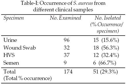
Isolation of Staphylococcus aureus from different clinical specimens: Of the 174 clinical samples examined, 51 (29.2%) were positive for Staphylococcus aureus. The highest isolation of S. aureus was from semen (66.7%) while the least was from urine (15.6%). (Table-I).
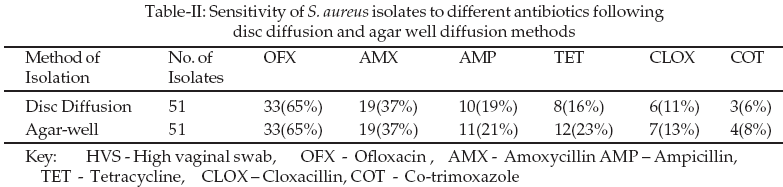
The highest sensitivity was recorded for Ofloxacin (65%) while the least was for Co-trimoxazole (6%). The inhibition zone diameters obtained ranged from 0 to 35mm (Table-III).

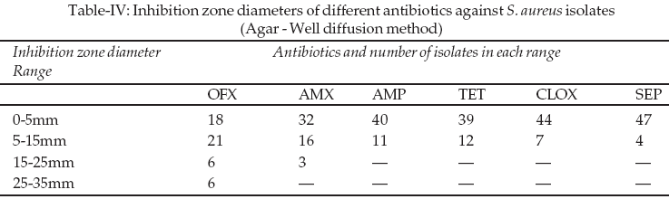
Sensitivity pattern of S. aureus isolates using the agar well diffusion Technique: The degree of sensitivity of S. aureus isolates was also analysed as in the case of disc diffusion. The highest sensitivity was recorded for Ofloxacin (65%) and the lowest for Co-trimoxazole (8%). (Table-II) The inhibition zone diameters obtained also ranged from 0 to 35mm (Table-IV).
DISCUSSION
In this study, out of the 174 samples analysed, 51 (29.2%) were positive for Staphylococcus aureus with 66.7% occurrence in semen, seconded by wound specimen (56.3%) and the least from urine samples (15.6%). This is in line with previous reports in which S. aureus was the most common organism isolated from semen
11,12 and wound samples.13 This picture was however different in Jos, Nigeria, where Oguachuba14 found Proteus species to be the most common isolate with occurrence rate of 41.9%, followed by S. aureus with 25.6%. The reason for this variation is not yet ascertained.The result of this study indicated that Ofloxacin had the highest sensitivity (65%) to the S. aureus isolates following both the disc diffusion and agar well diffusion techniques. This apparently high level of sensitivity to Ofloxacin appears to suggest that Ofloxacin could be a drug of choice for treating infections caused by S. aureus in the study area, especially at the present time, when S. aureus strains resistant to other commonly used antibiotics has been reported.
15 This finding is consistent with previous reports. For instance, 100% sensitivity of S. aureus isolates to Ofloxacin has been reported.16 In addition, high sensitivity of Ofloxacin against S. aureus isolates was also highlighted by Chalita et al.17 Low sensitivity however was reported among most strains of methicillin resistant Staphylococcus aureus (MRSA) isolated from patients with ocular infections.18Although low sensitivities of S. aureus isolates to ampicillin (37%) and cloxacillin (13%) were recorded in this study, enhanced susceptibility had been reported by previous workers. Uwaezuoke and Aririatu
8 reported 85.4% sensitivity of cloxacillin to S. aureus strains isolated from Owerri, Nigeria. Similarly, Farzana et al.,7 recorded 74% sensitivity of S. aureus isolates to ampicillin in Mullan city, Pakistan. The reason for the variation could most likely be attributable to strain differentiation. On the other hand, a number of prio investigations had reported high level of resistance of S. aureus to cloxacillin and ampicillin in line with the result of this study.6,19,20The 63% resistance of amoxycillin to S. aureus isolates as recorded in this work is in conformity with the findings of Astal et al.,
5 in which 73.6% amoxycillin resistant strains of S. aureus was reported. 100% resistance of S. aureus isolates to amoxycillin has also been reported.4Low sensitivities of S. aureus to tetracycline and co-trimoxazole as observed in this study is consistent with earlier studies. 98% and 69.6% resistance of S aureus isolates to tetracycline were reported by Obi et al.,
16 and Olayinka et al.,21 respectively. Also 87.5% and 68% resistance of S. aureus isolates to tetracyclines were also respectively reported by Uwaezuoke and Aririatu8 and Oyagade and Oguntoyinbo.19 Further, prio researchers including Farzana et al.,7 and Astal et al.,5 reported 81.8% and 66.1% resistance of S. aureus isolates to co-trimoxazole respectively. It is therefore less likely that tetracycline and co-trimoxazole will be desirable, as drugs of choice for the management of S. aureus infections in Abakaliki, area in Nigeria.There was a slight variation in the zone of inhibition obtained from disc diffusion and agar well diffusion methods respectively (Tables-III, IV). This was apparent with ampicillin, tetracycline, cloxacillin and co-trimoxazole. The reason for the apparently enhanced inhibition zone diameters recorded for agar well diffusion could be attributed to the possibility of losing some fractions of the antibiotics on the paper disc or its inability to express all absorbed drugs to the agar media. Nevertheless, the slight difference probably suggests that both techniques are appropriate for the determination of sensitivity of antibiotics to microbial agents.
This study therefore underscores the need for antibiotic sensitivity screening before the administration of any antibiotic for treatment of Staphylococcal infections in Abakaliki, Ebonyi State, Nigeria.
REFERENCES
1. Cheesbrough M. District Laboratory Practice in Tropical Countris. Part 2. Cambridge University press 2002;135-62.
2. Talaro KP, Talaro A. Foundations in Microbiology. 4th. Ed. McGraw Hill, New York 2002;544-52.
3. Mansouri S, Khaleghi M. Antibacterial resistance pattern and frequency of Methicillin resistant Staphylococcus aureus. Irn J Med Sci 1997;22:93.
4. Adewoye SO, Lateef A. Assessment of the Microbiological quality of Clarias gariepinus exposed to an Industrial effluent in Nigeria. Environmentalist 2005;24(4):249-54.
5. Astal Z. El-manama A, Sharif FA. Antibiotic resistance of bacteria associated with community acquired urinary tract infection in the southern area of Gaza Strip J Chemother 2002;14(3):259-64.
6. Chigbu CO, Ezeronye OU. Antibiotic Staphylococcus in Abia State, Nigeria. Afr J Biotech 2003;2(10):374-8.
7. Farzana K, Nisar S, Shah H, Jabeen F. Antibiotic resistance pattern against various isolates of Staphylococcus aureus from raw milk samples. J Research Science 2004;15(2):145-51.
8. Uwaezuoke JC, Aririatu LE. A survey of Antibiotic resistant Staphylococcus aureus strains from clinical sources in Owerri. J Appl Sci Environ Managt 2004;8(1):67-8.
9. Isu RN, Onyeagba RA. Basic principles in Microbiology 2nd Ed. Fasman Communication, Okigwe 2002;134-43.
10. Perez C, Pauli M, Bazerque P. An antimicrobial assay by the agar well diffusion method. Acta Biologae et Medicine Experimentalis 1990;15:113-15.
11. Jennings MG, McCowan MP, Baker HW. Is conventional Bacteriology useful in the management of male infertility? Clin Rep Fert 1986;4(6):359-66.
12. Umezurike E, Nwuzo AC, Onyeagba RA. Occurrence of Staphylococcus aureus in semen of men with fertility problems in Abakaliki, Ebonyi State, Nigeria. J Sci Engr Tech 2006;13(2):6809-15.
13. Onche II. Post operative wound infection in inplant surgery. Dissertation submitted to the National postgraduate medical college of Nigeria, Lagos.2000.
14. Oguachuba H. Wound infection in Orthopedic Traumatology Department of Jos University Hospital, Jos Nig Med J 1986;7:147-51.
15. Amadi ES, Nwofor GE, OgbuO, Ayogu TE, Ononiwu CE. Resistance of Staphylococcus aureus to commonly used antibiotic obtained from Different sources in Abakaliki. Afr J Sc 2007;8(1):1728-39.
16. Obi CL, Iyiegbuniwe AE, Olukoya DK, Babalola C, Igunbor EO, Okonta AA. Antibiogram and plasmids of Staphylococcus aureus and coagulase negative staphylococci isolated from different clinical sources. Cent Afr J Med 1996;42(9):258-61.
17. Chalita MK, Hofling-Lima, AL, Paranhos A, Schor P, Belfort R. Shifting trends in vitro antibiotic susceptibilities for common ocular isolates during a period of 15 years. Am J Ophthalmol 2004;137(1):43-51.
18. Ooishi M, Mayao M. Antibiotic sensitivity of recent clinical isolates from patients with ocular infections. Ophthalmologia 1997;21(suppl 1):15-24.
19. Oyagade JO, Oguntoyinbo FA. Incidence of antibiotic resistant Staphylococcus aureus strains among isolates from environmental and clinical Sources. Nig J Microbiol 1997;11:20-4.
20. Meremikwu MM, Nwachukwu EC, Asuquo AE, Okebe JU, Utsalo SJ. Bacterial isolates from blood cultures of children with suspected septicaemia in Calabar, Nigeria. BMC Infect Dis 2005;5:110.
21. Olayinka BO, Olayinka AT, Onadapo JA, Olurinola PF. Pattern of resistance to vancomycin and other antimicrobial agents in staphylococcal isolates in a University Teaching Hospital. Afr J Clin Expt Microbiol 2005;6(1):21-7.
HOME | SEARCH | CURRENT ISSUE | PAST ISSUES
Professional
Medical Publications
Room No. 522, 5th Floor, Panorama Centre
Building No. 2, P.O. Box 8766, Saddar, Karachi - Pakistan.
Phones : 5688791, 5689285 Fax : 5689860
pjms@pjms.com.pk