|
 |
|
Published
by : PROFESSIONAL MEDICAL PUBLICATIONS |
|
ISSN 1681-715X |
|
|
|
|
|
- |
|
ORIGINAL
ARTICLE |
|
- |
|
Volume 25 |
April
- June 2009 (Part-I) |
Number 2 |
|
|
|
Pattern of upper Gastaro Intestinal
malignancies in Northern Punjab
Durrani AA1, Nayyar Yaqoob2, Shahid
Abbasi3,
Masood Siddiq4, Shaheen Moin5
ABSTRACT
Objective: To record the number of cases of
carcinoma of stomach, geographical location and histological diagnosis
presenting to a hospital in northern Punjab
Methodology: This study was conducted at the
department of medicine (GI unit) Fauji Foundation Hospital Rawalpindi. All the
patients who underwent upper GI endoscopy and were found to have an upper GI
malignancy on histopathology were reviewed for part of the gut involved.
Patient demographics including age, sex, and place of residence, clinical
presentation, and subsequent histologic diagnosis were recorded.
Results: During the study period, 302 cases of
upper GI malignancy were seen at our institution, 83 (14.8%) were in patients
40 years of age or younger. Mean patient age was 58 years. The lesion was
distal in 43%, proximal in 20% and 7% involved the entire stomach. In 9.8% of
the cases, lesser curve was the site. The tumor was located in the upper
third, middle third and lower third in 33, 22 and 34.6% respectively. The
common presenting symptom in case of carcinoma of stomach was pain epigastrium.
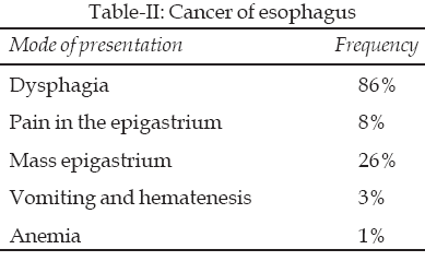
Dysphagia was the major subjective complaint in cases of esophageal carcinoma.
Conclusion: The common malignant tumor in males was
carcinoma of stomach and in females carcinoma of esophagus. Endoscopic
screening in subjects suspected of upper gastrointestinal malignancy results
in a significant yield of carcinoma. The gastric tumor in distal location (non
cardia cancer) is still more common. Asian race is no different from other
races as far as the ca stomach is concerned.
KEYWORDS: Gastric cancer, Cardia cancer, Upper
gastrointestinal cancer, Gastric adenocarcinoma, Esophageal adenocarcinoma,
Endoscopy.
Pak J Med Sci April - June 2009
Vol. 25 No. 2 302-307
How to cite this article:
Durrani AA, Yaqoob N, Abbasi S, Siddiq M, Moin S. Pattern of upper Gastaro
Intestinal malignancies in Northern Punjab. Pak J Med Sci 2009;25(2):302-307.
1. Durrani AA
2. Nayyar Yaqoob
3. Shahid Abbasi
4. Masood Siddiq
5. Shaheen Moin
Correspondence
Nayyar Yaqoob,
Fauji Foundation Hospital,
Rawalpindi - Pakistan.
E-mail: drnayyaryaqoob@gmail.com
* Received for Publication: October 7, 2008
* Revision Received: February 4, 2009
* 2nd Revision Received: February 11, 2009
* Final Revision Accepted: February 13, 2009
INTRODUCTION
Gastric and gastro-esophageal adenocarcinomas are a major
health burden globally. The incidence of upper gastrointestinal malignancies
varies widely based on geographic location, race, and socioeconomic class.
Gastric cancer remains the world�s third most common
malignancy.
1
A 2005 analysis of the global incidence and cancer related mortality revealed
that 934,000 cases of gastric cancer occurred in 2002 and approximately
700,000 patients died annually from this problem.2
In 1996, about 22,800 new cases of stomach cancer were registered in the
United States alone, and 14,000 succumbed to it. In industrialized countries,
mortality from gastric cancer has declined steadily over the years whereas
gastric cancer remains a leading cause of death from cancer in the developing
world.
Geographical residence and dietary habits may play a part
in the pathogenesis of gastric cancer. Social class and socioeconomic
conditions of the inhabitants confer a significantly increased risk of
developing gastric cancer. There is considerable intra-regional variation
within high-risk areas. Migration from high to low risk areas and adopting
dietary habits of the host country modifies the risk in second or third
generations. Screening gastric cancer has been advocated in geographic regions
where the prevalence of this disease is high. This study deals with upper GI
cancers in the predominantly rural socioeconomically disadvantaged population
of northern region of Pakistan.
METHODOLOGY
Fauji Foundation Hospital is a district level hospital
serving a well-defined segment of population (ex-servicemen and their
dependents). The population was mostly derived from rural areas. We analyzed
the data of these patients retrospectively. Cases of gastric cancer presenting
for the first time during the fourteen-year period 1992-20006 were recorded.
An open access endoscopy service has been available in our hospital for more
than 14 years. Mode of presentation, duration of symptoms, previous history
for dyspepsia and use of anti peptic ulcer drugs were documented. Alarming
symptoms and signs suggestive of underlying malignancy like unexplained recent
weight loss, dysphasia, haematemesis or melaena, anaemia, previous gastric
surgery, a palpable mass were investigated with upper GI endoscopy followed by
biopsy.
Demographic data including age, sex, and place of residence
were recorded. Dietary habits were inquired from the subjects. Exact tumor
site, sub site, and axial views were determined. The site of the tumor within
the stomach was classified as lesser curvature, greater curvature, anterior
wall, or posterior wall. Location was classified as distal type, cardia
cancer, cancer located in the corpus. In addition, the endoscopic and the
histological appearance of the tumor were analyzed. Cancers were classified by
the predominant histological type, as follows: Squamous carcinoma,
adenocarcinoma, scirrhous type, lymphoma, and further categorized into
differentiated type, moderately differentiated, undifferentiated type or
poorly differentiated carcinomas. Upper GI endoscopy was performed using video
endoscopes (Olympus TJ 140 and JF 20).
RESULTS
Of the 375 patients, 159 males and 143 females, who visited
the hospital GI clinic from March 1992 to Dec. 2006, with persistent upper
gastrointestinal symptoms underwent upper gastrointestinal endoscopy to
confirm the clinical suspicion of upper GI malignancies. The mean age of the
patients was 58 years and 5% of the patients were younger than 40 years. The
age range was 14 -85.
As regards geographical location of patients, one hundred
forty (46%) came from Rawalpindi, one hundred ten (36%) came from Kashmir and
fifty two (17%) were residents of Chakwal district.
Upper gastrointestinal cancer was reported histologically
in 302 patients. The most frequent site was the gastric antrum 92 (30.3 %)
followed by central esophagus 87 (28.7%), corpus 38 (12.5%), gastric cardia 45
(14.9%) distal oesophagus 18 (5.9%), gastro-oesophageal junction 14 (4.6%),
and proximal oesophagus 9 (2.9%). From axial views of the cardia, 1% and 2.2%
of tumours were found to originate from the lesser and greater curve,
respectively.
Out of a total of 128 histologically proved cases of
esophageal carcinoma, the histologic pattern was as follows: Squamous cell
carcinoma was noted in 69% adenocarcinoma 28% and esophageal lymphoma 3%.
There were 30 cases younger than 50 years. Ninety- eight cases were between 50
and 70 years. Out of a total of 175 histologically proved cases of gastric
carcinoma, the histologic pattern identified was as follows: Gastric
adenocarcinoma was reported in 85% gastric lymphoma 13%, gastric
leiomyosarcoma in 2%.
One hundred and twenty three cases were of intestinal type
(one hundred and two moderately differentiated; twenty one poorly
differentiated). Sixty-two cases were poorly differentiated diffuse type
carcinomas. There was an average of 6-month delay between the initial symptoms
and the diagnosis. The majority of upper GI tumours were advanced at the time
of presentation.
A total of 12 patients aged less than 30 had upper GI
malignancies. Out of which three patients had esophageal carcinoma, four had
gastric carcinoma and five were diagnosed to have lymphoma. Mode of
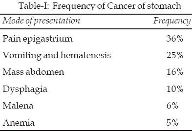
presentation is shown in Table-I. Nine patients had prior gastric surgery.
There were very few gastric ulcers. Most of the cases of carcinoma of stomach
had fungating, cauliflower or polypoidal growth like macroscopic appearance.
One hundred and forty-five of 302 cases of Upper GI cancer
detected during the fourteen-year period were aged less than 55. In two
patients, there was a delay in diagnosis of more than six months after first
presenting to a health care set up. Both these patients had significant
symptoms at presentation. Ninety-eight males and seventy females had carcinoma
of stomach and 72 females and 61 males had carcinoma of esophagus. The volume
of upper GI endoscopies was fairly adequate.
DISCUSSION
Oesophageal and gastric malignancies are considered as
separate disease entities. It is now generally recognized that cancer of the
oesophagus and stomach in 75% of cases is an adenocarcinoma located within 5cm
of the gastro-oesophageal junction rather than either purely Oesophageal or
purely gastric. The incidence of adenocarcinoma of the esophagus is rising
rapidly in Western Europe and North America.
3
It can be an aggressive disease and disseminates early.
Carcinoma of the stomach is an important cause of mortality
from cancer. Gastric cancer is the second most deadly malignant neoplasm all
over the world. Approximately 876,000 persons are diagnosed with this disease
every year and approximately 649,000 succumb to it.
In the UK alone, gastric cancer is the fourth most common
tumor that accounts for nearly 10000 deaths each year. Until 1985, gastric
cancer was the most common cancer globally with an annual incidence of nearly
700,000 cases. This has fallen over the recent years. Carcinoma of the stomach
is also common in the southern region of India.
4
A very high prevalence of gastric carcinoma was observed in Mizoram in one of
the studies from India.5
The mean age in our patients was 58
years and 14% of the patients were younger than 40 years.
Men are twice as likely to get stomach cancer as women.
This is borne out by our study as 67% of our patients with carcinoma stomach
were males and 15% were younger than 40 years. The most vulnerable group is
considered to be men over 50 years. The incidence increases incrementally
after age 40 and peaks in the seventh decade.
Gastric cancer can occur in any part of the stomach. The
location of the primary tumor has a bearing on the prognosis. Approximately 37
percent of gastric carcinomas in the United States originate in the upper
third of the stomach, whereas 20 percent originate in the middle third, and 30
percent in the lower third; 12 percent of gastric carcinomas involve the
entire stomach. The distal tumors have a five- year survival rate of
approximately 20 to 25 percent after resection. It is 10 percent for patients
with proximal tumors, and less than 5 percent for those whose entire stomach
is involved. The reduced survival of patients with proximal tumors is
reflective of a more aggressive and diffuse histologic disease.
Reviewing the site-specific distribution, the most
frequently reported site in the present study was the gastric antrum 30.3%
followed by the central esophagus. In another study, intestinal type of
adenocarcinoma was the commonest (55.9%) and the distal third was the most
common localization (88.4%).
6
Cancers of oesophagus and stomach
are also quite common in Indian administered Kashmir. A significant increase
in the proportion of patients with cardia carcinoma was also noted in the
period 1984 to 1993, rising from 14 percent between 1984 -1988 to 24 percent
between 1988 -1993 in Indian administered Kashmir as reported in an earlier
study from Kashmir region.7
The figure in our study was 14.9%.
The incidence of gastric cancer is highest in China, South
America and Eastern Europe. Japan has a high incidence of gastric cancer and
90% are located distal to the cardia. The gastric carcinoma is the most
frequent cancer being responsible for 20 to 30 percent of all cancers. The
occurrence of carcinoma of the stomach had a percentage rate of 2.7 young
adult Japanese patients with a male to female ratio of 1.0:0.88 in another
study.
In industrialized countries, the incidence of gastric
cancer has declined progressively since past many decades. In 1930 gastric
carcinoma was the foremost cause of cancer-related deaths among American males
and the third most common cause among women. Over the next fifty years, the
rate of gastric carcinoma in the United States dropped from 33 to 10 cases per
100,000 men and from 30 to 5 per 100,000 women in the United States. African
Americans, Hispanic Americans, and Native Americans are 1.5 to 2.5 times more
likely to have gastric carcinoma compared to white population.
The incidence of cancer at different anatomical sub sites
of the stomach in the Western countries in particular United States is
undergoing a marked change. A consistent upsurge has been witnessed in the
incidence of adenocarcinoma of the most proximal gastric cardia region and the
adjacent gastro-oesophageal junction, whereas the incidence of more distal
stomach cancer has remained largely unchanged or has gone down marginally over
the last 50 years.
8�9
Similar incidence trends in proximal gastric cancers have
been reported from Europe.
10
These findings suggest a common
pathogenesis for cancers in this location. This is
likely to be different from that of distal gastric tumours. In our study the
proximal gastric cancer comprised 20% of cases.
Northwestern region of Iran reportedly has a very high
incidence of upper gastrointestinal cancer especially cancer of cardia region.
The vast majority stem from the right side of the cardia.
11
In another study from Indian administered Kashmir, the
inhabitants of southern region had higher incidence of esophageal and gastric
cancer than northern Kashmir.
12
This study also reported a very high incidence of gastric cancer, which was
comparable to the current study. Unusual dietary and personal habits were
incriminated in its causation. These statistics were higher than most other
regions across India. The epidemiological features of esophageal cancer in
Kashmir were similar to that found in the Asian esophageal cancer belt.
Moreover, this study also reported different incidence rates for different
racial groups.
The effects of socio-economic/occupational factors on
gastric cancer at various sub sites are not well established and were
investigated in a study on the economically active Swedish population.
13
Manual workers and farmers had an increased risk of stomach cancer. Most of
our patients indulged in farming activities. The dietary habits of our
patients did not differ much from the general population. Somehow they may get
exposed to carcinogens in the fields. It could perhaps be an unrecognized
radioactive substance of some sort. This needs to be investigated. Otherwise
poor socioeconomic class can be considered a risk factor.
Carcinoma of esophagus is a distressing problem with a
dismal outcome. It is quite common in this part of the world. In an earlier
study from Pakistan, it constituted 25% of the total cardiothoracic operative
case load.
14
In another study from Balochistan region of Pakistan, carcinoma of esophagus
was the third most common tumor and comprised 11.3% of the total registered
cases.15
Esophageal malignancies are commonly reported in the sixth, seventh and eighth
decades of life and are relatively rare at a younger age because they require
a long period of carcinogenesis. This may account for its rarity in child
hood. One patient in the present study who was 18 years old presented with
dyspahagia and the subsequent histologic evaluation showed adenocarcinoma of
esophagus.
The prevalence of carcinoma of esophagus is unprecedentedly
high in the countries bordering esophageal cancer belt. But it varies
substantially depending upon the geographic location. High incidence areas
have been identified in the Caspian Littoral region of northern Iran, southern
republics of the former Soviet Union and northern China. The incidence
surpasses 100 per 100,000 individuals. Over 20% of all cause mortality has
been attributed to esophageal cancer. Northern part of Pakistan closely
borders these regions. The incidence of esophageal cancer ranges from 10 to 50
in Sri Lanka, India, South Africa, France and Switzerland.
The significance of tumor cell type on survival after
esophageal resection for carcinoma is not well documented. In one study,
squamous cell type gave a significant survival advantage in the longer term.
16
In the aforesaid study, squamous cell carcinoma made up 90% of cases while
adenocarcinoma only 7% of cases. Out of 832 diagnosed cases, 60% involved the
lower third of esophagus. Squamous cell carcinoma was noted in 81% of the
cases while adenocarcinoma was the second most common in another preceding
study from Aga Khan University Hospital, Pakistan.17
Over 75% of esophageal carcinomas among Asians/Pacific
Islanders were squamous cell carcinoma in an earlier study.
18
Adenocarcinoma made up less than 20%. The rate of esophageal squamous cell
carcinoma was 81% higher among Asian/Pacific Islander males than white males,
but it was 64% less than black males. The incidence of esophageal
adenocarcinoma was significantly lower among Asians/Pacific Islanders than
among both white and black males and females.
The majority of gastric carcinomas were noncardia
adenocarcinoma.
18
The rate of cardia adenocarcinoma was 23% lower among API males compared with
white males, but it was 26% higher compared with black males. In contrast, the
rate of noncardia adenocarcinoma among Asians/Pacific Islander were
approximately 3.7 times the rate among whites for both males and females and
33% higher than the rate for blacks. The race probably has an important
bearing on the incidence of Ca stomach. Asian race is perhaps equally prone to
Ca stomach like their counterparts in the west irrespective of their dietary
habits.
CONCLUSION
Targeted screening strategies using endoscopic examination
can detect gastric cancer at an early stage and can produce good therapeutic
outcome. In the absence of screening, patients present with advanced disease
and prognosis is unfavorable. Screening of stomach cancer in moderate to
high-risk population subgroups might prove cost-effective. Targeted screening
strategies for stomach cancer should be explored.
In the current study, cancer located in the distal part of
the stomach formed the major component of gastric cancer (43%) but the
incidence of adenocarcinoma of cardia region and adjacent gastro esophageal
junction is catching up despite varying dietary habits. The mean age for
gastric cancer was 52 in males and 49 in females. Open access endoscopy can
enhance the detection of disease in earlier stage. Most of the patients came
from Rawalpindi district. Gastric cancer without alarming features is
relatively rare below the age of 55.
REFERENCES
1. Whelan SL, Parkin DM, Masuyer E, eds. Trends in cancer
incidence and mortality. Lyon, France: IARC Scientific Publications, 1993. (IARC
scientific publications no. 102.)
2. Parkin DM, Bray FI, Devesa SS. Cancer burden in the year
2000. The global picture. Eur J Cancer 2001;37:S4-66.
3. Lagarde SM, ten Kate FJ, Reitsma JB, Busch OR, van
Lanschot JJ. Prognostic factors in adenocarcinoma of the esophagus or
gastroesophageal junction. J Clin Oncol 2006;24:4347-55.
4. Sambasivaiah K, Ibrarullah M, Reddy MK, Reddy PV,
Wagholikar JS. Clinical profile of carcinoma stomach at a tertiary care
hospital in south India. Trop Gastroenterol 2004;25:21-6.
5. Phukan RK, Narain, Zomawia E, Hazarika NC, Mahanta J.
Dietary habits and stomach cancer in Mizoram, India J Gastroenterol
2006;41:418-24.
6. Eskander H, Shoshtari S, Hossein M, Rahim M, Jalaj H,
Mehrdad A, et al. Clinical profile of gastric cancer in Khuzestan, Southwest
of Iran World J Gastroenterol 2006;12:4832-5.
7. Rumana M, Khan AR, Khurshid N, Seema Ali, Besina S, Lone
NA. The changing pattern of oeso-phagogastric cancer in Kashmir. JK-Practitioner
2005;12(4):189-90.
8. Crew KD, Neugut AI. Epidemiology of upper
gastrointestinal malignancies. Semin Oncol 2004;31:450-64.
9. Golematis B, Tzardis P, Hatzikostas P,
Papadimitriou K, Haritpoulos N. Changing pattern of distribution of
carcinoma of the stomach. Br J Surg 1990;77:63-4.
10. Blot WJ, Devesa SS, Kneller RW, Fraumeni JF Jr.
Rising incidence of adenocarcinoma of the esophagus and gastric cardia. JAMA
1991;265:1287-9.
11. Derakhshan MH, Yazdanbod A, Sadjadi AR, Shokoohi B,
McColl KEL, Malekzadeh R. High incidence of adenocarcinoma arising from the
right side of the gastric cardia in NW Iran. Gut 2004;53:1262-6.
12. Khuroo MS, Zargar SA, Mahajan R, Badday MA. High
incidence of esophageal and gastric cancer in Kashmir in a poulation with
special personal and dietary habits. Gut 1992;33:11-5.
13. Ji, Hemminki JK. Socio-economic and occupational risk
factors for gastric cancer: a cohort study in Sweden. Eur J Cancer Prev
2006;15:391-7.
14. Ziadddin AU, Saeedi I, Mehmood K. Geographical location
and histological presentation of gastric carcinoma in NWFP. J Postgraduate
Medical Inst 2003;17:111-5.
15. Roohullah, Ma K, Shah MA, Khan Z, SW H, Burdy GM, et
al. An alarming occurrence of esophageal cancer in Balochistan. Pakistan J Med
Res 2005;44:101-4.
16. Alexiou C, Khan OA, Black E, Field ML, Onveaka P, Beggs
L, et al. Survival after esophageal resection for carcinoma: the importance of
the histologic cell type Ann Thorac Surg 2006;82:1073-7.
17. Alidina A, Gaffar A, Hussain F, Islam M, Vaziri I,
Burney I, et al. Survival data and prognostic factors seen in Pakistani
patients with esophageal cancer. Annals of Oncology 2004;15:118-22.
18. Wu X, Chen VW, Ruiz B, Andrews P, Su LJ, Correa P. Incidence of
esophageal and gastric carcinomas among American Asians/Pacific Islanders,
whites, and blacks: Sub site and histology differences. Cancer
2006;106:683-92.
HOME
| SEARCH
| CURRENT
ISSUE | PAST
ISSUES
Professional
Medical Publications
Room No. 522, 5th Floor, Panorama Centre
Building No. 2, P.O. Box 8766, Saddar, Karachi - Pakistan.
Phones : 5688791, 5689285 Fax : 5689860
pjms@