|
 |
|
Published
by : PROFESSIONAL MEDICAL PUBLICATIONS |
|
ISSN 1681-715X |
|
|
|
|
|
- |
|
ORIGINAL
ARTICLE |
|
- |
|
Volume 25 |
April
- June 2009 (Part-I) |
Number 2 |
|
|
|
Properties and antimicrobial activity of
Apis Dorsata honey From Thailand
Chanpen Chanchao
ABSTRACT
Objectives: For Apis dorsata honey,
the basic properties, namely the pH, and the total proline, protein and invert
sugar contents, were determined. For proteins, the mass weight and partial
amino acid sequence of the three major proteins were assayed and homologs
screened for. A bioassay for antimicrobial activity was also performed.
Methodology: Proline content and the percentage of
invert sugar were evaluated, whilst the total protein content was assayed and
the major protein components were analyzed by reducing SDS-PAGE. From the
excised bands partial amino acid sequences deduced by MALDI-TOF MS and
homologs were searched for by Mascot database. Antimicrobial activity was
assayed by the agar well diffusion method.
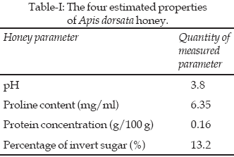
Results: The proline, invert sugar and total
protein contents were 6.35µg/ml, 13.2% (w/w) and 0.16% (w/w), respectively,
with three major protein bands of 50, 75 and 100 kDa. All tested (25-100%
(v/v)) dilutions of honey could inhibit the growth of S. aureus.
Conclusion: A. dorsata honey may contain an
epinecidin homolog, an antimicrobial peptide, and can inhibit the growth of
some bacteria. It suggests that honey could be used as an antimicrobial agent.
KEYWORDS: Apis dorsata, Honey, Proline, Invert
sugar, Epinecidin, Inhibition zone.
Pak J Med Sci April - June 2009
Vol. 25 No. 2 313-318
How to cite this article:
Chanchao C. Properties and antimicrobial activity of Apis Dorsata
honey From Thailand. Pak J Med Sci 2009;25(2):313-318.
1. Chanpen Chanchao,
Dept. of Biology, Faculty of Science,
Chulalongkorn University,
Bangkok 10330, Thailand.
Correspondence
Chanpen Chanchao,
E-mail: chanpen@sc.chula.ac.th
* Received for Publication: December 11, 2008
* Accepted: February 27, 2009
INTRODUCTION
Honey is modified from the nectar of plants by the function
of – glucosidase by bees as a storage food source, but is widely consumed by
humans (and other animals) as food. Honey is typically composed of fructose
and glucose (79% (w/v)), H
2O
(20% (v/v)), and minor acids such as gluconic acid (0.5% (w/v)), and minerals,
such as calcium, magnesium, potassium and phosphorus, together with vitamins,
such as riboflavin and niacin (0.5% (w/v)).1
The main components of honey are the two monosaccharides, fructose and
glucose; this allows it to be highly saturated and of a high osmotic potential
providing at least some of the antimicrobial properties in addition to the
sweet taste sought after by other animals, including bears and humans. In
contrast, gluconic acid provides the sour taste. The properties of different
honey batches will depend upon the nectar (and pollen) sources and, therefore,
the plants foraged. For example, different honey sources can differ in terms
of their taste, smell and color (from light yellow to darkish brown).2
Not only is honey nutritional, as a rich source of monosaccharides, trace
minerals and vitamins, but it also inhibits the growth of microbial pathogens.
It was recorded that Egyptians used honey in ancient medicine to prevent
localized bacterial infections by topical application in surgery and other
wounds.3
The antimicrobial activity of honey may principally be due to its low moisture
content and high osmotic pressure, which can desorb water out of microbe cells
(hypotonic condition) leading to either the death or the growth inhibition of
bacteria, fungi and yeasts.4-5
Nowadays, although antibiotics are widely used in the prevention of localized
and systemic microbial infections, there is still the increasing problem of
antibiotic resistance. Using honey for the growth inhibition of microorganisms
might be an alternative way in some suitable cases for topical application
and, perhaps, for some partially systemic infections following oral
administration.
Not only does the property of honey depend on the plant
nectar, but it also depends on the species of honeybee. This probably in the
main reflects the different flower nectar and pollen collecting preferences of
different bee species, as well as to some lesser extent the different
biochemical and microbiological components. In Thailand, as with most of
Southeast Asia, the species diversity of honeybees is high and consists of
four native Apis species, Apis dorsata, A. cerana, A. florea
and A. andreniformis, and one imported Apis species, A.
mellifera. In this research, A. dorsata was selected for study
since honey from this wild species is the most popular in local consumption
and sales. For the purposes of the present study, the properties of honey, in
terms of pH, proline and total protein and invert sugar contents, were
determined. The main proteins in honey were evaluated by reducing SDS-PAGE
resolution and the partial amino acid sequences of three of the main protein
bands were obtained. Furthermore, antimicrobial activity from various
dilutions of honey was observed on selected bacterial pathogens, yeasts and
fungi.
METHODOLOGY
Sample collection:
Honey of Apis dorsata was purchased from a bee farmer in the Samut
Songkram province.
Proline content: The proline content was determined as
described previously
6
with reference to a proline standard
curve constructed from 100, 200, 300, 400 and 500µg/ml. Honey was diluted in
water to 2% (v/v) and then 250 ml was mixed with formic acid (130 ml) and 3%
(w/v) Ninhydrin solution (500 ml). The mixture was boiled for 15 min, allowed
to cool, and mixed with isopropanol (2.5 ml). The absorbance was measured at
an incident light of 520 nm (1 cm path length).
Total protein content: The total protein content of
honey was evaluated as described previously
7
with reference to a Bovine Serum
Albumin (BSA) standard curve constructed from 0, 0.05, 0.10, 0.15, 0.20, 0.25
and 0.30 mg/ml. Honey was diluted in water to 5 % (v/v) and then 20 ml of this
was mixed with Bradford solution (200 ml), incubated at RT for five min, and
then the absorbance was measured at 595 nm (1 cm light path).
Sodium Dodecyl Sulfate Polyacrylamide Gel Electrophoresis (SDS-PAGE):
The principal protein component of honey was resolved and visualized by
separation on a discontinuous reducing SDS-PAGE gel, comprised of a 12% (w/v)
PA separating gel and 4% (w/v) PA stacking gel. The honey sample was mixed
with 5x loading dye, heated to 80
oC
for five min, and cooled on ice prior to loading. The electrophoresis was
performed at 100 V until the dye front reached the bottom of the gel. After
electrophoresis, the gel was coomassie blue-stained as summarized below.
Coomassie brilliant blue (CBB) staining and amino acid
sequencing: After electrophoresis, the SDS-PA gel was incubated in 1.25%
(w/v) CBB / 10% (v/v) acetic acid / 50% (v/v) methanol for 30 min, and then
destained (the solution of 10% (v/v) acetic acid and 10% (v/v) methanol) until
the background was clear. Major visible protein bands were cut from the gel
and sent to the Bioservice Unit (BSU) of Thailand for commercial evaluation of
the partial amino acid sequences by Matrix-Assisted Laser Desorption/Ionisation
Time-Of-Flight Mass Spectrometry (MALDI-TOF MS). The obtained mass
spectrometry was used to search for amino acid sequences in the MSDB protein
data base by Mascot searching.
Agar well diffusion method: Honey, at a dilution of 25,
50, 75 and 100 (v/v) % was made. Four microorganisms, Escherichia coli,
Staphylococcus aureus, Candida albicans and Aspergillus niger,
representative of human pathogens from Gram
+ve
and Gram-ve
bacteria, yeast and fungi, respectively, were selected for this study. The
microbe cultures were grown in LB broth for the two bacterial species and PDB
for the yeast and fungi, respectively. A culture of each of the above
microorganisms was inoculated into LB or PDB media, as appropriate, and grown
until the O.D. at 600 nm (1 cm light path) was 0.1. Then, 200 ml of culture
was spread onto each of triplicate 11 cm diameter LB or PDB agar plates, as
appropriate, per assay, and once adsorbed into the agar a hole was made in the
center using a cork borer (# 6), and 200 ml of the desired honey solution
(0-100 % (v/v)) was added in the hole. Three replications of each honey
dilution and of each microorganism were made. All samples were then incubated
at 37oC
for three days and the diameter of the clear inhibition zone was measured
daily. Comparison of the size of the inhibition zone induced by honey at the
different concentrations, for each of the various microorganisms, was analyzed
by One Way ANOVA (SPSS program).
Percentage of invert sugar: The method was followed as
described previously.
6
A 2% (v/v) honey solution (10 ml) was
mixed with Fehling’s solution (2.5 ml), boiled for 15 min, and then 0.2% (w/v)
methylene blue (500 µl) was added and diluted honey was titrated into the
mixture until the blue color was gone. The volume of added honey was recorded
(X ml). In a new flask, 2.5 ml Fehling’s solution was mixed with dd-H2O
(at the volume of 12.5–X ml), boiled as above and 0.2 % (w/v) methylene blue
(500 µl) was added. Diluted honey was then titrated into the mixture until the
blue color was gone and the volume of added honey was recorded (Y ml). The
percentage of invert sugar was then obtained from 25,000/[(gram of honey)(Y)].
RESULTS
Since honey is a supersaturated sugar solution, the amount
of invert sugar present in the honey was evaluated and found to be, in the
samples analyzed here, 13.2% (w/v). Other than simple monosaccharides, honeys
have been reported to be fairly complex in minor components, for example, one
sample is reported to be composed of at least 181 minor components.
8
Here, the total protein concentration was quantified as was the level of
proline, since this is one of amino acids that can influence the aroma of
honey. These results are summarized in Table-I.
After SDS-PAGE resolution of the honey and CBB staining,
five distinct bands were visible and located within the 50-150 kDa size range
(Fig-1). The two largest of these distinct bands were a close doublet and
rather indistinct and thus likely to be problematic for direct MALDI-TOF MS
analysis and so the three other smaller and more major proteins of 50, 75 and
100 kDa were excised as bands A, B and C, respectively, as indicated in Fig-1.
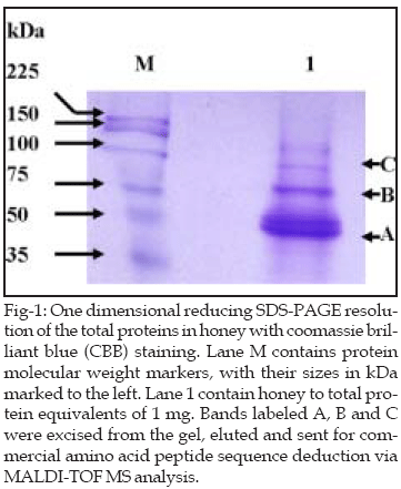
Partial amino acid sequences from these three major bands
(bands A-C, Figure 1) were obtained by Matrix-Assisted Laser Desorption/Ionisation
Time-Of-Flight Mass Spectrometry (MALDI-TOF MS) and Mascot Searching using the
MSDB database.
For the first of these proteins, the major band of an
apparent size of 50 kDa (A in Figure 1), the MALDI-TOF deduced peptide
sequences most closely matched the conserved immunoglobulin superfamily, with
the highest match being the immunoglobulin heavy chain variable region in
Mus musculus (Mouse), whose amino acid sequence in this region is shown
below with the matching peptides from the honey protein (band A) being shown
in bold.
1 CDGGSTYYPD TMERRFIISR DNTKKTLYLQ MSSLRSEDTA LYYCARRNGN
51 YVFAYWGQGT L
For the second protein band, a larger apparent MW protein
of ~75 kDa (B in Fig-1), the MALDI-TOF deduced peptide sequences most closely
matched the Drosophila pseudoobscura nsf attachment protein (snap) gene
(GA 19734-PA), and its homologs from related dipteran species including
Anopheles and Aedes. The fragment of the D. pseudoobscura
snap gene that matches the derived peptides of honey protein band B is shown
below with the bold text marking the matched peptides.
1 GDNEQKALQLMADAEKKLTQQKGFLGSLF GGGSNKVEDA IECYQRAGNM
51 FKMSKNWTKA GECFCEAATL HARAGSR HDA GTCYVDASNC
YKKVDVENAV
101 ACLMKSIDIY TDMGRFTMAA KHHQSIAEMY EADSNTLAQS
IQHYEQAADY
151 FKGEESVSSA NKCMLKVAQY AAQLEDYEKA ISIYEQVAAS
SLESSLLKYS
201 AKEYFFRAAL CHLSVDLLNA QHAIQKYAEQ YPAFQDSREF
KLIKILCEHL
251 EEQNIEGFTE AVKDYDSISR LDQWYTTILL RIKKAADEDP DLR
Finally, for the third protein, a band with an apparent MW
of ~100 kDa (band C, Fig-1), the MALDI-TOF deduced peptide sequences most
closely matched the antimicrobial 12 superfamily and in particular Epinecidin
(Epinecidin-1 prepropeptide precursor) in Epinephelus coioides
(Orange-spotted grouper). The complete sequence of this peptide is shown
below, with the matching MALDI-TOF MS peptides shown in bold.
1 MRCIALFLVL SLVVLMAEPG EGFIFHIIKG LFHAGKMIHG LVTRRRHGVE
51 ELQDLDQRAF EREKAFA
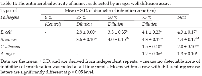
Analysis of the results of the antimicrobial activity shows
that neat honey is the most efficient concentration in order to inhibit the
growth of all four tested microorganisms (Table-II), and indeed only honey at
concentrations of >75% (v/v) were able to cause any detectable inhibition of
proliferation of the tested yeast and fungi isolates. Within the two bacteria,
S. aureus was more sensitive than E. coli, yielding an overall
sensitivity to proliferation inhibition of S. aureus > E. coli >
C. albicans > A. niger (Table-II).
DISCUSSION
Although largely composed of two monosaccharides (fructose
and glucose) and some invert sugar, honey also contains a diverse array of
minor components, the actual composition of which will vary from hive to hive
and is likely to be principally varied by the plant species foraged and,
therefore, bee species, geographical location and season, but also other
factors like climate and environmental conditions will likely have an
influence. Here, polyfloral honey of A. dorsata was assayed since the
local Thai people consume and use polyfloral honey much more than unifloral
honey. Four parameters were measured and reported (Table-I). Three of these
parameters were expected to be involved in antimicrobial activities. The first
parameter was the honey pH, which is the degree of acidity. A. dorsata
honey was found to be fairly acidic (pH 3.8), but within the reported pH range
of other honey types such as pH 3.4-5.4 for A. mellifera honey from
Switzerland.
1
The second parameter was the percentage of invert sugar and proline. Compared
to the honey from Apis mellifera in Thailand, both the proline content
and the percentage of invert sugar of A. dorsata honey were lower.9
Sugar was one of the interests in this research since it provides a strong
osmotic effect that is likely to play an important role in an antibacterial
action.
Finally, the third component was total protein, which can
contain anti-microbial. Anklam reported that honey contained about 0.2% (w/v)
protein.
10
In this research, the total protein content of A. dorsata honey was
slightly but not dramatically lower at 0.16% (w/v), and indeed was the same as
that reported for heterofloral honey and close to that of chestnut honey
(0.17% (w/v)).4
At present, the continued use, especially common misuse of
antibiotics in some disease treatments may lead to an increasing resistance
frequency in pathogen populations to unacceptable levels, necessitating
alternative treatments. Honey, as a natural product, seems to be one possible
option, and has long been used in traditional medicine for both topical
applications and as an orally administered systemic prophylactic.
11-13
The microbial inhibition ability of A. dorsata honey revealed
significant differences between the different dilutions of honey and the type
of pathogens. A. dorsata honey was most effective against S. aureus,
a representative of Gram+ve
bacteria, which agrees well with the previous results of Chanchao et al. and
Marcucci et al.9,14
Certainly, the flavonone pinocembrin, the flavonol galangin, and caffeic acid
phenethyl ester have been shown to be able to inhibit the function of
bacterial RNA polymerase.15
In addition, Mirzoeva et al. reported both that there was quercetin in honey
that it could increase the membrane permeability preventing bacterial ATP
synthesis, membrane transport and mobility.16
Moreover, galangin, one of the
flavonoids in honey, can degrade the bacterial cytoplasm membrane.17
Since the determination of
antibacterial activity can be measured quantitatively, it can be used as an
additional quality criterion for honey.
CONCLUSION
In the honey of Apis dorsata, three new findings
relating to antimicrobial activity were reported in this study. The first one
is the acidity (pH 3.8) of honey. The second is the presence of the
antimicrobial peptide epinecidin (about 76 kDa) in honey as one of the three
major proteins. In addition, although another of the three major protein bands
(fragment of immunoglobulin heavy chain variable region) is not an
antimicrobial peptide, it is still related to immune system. The last novel
observation was the antimicrobial activity of this A. dorsata honey.
S. aureus was the most susceptible to honey at all tested dilutions
(25-100% (v/v)), whilst C. albicans and A. niger were not
susceptible to honey at 50% (v/v) and lower concentrations. It could be
concluded that honey at different dilutions performs different inhibition
activities and or that different microorganisms show different responses to
honey. Due to the data mentioned above, A. dorsata honey from Thailand
could be a good source for antimicrobial agents that can be used to protect
health and fight against some diseases.
ACKNOWLEDGMENTS
I would like to thank Ms. Phansaang Wannakun for help and
suggestions. This research was financially supported by grants from the
Thailand Research Fund (grant # RMU5180042), the Asian Research Center, the
Korea Foundation for Advanced Studies and Cerebos (Thailand) Ltd. Also, I am
thankful to Dr. Robert Butcher of the Publication Counseling Service, Faculty
of Science, Chulalongkorn University, for manuscript preparation.
REFERENCES
1. Bogdanov S. Nature and origin of the antibacterial
substances in Honey. Food Sci Technol-Leb 1997;30:748-53.
2. Wongsiri S, Chanchao C, Deowanish S, Aemprapa S,
Chaiyawong T, Peterson S, et al. Honeybee diversity and beekeeping in
Thailand. Bee World 2000;81:20-29.
3. Mizrahi A, Lensky Y. Bee Product: Properties,
Applications, and Apitherapy. 1997. Plenum Press, New York.
4. Kucuk M, Kolayli S, Karaoglu S, Ulusoy E, Baltaci C,
Candan F. Biological activities and chemical composition of three honeys of
different types from Anatolia. Food Chem 2007;100:526-34.
5. Noori SA. Mixture of honey, beeswax, and olive oil
inhibits growth of Staphylococcus aureus and Candida albicans.
Arc Med Res 2005;36:10-13.
6. Sidney W. Official Methods of Analysis. 1984. 14th ed.,
Association of Official Analytical Chemists, Inc., Virginia.
7. Bradford MM. A rapid and sensitive method for the
qualitatively of microgram quantities of protein utilizing the principle of
protein – dye binding. Anal Biochem 1976;72:248-54.
8. Gheldof N, Wang XH & Engeseth NJ. Identification and
quantification of antioxidant components of honeys from various floral
sources. J Agric Food Chem 2002;50:5870-7.
9. Chanchao C, Sintara K, Wongsiri S. Comparison of
antibiotic and organoleptic properties of honey from various plant sources in
Thailand. J Apicult Sci 2006;50(2):59-64.
10. Anklam E. A review of the analytical methods to
determine the geographical and botanical origin of honey. Food Chem
1998;63:549-62.
11. Snow MJ, Manley-Harris M. On the nature of non-peroxide
antibacterial activity in New Zealand manuka honey. Food Chem 2004;84:145-7.
12. Simon A, Traynor K, Santos K, Blaser G, Bode U, Molan
P. Medical honey for wound care – still the ‘latest resort’? eCAM Advance
Access published on January 7, 2007.
13. Viuda-Martos M, Ruiz-Navajas Y, Fernandez-Lopez J,
Perez-Alvarez JA. Functional properties of honey, propolis, and royal jelly. J
Food Sci 2008;73(9):R117-R124.
14. Marcucci MC, Ferreres F, Garc a-Viguera C, Bankova VS,
Castro SL, Dantas AP, et al. Phenolic compounds from Brazilian propolis with
pharmacological activities. J Ethnopharmacol 2001;74:105-112.
15. Takaisi NB, Scjoncjer H. Electron microscopy and
microcalorimetric investigations of the possible mechanism of the
antibacterial action of a defined propole provenance. Planta Med
1994;60:222-7.
16. Mirzoeva OK, Grisjanin RN, Calder PC. Antimicrobial
action of propolis and some of its components: the effects on growth, membrane
potential and motility of bacteria. Microbiol res 1997;152:239-46.
17. Cushnie TPT, Lamb AJ. Detection of galangin-induced cytoplasmic
membrane damage in Staphylococcus aureus by measuring potassium loss. J
Ethnopharmacol 2005;101:243-8.
HOME
| SEARCH
| CURRENT
ISSUE | PAST
ISSUES
Professional
Medical Publications
Room No. 522, 5th Floor, Panorama Centre
Building No. 2, P.O. Box 8766, Saddar, Karachi - Pakistan.
Phones : 5688791, 5689285 Fax : 5689860
pjms@