|
|
||||
|
Published by : PROFESSIONAL MEDICAL PUBLICATIONS |
||||
|
ISSN 1681-715X |
||||
|
||||
|
- |
||||
|
CASE REPORT |
||||
|
- |
||||
|
Volume 25 |
April - June 2009 (Part-I) |
Number 2 |
||
|
|
||||
|
||||
|
|
||||
|
Published by : PROFESSIONAL MEDICAL PUBLICATIONS |
||||
|
ISSN 1681-715X |
||||
|
||||
|
- |
||||
|
CASE REPORT |
||||
|
- |
||||
|
Volume 25 |
April - June 2009 (Part-I) |
Number 2 |
||
|
|
||||
|
||||
Sternalis: A Clinically Important Variation
Walid Abbas Zaher1, Hasem Hasan Darwish2,
Ahmed Magzoub Elhag Abdalla3,
Muhammad Saeed Vohra4, Muhammad Mujahid Khan5
ABSTRACT
Sternalis muscle is an anatomical variation that is considered as a dilemma for surgeons and radiologists whereas a matter of interest for anatomists. It is found occasionally in the human on the anterior aspect of the thorax, superficial to the pectoralis major muscle. During routine cadaveric dissection for undergraduate medical students, presence of sternalis muscle was detected in a 60 years old male cadaver. This rare anomaly has puzzled radiologists and surgeons in confirming diagnosis, missing it completely or mistaking it for a tumor on mammography or CT scan as it may mimic a malignant mass lesion on the medial aspect of craniocaudal mammograms. A detailed description of this important variation is needed in the text books for radiologists and surgeons.
KEY WORDS: Chest, Intercostal, Mammography, Muscle, Pectoralis major.
Pak J Med Sci April - June 2009 Vol. 25 No. 2 325-328
How to cite this article:
Zaher WA, Darwish HH, Abdalla AME, Vohra MS, Khan MM. Sternalis: A Clinically Important Variation. Pak J Med Sci 2009;25(2):325-328.
1. Walid Abbas Zaher,
2. Hasem Hasan Darwish,
3. Ahmed Magzoub Elhag Abdalla,
4. Muhammad Saeed Vohra,
5. Muhammad Mujahid Khan
1-5: Department of Anatomy,
College of Medicine,
King Khalid University Hospital,
King Saud University,
Riyadh,
Saudi Arabia.
Correspondence
Dr. Muhammad Mujahid Khan
Assistant Professor,
Dept. of Anatomy, (28),
College of Medicine,
King Khalid University Hospital,
King Saud University,
P.O. Box 2925.
Riyadh 11461,
Kingdom of Saudi Arabia.
Email: dralkhan@hotmail.com
* Received for Publication: July 8, 2008
* Accepted for Publication: Febraury13, 2009
INTRODUCTION
Sternalis muscle (also known as episternalis, presternalis, sternalis brutorum, rectus thoracic, rectus sterni and superficial rectus sterni) is a mysterious muscle that is rarely found on the anterior thoracic wall. This variant muscle is a dilemma for surgeons and radiologists whereas a matter of interest for anatomists. Its size varies from a short thin slip to a long broad band of muscle. Sternalis is described as a superficial vertical slip (or slips) that may ascend from the lower costal cartilages and rectus sheath to blend with sternocleidomastoid muscle or to attach to the upper sternum or costal cartilages.
1Turner
2 describes sternalis as an occasional occurrence of a muscular slip on the anterior aspect of the thorax, superficial to the pectoralis major muscle. He further mentions that Bartolemen Cabrolio first noticed this muscle in the year 1604 and a proper description was first made by Du Puy in 1726.2 The occasional presence of the Musculus sternalis in human has always been a subject of great interest to anatomists.3 This muscle occurs in 3-5% of humans,4 found slightly more frequently in females than males,5 8.7% and 6.4% in females and males respectively.6 It was also noted that the incidence varies in different ethnic groups and ranges from 2.96% in Balgarians,7 7.1% in Africans8 and 17.3% in Chinese.9 In Japanese alone its frequency ranges between 2.4%10 to 10.4%.11 This muscle, if present, covers the medial border of the pectoralis major and runs parallel to the sternum and extends from the jugular notch to the costal region.12,13 This rare anomaly has puzzled radiologists and surgeons in confirming diagnosis, missing it altogether or mistaking it for a tumor on mammography or CT scan,12 as it may mimic a malignant mass lesion on the medial aspect of craniocaudal mammograms.14CASE REPORT
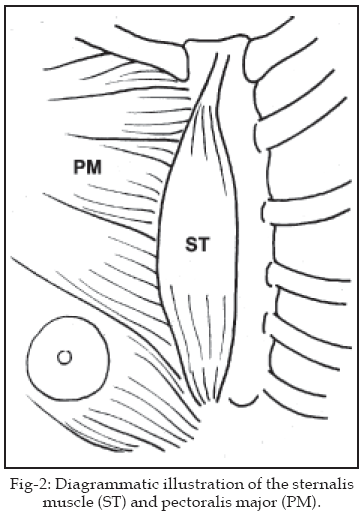
During the routine cadaveric dissection for undergraduate medical students at College of Medicine, King Saud University Riyadh, an anterior thoracic wall muscular variation (sternalis) was detected in a 60 years old male cadaver. Sternalis is regarded as an accessory muscle of the chest wall, its longitudinal fusiform muscular band is covered by a superficial fascia and overlies the right margin of the sternum and sternal end of the pectoralis major (Fig-1).
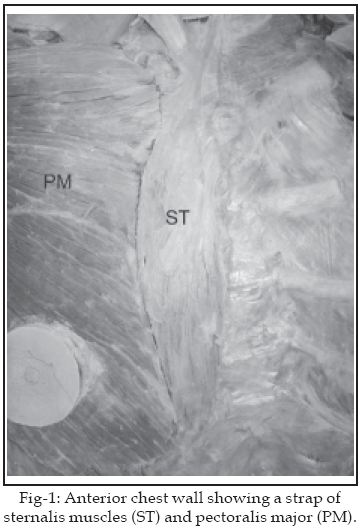
Caudally it was attached to the 5
th and 6th costal cartilages, and extends 17cm upward to be attached cranially to the manubrium sternum, 0.5cm below the jugular notch. It was measured 3.1cm wide at the broadest part at the level of 4th intercostal space, whereas thickness was 1.5mm. The anterior chest wall was carefully dissected and the lateral margin of the muscle was raised to trace the nerve supply, it was found to be innervated by twigs from the 3rd and 4th intercostal nerves which penetrated the deep surface of the muscle. Pectoralis major and minor muscles were normally placed.
DISCUSSION
Sternalis is a slender muscle, looks like a band of fibers running parallel to the sternum and lies superficial to the pectoral fascia. Several reports are available about its attachment. Caudally is attached to the cartilages of the lower true ribs into which rectus abdominis was inserted, but in the great number of cases it blends with the external oblique muscles,
2 to the costal cartilages of the 6th rib and takes extra slip from the 3rd, 4th and 5th costal cartilages, and also to the right half of the anterior aspect of the sternum,15 attached to the 5th, 6th and 7th ribs immediately external to their cartilages and external oblique muscle,4,16-18 also takes fibers from the rectus abdominis muscle or rectus sheath.6,19,20 Cranially it terminates in the pectoral fascia, sternal body, costal cartilages or the sternal head of the sternocleidomastoid muscle.4,6,16-20 In the present case it is caudally attached to the 5th and 6th costal cartilages while cranially to the manubrium sternum (Fig-1).Nerve supply to the sternalis is variable, it maybe innervated through the intercostal nerves
3,9,15-17,21,22 or by the pectoral nerves.23,24 From these reports, different theories are assumed about its source, and variability of its nerve supply appears to indicate that the muscle is not constantly derived from the same source.25 In our case sternalis is innervated by 3rd and 4th intercostal nerves. It occasionally has tendinous intersections and when a well developed platysma coexists with it, the platysma is always on a plane superficial to it.4 Turner2 also considered that the evidence was in favor of classifying it with the platysma.In general, when a muscle contracts, the insertion is pulled towards its origin. Therefore, because of its particular location, it has been suggested that the contraction of the sternalis muscle is thought to play a role elevating the lower part of the chest,
26 but absolute proof of such a function may be difficult to obtain. Natsis and Totlis18 described it to have a little functional significance, or it may function primarily as a proprioceptor sensing and signaling the movements of the thoracic wall. Ruge27 anticipated that sternalis represents a vestige of the circular muscle of mammals that constitutes the great subcutaneous muscle of the trunk, but Clemente28 described it to be a misplaced pectoralis major or is a part of the ventral, longitudinal column of muscle arising at the ventral tips of the hypomeres. This column is represented by the rectus abdominis muscle in the abdominal region and by the infrahyoid musculature in the cervical region. In the thorax, the longitudinal muscle normally disappears but is occasionally represented by the sternalis muscle.29Musculus sternalis is a rare anatomic variation of the chest wall that may be included in the projection of the breast tissue.
30 Either insufficient description of sternalis muscle is given or not mentioned at all in the famous text books of Anatomy.Presence of sternalis causes a dilemma for the radiologists and surgeons, particularly in the diagnosis of the breast cancer. It is particularly important for radiologists and surgeons to have a sufficient knowledge of such a variant muscle to avoid misdiagnosis of malignant tumors of the chest wall. With the use of sophisticated diagnostic and therapeutic tools, there is a need to record and discuss unusual anatomical variants, such variations may appear pathological, making the diagnosis difficult for the physicians.
12 A detailed description is needed in the text books for this important variation for radiologists and surgeons.ACKNOWLEDGEMENT
The authors are thankful to Mr. V. Salvador for drawing the illustrations.
REFERENCES
1. Williams PL, Bannister LH, Berry MM. Gray’s anatomy. 38th Ed. New York; Churchill Livingstone; 1995;838.
2. Turner WM. On the Musculus sternalis. J Anat Physiol 1867;1:246-378.25.
3. Cunningham DJ. The Musculus sternalis. J Anat Physiol 1884;18:208-10.
4. Parson FG. On the morphology of the musculus sternalis. J Anat Physiol 1893;27:505-7.
5. Carol EH, Scott-Conner, Al-Jurf AS. The sternalis muscle. Clin Anat 2002;15:67-9.
6. Tountas CP, Bergman RA. Anatomic variations of the upper extremity. Churchill Livingstone; 1993;87.
7. Jelev L, Georgiev G, Surchev L. The sternalis muscle in the Bulgarian population: classification of sternalis. J Anat 2001;199:359-63.
8. Le Double A. Traite des variations du systeme musculaire de L Homme. Paris: Schleicher Freres; 1897;275-86.
9. Jeng H, Su SJ. The sternalis muscle: an uncommon anatomical variant among Taiwanese. J Anat 1998;193:287-8.
10. Watanabe R. The sternalis muscle of the school boys. Juzenkai Zasshi 1942;47:2040-44.
11. Adachi B. On the examination of the musculus sternalis in the living persons. Tokyo Igakkai Zasshi 1897;11:57-65.
12. Kumar H, Rath G, Sharma M, Kohli M, Rani B. Bilateral sternalis with unusual left sided presentation: A clinical perspective. Yonsei Med J 2003;44:719-22.
13. Kitamura S, Yoshioka T, Kaneda M, Matsuoka K, Chen KL, Sakai A. A case of the congenital partial defect of the pectoralis major accompanied by the sternalis with enormous size. Kaibogaku Zasshi J Anat 1985;60:728-32.
14. Goktan C, Orguc S, Serter S, Ovali GY. Musculus sternalis: A normal but rare mammography finding and magnetic resonance imaging demonstration. The Breast J 2006;12:488-9.
15. Wallace D. Note on the nerve supply of the Musculus sternalis. J Anat Physiol 1886;21:153-4.
16. Shepherd FJ. The musculus sternalis and its nerve supply. J Anat Physiol 1889;23:303-7.
17. Patten CJ. Right sternalis muscle, narrow and spindle-shaped. J Anat 1934;68:426.
18. Natsis K, Totlis T. A rare accessory muscle of the anterior thoracic wall. Clin Anat 2007;20:980-1.
19. Huntington GS. The derivation and significance of certain supernumerary muscles of the pectoral region. J Anat Physiol 1905;39:1-54.
20. Bergman RA, Afifi AK, Miyauchi R. Axillary arch, pectoralis quartus, and sternalis. In: "Illustrated encyclopedia of human anatomic variation: 1992;Part I. Muscular system, Alphabetical listing of muscles, pectoralis major and pectoralis minor."http//www.vh.org/providers/Textbooks/Anatomicvariants/AnatomyHP.html.
21. Lamont JC. Note on the nervous supply of the musculus sternalis. J Anat Physiol 1887;21:514-5.
22. O’Neill MN, Folan-Curran. Case report: Bilateral sternalis muscles with a bilateral pectoralis major anomaly. J Anat 1998;193:289-92.
23. Kita MY, Kudoh H. Innervation of the sternalis muscle accompanied by congenital partial absence of the pectoralis major muscle. Okajima Folia Anat Jpn 1991;67:449-55.
24. Kida MY, Izumi A, Tanaka S. Sternalis muscle: topic for debate. Clin Anat 2000;13:138-40.
25. Harper WF. The sternalis muscle in the anencephalous foetus. Anat Notes 1936;317-20.
26. Arraez-Aybar LA, Sobrado-Perez J, Merida-Velasco JR. Left musculus sternalis. Clin Anat 2003;16:350-4.
27. Ruge G. Der Hautrumfmuskel der Saugetiere: der M. sternalis and der Achselbogen des Menschen. Morph Jahrb 1905;33:379-531.
28. Clemente CD, editor. Gray’s anatomy. 30th Ed. Philadelphia: Lea and Febiger; 1985;520.
29. Sadler TW. Langman’s medical embryology. 10th Ed. Maryland; Lippincott Williams and Wilkins; 2006;146.
30. Bradley FM, Hoover HC jr., Hulka CA. The sternalis muscle: An unusual normal finding seen on mammography. Am J Rad 1995;166:33-6.
HOME | SEARCH | CURRENT ISSUE | PAST ISSUES
Professional
Medical Publications
Room No. 522, 5th Floor, Panorama Centre
Building No. 2, P.O. Box 8766, Saddar, Karachi - Pakistan.
Phones : 5688791, 5689285 Fax : 5689860
pjms@pjms.com.pk