|
|
||||
|
Published by : PROFESSIONAL MEDICAL PUBLICATIONS |
||||
|
ISSN 1681-715X |
||||
|
||||
|
- |
||||
|
ORIGINAL ARTICLE |
||||
|
- |
||||
|
Volume 23 |
April - June 2007 (Part-II) |
Number 3 |
||
|
|
||||
|
||||
|
|
||||
|
Published by : PROFESSIONAL MEDICAL PUBLICATIONS |
||||
|
ISSN 1681-715X |
||||
|
||||
|
- |
||||
|
ORIGINAL ARTICLE |
||||
|
- |
||||
|
Volume 23 |
April - June 2007 (Part-II) |
Number 3 |
||
|
|
||||
|
||||
Resistance to vancomycin in enterococcus
faecium and faeca lis clinical isolatesA. Aleyasin1, A.M. Mobarez2, M. Sadeghizadeh3, R. Hosseini Doust4, N. Khoramabadi5
ABSTRACT
The aim of this study was to investigate antibacterial resistance among enterococci species isolated in Tehran Baghyatallah Hospital. It consisted of 126 isolates of E. faecalis (86%), E. faecium (9%) and other Enterococus Spp. (5%) isolated from urine (34.92%), blood (27.77%), wound swabs (19.84), stool (5%) endotracheal secretions (3.37%), abscess (3.4%), dialysis fluids (1.7%) and catheter (4%). Twelve (9.5%) isolates were resistant to vancomycin. The VRE isolates were resistant to ampicillin (75%), erythromycin (50%), tetracycline (58%), ciprofloxacin (41.6%), chloramphenicol (33.3%) and gentamicin (41.6%). Two (16.66%) of VRE isolates were multidrug resistant. Eight (66.6%) of the vancomycin-resistant strains and all of the MDR strains carried the vanA phenotype and genotype. The MIC of VRE isolates were between 32-512µg/ml. Our results show that most glycopeptide resistant E. faecalis and E. Faecium carried vanA. It is also possible that frequency of infections caused by glycopeptide-resistant enterococci will increase in our geographical area.
KEY WORDS: Enterococcus, Clinical Samples, VanA, Vancomycin-resistant Enterococci, Iran.
Pak J Med Sci May - June 2007 Vol. 23 No. 3 390-393
1. A. Aleyasin,
2. Dr. A. M. Mobarez,
3. Dr. M. Sadeghizadeh,
Department of Genetic, Faculty of Basic Sciences,
4. Dr. R. Hosseini Doust,
Dept. of Microbiology &
Research Center of Molecular Biology,
Baghyatollah University, Tehran – Iran.
5. N. Khoramabadi,
1,2,5: Department of Bacteriology,
Faculty of Medical Sciences,
1,3,5: University of Tarbiat Modares,
Tehran – Iran.
Correspondence
Dr. A. M. Mobarez,
E-mail: mobares80@yahoo.com
* Received for Publication: September 26, 2006
* Accepted: January 28, 2007
INTRODUCTION
Enterococci are important nosocomial pathogens recovered often from the patients with urinary tract infections, wounds, bacteremia, endocarditis or meningitis.1 Detection of vancomycin-resistant enterococci (VRE) in the mid-1980s was therefore a major therapeutic concern. VRE were first isolated in France and England2,3 but the strains have subsequently been reported on a worldwide scale.2,4,5 Acquired resistance to vancomycin by Enterococci greatly reduces the number of treatment options for disease management2-6 and the problem is further compounded by the fact that resistance genes can potentially be transferred to other pathogenic organisms, such as Staphylococcus aureus and Streptococcus species.2,3 To date seven genotypes (vanA, vanB, vanC1, vanC2/3, vanD, vanE and vanG) of vancomycin resistance have been reported for enterococci.7 Strains with the vanA genotype are characterized by high-level vancomycin and teicoplanin resistance, whereas, those with the vanB genotype exhibit moderate to high resistance to vancomycin.1-4 The aim of the present study was to use PCR for detection of vanA isolates of E. faecium and E. faecalis from the patients in Tehran Baghyatallah hospital within a specific time-period in 2004-05.
MATERIAL AND METHODS
A total of 126 isolates were obtained from different clinical samples between March 2004 and December 2005 in Bagyatallah hospital in Tehran. They were cultured from urine, wound swabs, blood, endotracheal secretions, dialysis fluids, abscess and stool. Stool samples were inoculated into enterococcal broth, incubated overnight at 35°C and subcultured onto brain-heart infusion agar (BHIA) containing 6µg vancomycin ml-1 8,9 and onto BHIA without vancomycin to recover vancomycin-susceptible isolates. The isolates were identified to the genus and species level by cultural characteristics, Gram’s stain, catalase test, bile aesculin reactions and arabinose, sorbose and manitol fermentation.
Susceptibility testing: Disk diffusion method on Mueller–Hinton agar10 was used to detect resistance to tetracycline, erythromycin, chloramphenicol, ampicillin, ciprofloxacin, gentamicin, kanamycin, streptomycin, linezolid, vancomycin and teicoplanin. The MICs for vancomycin and teicoplanin were determined by the agar dilution method.
Detection of vancomycin-resistance determinants: The presence of the vanA resistance gene was assessed by PCR analysis describes by Khan et al.11 The primers were ( vanA Forward; 5'-AAT ACT GTT TGG GGG TTG CTC-3' and vanA Reverse, 5'-CTT TTT CCG GCT CGA CTT CCT-3'). The amplification mixture consisted of 5µl of 10 X PCR buffer (100mM Tris / HCl, pH 8.4, 500 mM KCl, 20mM MgCl2 ), 220µM each dNTP, 22 U recombinant Taq DNA polymerase ml-1, 5µl bacterial DNA and 5µl primer, 6µl H2O. An Ependroff thermocycler was programmed for 30 cycles with the following parameters: denaturation at 97°C for 1 min, annealing at 52°C for 55 seconds, extension at 72°C for 1.5min and final extension at 72°C for 10 min. Amplified products were detected by agarose gel electrophoresis using 1.5% agarose (w/v) in TAE buffer for 2h at 70 V. E. faecium, ATCC 51559 (kindly provided by Dr M. feyzabady) were used for standardizing the PCR amplification of vanA. A vancomycin-sensitive strain, Enterococcus faecalis ATCC 29212, was used as the negative control.RESULTS
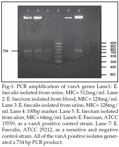
One hundred twenty six bacterial isolates from urine (34.92%), blood (27.77%), wound swabs (19.84%), dialysis fluids (1.7%), stool (5%), abscess (3.4%) endotracheal secretions (3.37%), and catheter (4%) were studied in the years 2004 - 2005 in Tehran Baghytallah Hospital. Of the 126 samples, 59% collected in medical ward, 19% in intensive care facilities and 17% in surgical wards while 5% could not be classified. The strains with vancomycin MICs>16mg/ml were considered as vancomycin resistant. Special emphasis was given to detection of vancomycin resistance marker vanA (Fig1), because vancomycin is considered as one of the antibiotics to be used as a last resort12 used.
To test the presence of vanA, we set up a PCR assay11 for detecting the vanA (734-bp) marker (Fig.1). When the individual PCR reactions were carried out, specific and predicted size amplicon of vanA (734bp) was observed (Fig-I, lane 1, 2, 3, and 5). No PCR product was observed when template DNA from a vancomycin-sensitive E. faecalis strain, ATCC 29212, was used (Fig.1, lane6). As expected, in strain with MICs < 16mg/ml, no vanA was detected. Four of Twelve VRE isolates did not possess any vanA genes, even though they were resistant to vancomycin (MICs between 16-32mg/ml) (Table-I).
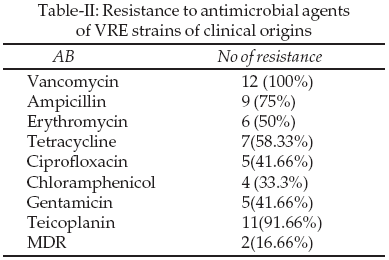
Most of the VRE strains were resistant to 6-8 other antibiotics (Table-II) indicating either previous exposure to these antibiotic or the acquisition of resistance markers. Vancomycin resistance was detected in 12 (9.5%) isolates consisting of five (41.6%) E. faecium and seven (58.33%) E.faecalis (Table-I). Most of the VRE isolates studied were also resistant to teicoplanin (91.6%), ampicillin (75%), erythromycin (50%), tetracycline (58%), ciprofloxacin (41%), chloramphenicol (33%), and gentamicin (41%) (Table-II). Two (16.66%) of the VRE isolates were multidrug resistant. Eight (66.6%) of the vancomycin-resistant and all multidrug resistant strains carried the vanA phenotype and genotype. The MIC of multidrug resistance isolates were between 128-512mg/ml. One of the seven vancomycin-resistant E. faecalis and two vancomycin-resistant E. faecium isolates had MIC values>128µg ml-1 for vancomycin (Table-II). Two multidrug resistant E. faecium from urine and blood, with MICs>128-512µg/ml were isolated.
DISCUSSION
Vancomycin-resistant enterococci have been increasingly reported worldwide since first described in 1987, although the epidemiology of these microorganisms varies widely in different geographical areas.13,14 The present study documents the phenotypic and genotypic characterization of 12 vancomycin-resistant enterococci, isolated over 1-year period (2004-2005) from different clinical samples at Tehran Baghyatallah Hospital. The predominant species were E. faecium (9%) and E. faecalis (85%). The prevalence of E. faecium in this study was 9% lower than 29% prevalence reflected in a similar study from Cincinnati.15 Of the 12 VRE isolates, 75, 50 and 58% were resistant to ampicillin, erythromycin and tetracycline respectively. All of the isolates were susceptible to linezolid. High-level resistance to glycopeptide antimicrobials was first reported in Europe in 1986 and the United states in 1987. Between the years 1989 and 1993, the rate of vancomycin resistance in the United States increased from 0.3% to 7.9%.16 In this study, we found that 9.5 % of the enterococcal isolates were resistant to vancomycin. Of the 12 VRE isolates, 7(58.3%) were E. faecalis and 5(41.7%) were E. faecium, respectively. Vancomycin-resistance phenotypes in enterococci were classified as vanA, vanB, vanC, vanD and vanE based on levels of resistance.17 The vanA determinant was carried on the transposon Tn1546 or its close relatives that are transferable in conjugation experiments.17 Eight of the 12 vancomycin-resistant isolates in this study expressed vancomycin-resistance patterns compatible with the vanA phenotype and all the multidrug resistant strain gave positive results in PCR experiments for the vanA genotype. The vanA phenotype is responsible for approximately 70% of the VRE isolates. In our study, vanA was found in 66.6% of the isolates and all multidrug isolates were vanA positive, respectively. The detection of gentamicin resistance in 41% of the VRE isolates was cause for concern, as it may signify the beginning of a major resistance problem. The prevalence of glycopeptide resistance among the studied isolates, their presence together with aminoglycoside resistance calls for regular surveillance of antibacterial susceptibilities to detect the emerging resistance and prevent the establishment and spread of multidrug antibacterial-resistant strains.
ACKNOWLEDGMENTS
We would like to acknowledge Dr. Feyzabady (Tehran University of Medical Science) and Dr. Soltanpour (Head of Medical Laboratory at Bagyatallah hospital) for their assistance with the collection of enterococcal isolates.
REFERENCES
1. Murray PR, Baron EJ, Pfaller MA, Tenover FC, Yolken RH. Manual of Clinical Microbiology, 7th Ed. Washington, DC: American Society for Microbiology 1999.
2. Hayden MK. Insights into the epidemiology and control of infection with vancomycin resistant Enterococci. Clin Infect Dis 2000; 31:1058-65.
3. Murray BE. Vancomycin - resistant enterococcal infections. N Engl J Med 2000;342:710-21.
4. Cetinkaya Y, Falk P, Mayhall CG. Vancomycin - resistant enterococci. Clin Microbiol Rev 2000;13:686-707.
5. Goossens H. Spread of vancomycin-resistant enterococci: differences between the United States and Europe. Infect Control Hosp Epidemiol 1998;19:546-51.
6. Mundy LM, Sahm DF, Gilmore M. Relationships between Enterococcal virulence and antimicrobial resistance. Clin Microbiol Rev 2000;13:513-22.
7. McKessar SJ, Berry AM, Bell JM, Turnidge JD, Paton JC. Genetic characterization of vanG, a novel vancomycin resistance locus of Enterococcus faecalis. Antimicrob Agents Chemother 2000;44:3224-8.
8. Jordens JZ, Bates J, Griffiths DJ. Fecal carriage and nosocomial spread of vancomycin- resistant Enterococcus faecium. J Antimicrobial Chemother 1994;34:515-28.
9. Ieven M, Vercauteren E, Deschemaeker P, Van laer F, Goosens H. Comparison of direct plating and broth enrichment culture for the detection of intestinal colonization by glycopeptide - resistant enterococci among hospitalized patients. J Clin Microbial 1999;37:1436-40.
10. National Committee for Clinical Laboratory Standards. Performance Standards for Antimicrobial Disk Susceptibility Tests Approved Standard, 7th Ed (M2-A7). Villanova: National Committee for Clinical Laboratory Standards 2000.
11. Khan SA, Nawas MS, Khan AA, Hopper SL, Jones RA, Cerniglia CE. Molecular characterization of multidrug- resistant Enterococcus spp. From poultry and dairy farms: detection of virulence and vancomycin resistance gene markers by PCR. Molecular and Cellular Probes 2004;20:1-8.
12. Schentag JJ. Antimicrobial management strategies for gram-positive bacterial resistance in intensive care unit. Crit Care Med 2001;29:N100-7.
13. Shepard BD, Gilmore MS. Antibiotic resistant enterococci: the mechanisms and dynamics of drug introduction and resistance. Microbs Infect 2002;4:215-24.
14. Gossens H, Habes D, Rossi R, Lammens C, Privitera G, Courvalin P. European survey of vancomycin resistant enterococci in at- risk hospital wards and in vitro susceptibility testing of ramoplanin against these isolates. J Antimicrob Chemother 2003;51(Suppl .S3) iii5-iii 12.
15. Prelada DE, George SA, Cushion MT. Molecular epidemiology and antibiotic susceptibility of Enterococci in Cincinnati, Ohio: a prospective citywide survey. J Clin Microbiol 1997;35:2342-7.
16. Centers for Disease Control and Prevention. Nosocomial Enterococci resistant to vancomycin-United States, 1989-93. Morbidity and Mortality Weekly Report 1993;42:597-9.
17. Arthur M, Courvalin P. Genetics and mechanisms of glycopeptides resistance in Enterococci. Antimicrob Agents Chemother 1993;37:1563-71.
HOME | SEARCH | CURRENT ISSUE | PAST ISSUES