Published by : PROFESSIONAL MEDICAL PUBLICATIONS
ISSN 1681-715X
HOME | SEARCH | CURRENT ISSUE | PAST ISSUES
-
ORIGINAL ARTICLE
-
Volume 22
January March 2006 Number 1
Clinical profile of epilepsy during the
first two years of LifeWael Hayel Khreisat1
ABSTRACT:
Objective: This study aims at identifying the different presentations of infantile epilepsy with their E.E.G. and neuro-radiological patterns.
Patient & Methods: Patients included in this study were children suffering from epilepsy starting below 2 years of age. They were attending the outpatient clinic or were from the in-patient departments in Prince Ali – Ben Al - Hussein hospital. The study included 100 children with ages ranging from three month to two years. All patients were subjected to: careful history taking, general and neurological examination including fundus examination, E.E.G. tracing, CT were done for all cases. Febrile convulsions and convulsions due to metabolic disturbances were excluded from the study.
Results: Patients were divided clinically into 2 groups: Group A (symptomatic) group-80 cases (80%): included those who had evident etiology, evident neurological deficit or developmental delay of significant degree prior to onset of seizures while Group B (cryptogenic) group-20 cases (20%). male to female ratio was 1.5: 1. The etiological factors encountered were perinatal asphyxia (55%) while heredofamilial disorders were least common 44% of patients had normal clinical examination as well as development while 27 % were mentally handicapped. The generalized tonic-clonic seizure was the commonest in both group while infantile type was the least, EEG was normal in 18 % of cases while CT scan lesion were present in 70% of the patients.
Conclusions: Careful history taking is extremely important for accurate diagnosis of epilepsy. The investigation of choice is the E.E.G. Computerized tomography should be considered an accurate neurodiagnostic test. It provides a good anatomic description of the brain.
KEY WORDS: Infantile epilepsy, presentations, E.E.G.
Pak J Med Sci January-March 2006 Vol. 22 No. 1 55-59
1. Dr. Wael Hayel Khreisat MD.JB
Pediatrician,
Pediatric Department,
Prince Ali Ben Al-Hussein Hospital,
Karak, Jordan.
Correspondence:
Dr. Wael Hayel Khreisat
P.O. Box: 211133,
Amman-11121, Jordan
Email: wael_khreisat@yahoo.com
* Received for Publication: January 26, 2005
Revision Received: May 28, 2005
Revision Accepted: August 22, 2005
Introduction
Seizure disorder is one of the common childhood neurological illnesses occurring in 4 - 6 per 1000 children in the general population.
1 The degree and type of central nervous system damage existing at the onset of the seizure is decisive for the final out come, but the cessation of the seizures also improves the child’s developmental possibilities.2Determining the type of seizure, and whether it constitutes part of a specific epilepsy syndrome, is important for purposes of work-up, treatment and prognosis. The correct diagnosis of epilepsy leads to an appropriate treatment. It is visible by identifying the etiology or basic disorders underlying the epilepsy by physical and neurological examinations, laboratory tests, including EEGs and neuro-radiological examinations.
3This study aims at identifying the different presentations of infantile epilepsy with their E.E.G. and neuro-radiological patterns to help in their classification and identifying whether the condition is cryptogenic or secondary to intracranial pathology. This might be of value in the management of the cases and their outcome.
patients and Methods
Patients included in this study were children suffering from epilepsy starting below 2 years of age. They were attending the outpatient clinic or were from the in-patient departments in Prince Ali – Ben Al - Hussein hospital. The study included 100 children with ages ranging from three month to two years. All patients were subjected to: careful history taking, general and neurological examination including fundus examination, E.E.G. tracing and CT was done in all cases. Febrile convulsions and convulsions due to metabolic disturbances were excluded from the study.
Results
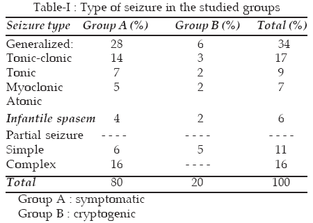
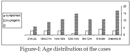
Patients were divided clinically into 2 groups: Group A (symptomatic) 80 cases (80%): included those who had evident etiology, evident neurological deficit or developmental delay of significant degree prior to onset of seizures a Group B (cryptogenic) group-20 cases (20%): this comprised children who had apparently no antecedent etiology, no neurological disorder and who had normal development until the onset of seizures. the age and six distribution of the patients is presented in Figure-I & II.
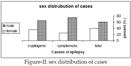
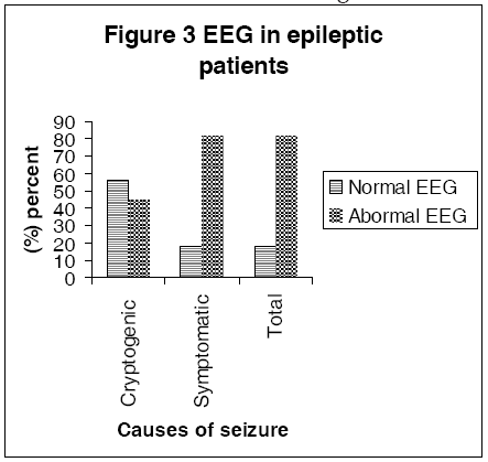
In all patient male to female ratio was 1.5: 1 male preponderance appear more evident of symptomatic group 3:1 as shown in the Figure-II. The etiological factors encountered were perinatal asphyxia (55%); prematurity (5%), CNS. infections (15%), head injuries (8%), congenital anomalies of central nervous system (9%) and heredofamilial disorders (8%). The generalized tonic-clonic seizure was the commonest in both groups while infantile type was least common (5%) Table-I. EEG was normal in 18 % of cases (abnormal 82.2 % in group A and 56.7 % in group B) as showed in Figure-II. 44% of patients had normal clinical examination as well as development while 27 % were mentally handicapped. The head circumference in group A was between 5th and 9th percentile in 27 cases (33.75%) above 95th percentile in 15 % of cases. In group B, it was between 5th percentile and 95th percentile in all cases.
Discussion
Perinatal asphyxia is an important determinant of infant neurological outcome
4, in our study perinatal asphyxia was the most frequently observed cause accounting for 55 % while other studies have repotted 35%.4 It was also reported that seizures are common squeal to sever perinatal asphyxia.5,6 In another study it was found that perinatal factors were the most common cause of epilepsy.7-9 Seizure types were partial in 45% of the children and generalized in 55%.Prematurity was the relevant finding in the history of 5% of the studied cases. Motor and mental disability was present in 75% of them while other studies have reported 33%.
10 They all had generalized seizures. The fits were tonic clonic in 33.3% of cases, myoclonic in 20 %while tonic in others. Brain CT scan showed cortical atrophy in all cases.CNS infection was an etiological factor for seizures in 15% of cases. This reflects a high incidence of infection among the studied group while earlier studies have reported a lower incidence.
11 They found that CNS infection was an etiological factor in 10% of their cases. Some reported an incidence of seizures of 2-8% in a prospective study of 50 infants who recovered from H. influenza meningitis.12Seizures of post traumatic epilepsy may aggravate the brain dysfunction due to primary trauma and surgical treatment often yields good result and lessens the toxic and adverse effect of antiepileptic drugs.
13 It was reported that post-traumatic epilepsy represents 4% of the prevalence of the disorder and is one of the sequels which is most difficult to prevent.14 In our study head injuries formed (8%) which is much lower incidence of early post-traumatic seizures (1.6%) as noted by others.15 5% of patients with post-traumatic seizures had normal brain CT –scan as compared to other studies 5.6%.16,17Neuroimaging examines the relationship between abnormalities of brain function in epilepsy patients (seizures, impaired cognitive function, psychiatric co-morbidity etc.) and focal or more widespread brain pathology.
18 Computed Tomography (CT) is an important tool for neuroimaging as it offers an opportunity to investigate structural lesions as a cause of seizures with little morbidity.19 CT scan lesion were visible in 70% in the present study, which is comparable to other studies from the subcontinent where it was reported between 50 - 75% but is higher than the prevalence reported in western literature.20-.23In our study, C.T. abnormalities were present in 70% of group A (it was normal in 30% of cases of group A: 17% of patients had history of birth asphyxia, 5% with t metabolic disorders, 5% had history of meningitis and 3% had history of head trauma. All patients had normal brain CT scan). In patients with generalized seizures, CT abnormalities were detected in 50%. In those with evident neurological manifestations, CT abnormalities were noted in 78%, compared to 25% abnormal scans with generalized seizures and normal neurological findings. In some studies CT findings in children with all varieties of seizures and found an overall abnormality in 33.5%.
24Diagnosis of epilepsy is based upon clinical data. But the electroencephalogram (EEG) remains absolutely necessary for proper management of epileptic patients by the neurologist both in consultation and in hospital practice.
25 Normal E.E.G. records were found in (18%) while other studies reported it to be 14%.26 Generalized tonic-clonic were the dominant types of seizures observed in this study (34%) Table-I. It was observed that despite many unusual types of seizures in childhood, the major tonic-clonic convulsions were the commonest manifestation of childhood epilepsy.27 Myoclonic seizures were observed in 9% of patients in our study while in others it was reported to be 7.5%.27The work up of 8% cases in the symptomatic group led us to the diagnosis of degenerative brain disease. They all showed progressive mental deterioration associated with motor dysfunction affecting the tone, power and reflexes. Macrocephaly was present in two of them. They were offspring of consanguineous marriages of the first degree.
The recent intense focus of attention on further characterization and management of infantile spasms is due, in part, to the resistant nature of these seizures and the frequently poor cognitive outcome, even when the seizures are controlled.
28Six cases 6% (2% cryptogenic and 4% symptomatic) presented with infantile spasms, usually the onset of their seizures was in the neonatal period. Burst suppression pattern and hypsarrhythmias are age-related EEG patterns that present in infancy and may get modified later on.
29 E.E.G. tracings showed hypsarrhythmias and burst suppression which was detected by others who found that perinatal insult was the commonest etiology.30,31All patients of partial seizures in our study (27%) showed motor seizures while others have reported 48.5 %.
7 E.E.G. tracing was abnormal in 67% of patients. It was found that the 80% of patients had abnormal neurological examination with partial seizures32 while in our study it was 75%. Other studies have reported partial seizures and abnormal neurological examination in 95% of patients.11Conclusion
Careful history taking is extremely important for accurate diagnosis of epilepsy. The most important and useful investigation for epilepsy is the E.E.G. Computerized tomography is to be considered an accurate neuro-diagnostic test. It provides a good anatomic description of the brain. It is especially important in the symptomatic group. Neuroimaging should be considered in any child with a first seizure who does not have an idiopathic form of epilepsy. New techniques such as PET and MRI may provide additional information of the functional disturbances in epilepsy.
References
1. Cowan L, Bodeasteiner J, Leviton A. Prevalence of epilepsies in children and adolescent. Epilepsia 1989; 30: 94-100.
2. Czochanaska J, Langner-tyszka B,losiowski Z. Children who develop epilepsy in the first year of life : a prospective study . Deve Med Child Neurol 1994; 36 (4): 345-50.
3. Oguni H, Diagnosis and treatment of epilepsy, Epilepsia 2004; 45 Suppl 8:13-6.
4. Sridhar K, Kumar P, Katariya S. Postasphyxial encephalopathy in pretermneonates. Indian J Pediatr 2001; 68(12): 1121-5.
5. de souza, SJW. & richards, B. Neurological sequelae in newborn babies after perinatal asphyxia. Arch Dis Child 1978; 53: 564.
6. de Menezes MA, Rho JM. Clinical and electrographic features of epileptic spasms persisting beyond the second year of life Epilepsia. 2002; 43(6): 623-30. Erratum in: Epilepsia 2002; 43(9): 1111-2.
7. Kwong KL, Chak WK, Wong SN: Epidemiology of childhood epilepsy in a cohort of 309 Chinese children Pediatr Neurol 2001; 24(4): 276-82.
8. Miller SP, Weiss J, Barnwell A: Seizure-associated brain injury in term newborns with perinatal asphyxia. Neurology. 2002; 58(4): 542-8.
9. Gunn AJ, Gunn TR. Changes in risk factors for hypoxic-ischaemic seizures in term infants. Aust N Z J Obstet Gynaecol 1997; 37(1): 36-9.
10. Holcroft CJ, Blakemore KJ, Allen M. Association of prematurity and neonatal infection with neurologic morbidity in very low birth weight infants. Obstet Gynecol 2003; 101(6): 1249-53.
11. Scrapa arid Carassini - Partial epilepsy in childhood Clinical and E.E.G. study of 261 cases. Epilepsia 1982; 23: 333.
12. Sell, SHW. Long-term sequelae of bacterial meningitis in children. Pediatr. Inf Dis 1983; 2: 90.
13. Qi ST, Qiu BH, Ouyang H. Clinical features and surgical treatment of posttraumatic epilepsy Di Yi Jun Yi Da Xue Xue Bao 2004; 24(4): 472-4.
14. Carvajal P, Almarcegui C, Pablo MJ. Post traumatic partial seizures Rev Neurol. 2001; 33(8): 737-9.
15. Snock, JW. Menderloud, J. Delayed deterioration following mild head injury in children. Brain 1984; 107: 15.
16. Ong LC, Dhillon MK, Selladurai. Early poat-traumatic seizures in childern: clinical and radiological aspects of injury. J Paediatr Child Health 1996; 32(2): 173-6.
17. Frey LC. Epidemiology of posttraumatic epilepsy: a critical review. Epilepsia 2003; 44 Suppl 10:11-7.
18. Wormann FG. The value of neuroimaging in diagnosis of epilepsy. Ther Umsch 2001; 58(11): 645-9.
19. Obajimi MO, Fatunde OJ, Ogunseyinde AO, et al. Computed tomography and childhood seizure disorder in Ibadan. West Afr J Med 2004; 23(2): 167-72.
20. Singhi S, Singhi PD, Walia BNS. Clinical profile and etiology of partial seizures in Chandigarh children. Epilepsia 1990; 31S: 666-7.
21. Bajaj S, Barthwal SP, Dwivedi NC, et al. Contrast computed tomography in epilepsy with special reference to intracranial ring or disc enhancing lesions. J Assoc Physicians India 1991; 39: 543-5.
22. Garg RK, Nag D. Single enhancing CT lesions in Indian patients with seizures: clinical and radiological evaluation and follow-up. J Trop Pediatr 1998; 44: 204-10.
23. Kramer U, Nevo Y, Reider-Groswasser I et al. Neuroimaging of children with partial seizures. Seizure 1998; 7: 115-18.
24. Yong PJ, Berger PE, Cohen ME. Computed tomography and childhood seizure disorders. Neurology, 1979; 29: 1084.
25. Szurhaj W, Derambure P. The role of the EEG in epilepsy management. Rev Neurol 2004; 160(11): 1113-9.
26. Waaler PE, Blom BH, Skeidsvoll H, Mykletun A Prevalence, classification, and severity of epilepsy in children in western Norway. Epilepsia 2000; 41(7): 802-10.
27. Kramer U, Phatal A, Neufeld MY, et al Outcome of seizures in the first year of life. Eur J Paediatr Neurol. 1997; 1:165-71.
28. Chugani HT. Infantile spasms . Curr Opin Neurol 1995; 8(2): 139-44.
29. Duchowny M, Harvey AS. Pediatric epilepsy syndromes: an update and clinical review. Epilepsia 1996; 37: S26-36.
30. Bauer G, May U, Pallua. Computerized axial tomography in chronic partial seizures. Epilepsia 1980; 2(1): 227.
31. Liou HH, Oon PC, Lin HC. Risk factors associated with infantile spasms: a hospital-based case-control study in Taiwan. Epilepsy Res 2001; 47: 91-8.
32. Okumura A, Watanabe K. Clinico-electrical evolution in pre-hypsarrhythmic stage: towards prediction and prevention of West syndrome. Brain Dev 2001; 23(7): 482-7.
HOME | SEARCH | CURRENT ISSUE | PAST ISSUES
Professional
Medical Publications
Room No. 522, 5th Floor, Panorama Centre
Building No. 2, P.O. Box 8766, Saddar, Karachi - Pakistan.
Phones : 5688791, 5689285 Fax : 5689860
pjms@pjms.com.pk