|
|
||||
|
Published by : PROFESSIONAL MEDICAL PUBLICATIONS |
||||
|
ISSN 1681-715X |
||||
|
||||
|
- |
||||
|
Brief Communication |
||||
|
- |
||||
|
Volume 23 |
January - March 2007 |
Number 1 |
||
|
|
||||
|
|
||||
|
|
||||
|
Published by : PROFESSIONAL MEDICAL PUBLICATIONS |
||||
|
ISSN 1681-715X |
||||
|
||||
|
- |
||||
|
Brief Communication |
||||
|
- |
||||
|
Volume 23 |
January - March 2007 |
Number 1 |
||
|
|
||||
|
|
||||
An analysis of hydatid cyst surgeries in
Tehran Milad Hospital, Iran, during 2001- 2004
A. Pezeshki1, E.B. Kia2, A. Gholizadeh3, A.Koohzare4
ABSTRACT
Hydatidosis is a zoonotic disease which is due to infectivity with larval stage of dog tapeworm,"Echinococcus granulosus". The disease is chronic and cysts can be lodged in different organs of the intermediate hosts. It has cosmopolitan distribution and impact health and economical challenges for the many countries throughout the world. In Iran, human cases are constantly reported from different medical centers, Therefore, accurate information on the distribution of the disease is first step for the control and prevention. In this descriptive study, demographic information ( sex, age, occupation ) about patients who underwent hydatid cyst surgeries during 2001-2004 in Tehran Milad Hospital were collected and analyzed.
Among 78 patients who had hydatid cyst operations in Milad hospital, 56.5% were female and 43.5% male. Liver was the most commonly invovled organ. According to the result of this study, females were found more infected with hydatid cysts than males.
KEY WORDS: Hydatid cyst, surgery, Iran.
Pak J Med Sci January - March 2007 Vol. 23 No.1 138-140
1. Dr. A. Pezeshki
2. Dr. E.B. Kia
3. Dr. A. Gholizadeh
4. Dr. A.Koohzare,
Tehran Milad Hospital,
Tehran, Iran.
1-3: Department of Medical Parasitology &
Mycology,
School of Public Health &
Institute of Health Research,
Tehran University of Medical Sciences,
P.O.Box : 14155 � 6446,
Tehran, Iran.
Correspondences:
Dr. A. Pezeshki
E-Mail: pezeshkii@yahoo.com
* Received for Publication: March 2, 2006
* Accepted: September 30, 2006
INTRODUCTION
Hydatid disease is a zoonotic infection caused by Echinococcus species and is divided into two histopathologic forms: cystic and alveolar form. The cystic form of the disease is more common and usually characterized with large, fluid � filled, uniloculer cysts in which Echinococcus granulosus is the causative agent.
1 Hydatid cyst is found in human and some other mammalian tissues, especially liver and lungs and causes echinococcosis.2 The disease poses an important public health problem in many areas of the world, particularly among populations that practice sheep husbandry.3 Cystic echinococcus is endemic in Iran, and is maintained in three distinct cycles, a livestock / dog domestic cycle, a desert cycle between dogs and camels, and a cylvatic cycle between wild carnivores and wild ruminants.4The E . granulosus cycle is typically a dog-sheep cycle and implies contamination of a sheep through the feces of an infected dog. Humans accidentally take the place of the sheep in the parasite cycle through close contact with an infected dog.
5 Humans are usually a "dead- end" for the parasite.6 Serological methods, used for the diagnosis of hydatid disease are direct hemagglutination, latex agglutination, immunoelectrophoresis, skin tests and ELISA.7 Radiological imaging methods such as computed tomography (CT) and MR imaging play an important role in the diagnosis.8,9Therapy for echinococcosis is based on the size, location, and symptomatic complications of the cysts. Surgery is the treatment of choice.
10-12 Preventive strategies would, therefore, involve health education, reduced contacts with dogs and sheep and with effective disposal of their wastes.13In Iran, human cases are constantly reported from different medical centers. Milad hospital is located in Tehran, capital city, and people from different parts of the country are referred here for hydatid surgery. In this retrospective study the hydatid surgeries in this hospital during five years were analyzed.
PATIENTS AND METHODS
Milad hospital is a private sector healthcare facility in Tehran which attract patients from all parts of the country who are reffered here for hydatid surgery. In this descriptive study, demographic information (sex, age, occupation) about patients who underwent hydatid cyst surgeries during 2001-2004 in Tehran Milad Hospital were collected from hospital files and analyzed.
RESULTS
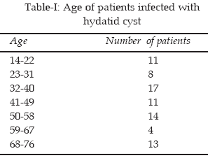
A total of seventy eight patients 34 (43.5%) male and 44 (56.5%) female were enrolled in this study. Majority of the patients 31.5% were in the group of 32-40 years.(Table-I)
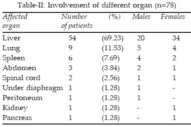
Liver was the most common organ invovled i.e (69.23%). The involvment of other organs was lung (11.53%), spleen (7.69%), abdomen (3.84%), spinal cord (2.56%), under diaphragm (1.28%), peritoneum (1.28%), kidney (1.28%) and pancreas (1.28%) (Table-II).
DISCUSSION
Hydatid disease is a medical and public health problem, especially in endemic parts of the world. The transmission of the disease mostly depends on the close relationship between human beings and canine carnivores. Humans are intermediate hosts and are being exposed to the parasite by fecal-oral and hand-to- mouth spread ways.
1 Iran is an important endemic focus of cystic hydatid disease.2 Investigations on echinococcosis, which have been performed in this country showed the presence of infection among human.14-19 According to the result of this study, females were found more infected with hydatid cysts than males. Different organs are involved with cyst, however, liver is the most affected organ. During a study carried out by Fattahi et al. between 1991-1997.20 In Markazi province women had more hydatid surgeries than men and the most affected organ was liver. Nilforoushan et al21 In Isfahan and Moradi & Majidpoor22 in Kordestan reported similar results. However, Adibpoor et al.23 in Tabriz found more hydatid surgeries in men than women and lung was the most affected organ. In conclusion, echinococcosis is still an important health problem in Iran that needs further studies. Therefore, accurate information on the distribution of the disease is first step for the control and prevention. Moreover, it is necessary that in each province the role of different intermediate hosts and the strains of E .granulosus in human and animals be investigated.REFERENCES
1. Smith DM, Rickman LS. Echinococcosis: A drop of water � review on human disease, diagnosis, and managemant. Infect Disease Clin Practice 2001;10:355-9.
2. Harandi MF, Hobbs RP, Adams PJ, Mobedi I, Morgan � Ryan UM, Thompson RC. Molecular and morphological characterization of Echinococcus granulosus of human and animal origin in Iran. Parasitol 2002;125( 4):367-73.
3. Anonymous. Guidelines for surveillance, prevention andcontrolofechinococcosis /hydatidosis.WHO/FAO/UNEP/8128. 1981.
4. Dalimi AGH, Motamedi M, Hosseini B, Mohamm-adian H, Malaki Z, Ghamari, et al. Echinococcosis / hydatidosis in western Iran. Vet. Parasitol 2002;105:161-71.
5. Horchani AY, Nouria, Kabier I. Hydatid cyst of the kidney: A report of 147 controlled cases. Eur Urol 2000;33:461-7.
6. Macpherson CNL. An active intermediate host role for man in the life cycle of Echinococcus granulosus in Turkana, Kenya. Am J Trop Med Parasitol 1983;31:397-404.
7. Romos G, Orduna A, Garcia-yuste M. Hydatid cyst of the lung: Diagnosis and treatmant. World J Surg 2001;25:46-57.
8. Tuzun M, Hekimoglu B. Various locations of cystic and alveolar hydatid disease. CT Appearances. J Comput Assist Tomogr 2001; 25: 81-7 .
9. Akhan O, Ozmen MN, Dincer A, Sayek I, Gocmen A. Liver hydatid disease: long-term results of percutaneous treatment. Radiology 1996;198:259-64.
10. Kammerer WS, Schantz PM. Echinococcal disease. Infect Dis Clin North Am 1993;7:605-18.
11. Ammann RW, Eckert J, Cestods. Echinococcus Gastrol Clin North Am 1996;25:655-89.
12. Garcia LS, Bruckner DA. Tissue cestodes: larval form. In: Diagnostic medical Parasitology. 4ed. Washington, DC. ASM press 2001;387-403.
13. Palmer SR, Biffin AH, Crig PS, Walters TM. Control of hydatid disease in Wales. BMJ 1993;12:674-5.
14. Bastani B, Dehdashti F. Hepatic hydatid disease in Iran with review of the literature. Mt Sinai J Med 1995;62-9.
15. Kelsey DS, Shaffner LS. Pulmonary echinococcosis: the geographic history scores again. NC Med J 1984; 45 (4):218-19.
16. Panahi F. Costal echinococcosis. Report of a case and review of the literture. Sem. Hop 1978;8-15(43-44):1389-92.
17. Lomheh F. Immunofluorescence in the serodiagnosis of human hydatidosis. Acta Med Iran 1997;20(1-2):27-35.
18. Sabbaghian HN, Hoghochi, Ghaderian E. A survey on the prevalence of echinococcosis in shahrkord Iran. Bull Soc Pathol Exot Filials, 1975;68(6):547.
19. Nasseh GA, Khadivi B. Epidemoilogical and clinical aspects of echinococcosis in east Iran. J Trop Med Hyg. 1975;78(6):120-22.
20. Fattahi F, Davami M, Mosayyebi M, Study on hydatid cysts surgeries in Markazi province, Iran. Proceedings of 3rd National Congress of Medical Parasitology, IRAN, 27 Feb.-1 Mar. 2001; SARI, IRAN.
21. Nilforoushan MR, Davoodi VS, Kia I EB. Mobedi, Study on Hydatid cysts surgeries in Isfahan Educational centers during 1996-2001. 13th Iranian congress on Infectious Disease and Tropiacal / Medicine 11-15 Dec, 2004.
22. Moradi G, Majidpoor MS. Epidemiological study on Hydatid cysts in Kordestan province, Iran. during 2000-2004. 13th Iranian congress on Infectionus Disease and Tropical / Medicine 11-15 Dec. 2004.
23. Adibpoor MR, Jamali, Sarkarty F. Survey on hydatid cysts surgeried in Tabriz (Iran) Hospitals during 1991-1997. Proceedings of 3rd National Congress of Medical Parasitlogy, IRAN, 27 Feb. 1Mar. 2001; SARI, IRAN.
HOME | SEARCH | CURRENT ISSUE | PAST ISSUES
Professional
Medical Publications
Room No. 522, 5th Floor, Panorama Centre
Building No. 2, P.O. Box 8766, Saddar, Karachi - Pakistan.
Phones : 5688791, 5689285 Fax : 5689860
pjms@pjms.com.pk