|
||||
|
Published by : PROFESSIONAL MEDICAL PUBLICATIONS |
||||
|
ISSN 1681-715X |
||||
|
||||
|
- |
||||
|
ORIGINAL ARTICLE |
||||
|
- |
||||
|
Volume 24 |
January - March 2008 |
Number 1 |
||
|
|
||||
|
|
||||
Shoulder Instability: The Role of MR Arthrography
in
Diagnosing Anteroinferior Labroligamentous Lesions
Our experience at King Hussein Medical Center
Asem Al Hiari1
ABSTRACT
Objective: To determine the reliability and accuracy of magnetic resonance arthrography of the shoulder for the diagnosis of anteroinferior labroligamentous lesions in patients with gleno-humeral joint instability.
Methodology: This retrospective study was performed at King Hussein Medical Center in Jordan. Twenty eight patients who underwent shoulder MR arthrogram and arthroscopy during a 22-month period were reviewed. All the twenty eight patients had history of previous shoulder dislocation and clinical suspicion of anteroinferior labroligamentous lesions and glenohumeral joint instability. The series included 24 males and 4 females. The mean average age of the patients was 29 years. All patients underwent shoulder MR arthrogram and the results of MR arthrogram were compared with the arthroscopic findings which were used as the reference standard. MR arthrograms were analyzed for the presence and type of labroligamentous injuries which include (Bankart, anterior labral periosteal sleeve avulsion [ALPSA], Perthes, glenolabral articular disruption [GLAD], or nonclassifiable lesion). Sensitivity, specificity & accuracy for the detection & classification of anteroinferior labroligamentous lesions with MR arthrography were calculated.
Results: At arthroscopy, 21 anteroinferior labroligamentous lesions were diagnosed, including 15 Bankart lesions, three ALPSA lesions, two Perthes lesions and one GLAD lesion. Seven labral lesions were nonclassifiable at arthroscopy, all of which occurred after a history of chronic instability. When compared with arthroscopic findings, Shoulder MR Arthrography had two false-negative results (sensitivity, 92.8%) and no false-positive results. The sensitivity of shoulder MR Arthrography in detecting anteroinferior labroligamentous lesions was 92.8% (26/28) and specificity was (100%). The overall accuracy of Shoulder MR Arthrography in detecting labroligamentous lesions in this study was 90.5% (19/21).
Conclusion: MR arthrography of the shoulder is reliable and accurate in classification of acute and chronic anteroinferior labroligamentous injuries.
KEY WORDS: Magnetic resonance (MR), Arthrography, Shoulder, Arthrography.
Pak J Med Sci January - March 2008 Vol. 24 No. 1 6-11
1. Dr. Asem A. AL-Hiari, DMRD, FRCR,
Consultant Musculoskeletal Radiology,
Radiological Department,
King Hussein Medical Center, Jordan.
Mailing Address: P.O. Box 961802,
Amman 11196, Jordan.
Correspondence
Dr. Asem A. AL-Hiari,
E-mail: ahiari@hotmail.com
* Received for publication: August 11, 2007
* Revision Received: November 15, 2007
* Revision Accepted: November 22, 2007
INTRODUCTION
The shoulder joint is a simple structure that provides complex function. It is the most mobile joint of the body, and it is also the joint that is most frequently dislocated. Normal shoulders have a certain degree of laxity due to minimal bony restraint of the joint, which in turn allows the widest range of motion of any joint in the body. When shoulder dislocation occurs in adolescents and children, it may be a very bad experience for the patient. Although certainly not life threatening, recurrent subluxation or dislocation is clearly lifestyle threatening and can effectively disable an otherwise active individual. The rate of recurrence in later years is at least 70%.
1 One fourth of all patients with dislocations present with a family history of the same problem. 95% of shoulder dislocations are anterior which often lead to recurrent anterior glenohumeral joint instability.1-5Anteroinferior dislocation is the most frequent cause of shoulder instability. The glenohumeral ligaments, particularly the inferior glenohumeral ligament, are currently believed to represent the major passive stabilizers of the shoulder.
2-8 The glenoid labrum functions more as a site of ligamentous attachment than increasing the depth to the glenoid fossa. A number of variants of anteroinferior labroligamentous lesions (Bankart and Bankart variant lesions) have been described.1-10 The classical Bankart lesion is described as detachment of the anteroinferior labrum with its associated glenohumeral ligament complex. Neviaser described the anterior labral periosteal sleeve avulsion (ALPSA) lesion as a tear of the anteroinferior labrum without rupture of the anterior scapular periosteum.11 The Perthes lesion is a labroligamentous avulsion, as well, but with medially stripped intact periosteum. The glenolabral articular disruption (GLAD) lesion represents a superficial tear of the anterior labrum attached to a fragment of articular cartilage without associated capsuloperiosteal stripping.1,3-6,9,11-13Since different types of anterior labroligamentous lesions require different surgical procedures, preoperative determination of lesions is of great importance. The purpose of this study was to retrospectively evaluate the accuracy of magnetic resonance (MR) arthrography in the diagnosis and classification of anteroinferior labroligamentous injuries by using arthroscopy as the reference standard.
METHODOLOGY
Patients: Twenty eight patients were studied retrospectively during a 22 months period at King Hussein Medical Center, Amman, Jordan. The criteria used to include the patients were (a) clinical suspicion of anterior labroligamentous lesions in patients with history of shoulder dislocation, (b) MR arthrography of the shoulder performed at our department, and (c) arthroscopy done at our hospital by an expert shoulder orthopedic surgeon. The study included 24 males, and 4 females. The mean average age of the patients was 29 years. All patients underwent shoulder MR arthrogram and the results of MR arthrogram were compared with the arthroscopic findings which were used as the reference standard. Arthrogram and MR Imaging Studies Injection of contrast media for MR arthrography was performed under fluoroscopic guidance. An anterior approach was used by a 22-gauge needle. The Intraarticular positioning of the needle was confirmed by injecting small amount of iodinated contrast media. Then 10-15ml of a prepared solution of normal saline and Gadolinium with a concentration of 2.5mmol/L was injected. MR imaging was initiated within 30 minutes after contrast injection. MR imaging was performed with a 1.5-T scanner (Symphony; Siemens Medical Systems, Germany). Shoulder coil was used with the arm in a neutral position. Our MRI protocol for shoulder arthrogram includes (Coronal oblique, sagittal oblique and transverse T1-weighted spin-echo sequences and a coronal oblique T2-weighted fast spin-echo along with coronal oblique T1-weighted fat saturation sequences). For all pulse sequences, parameters were a section thickness of 4mm with a 0.2-mm intersection gap, a matrix size of 256 x 256 and a field of view of 14–16cm. MR arthrograms were analyzed for the presence and type of labroligamentous injuries which include (Bankart, anterior labral periosteal sleeve avulsion ALPSA, Perthes, glenolabral articular disruption GLAD, or nonclassifiable lesion).
Data Analysis: We studied the presence or absence of Bankart and Bankart variant lesions were studied and then compared the result with the surgical findings in each patient and determined the number of true-positive, true-negative, false-positive and false-negative imaging results. The sensitivity, specificity, and accuracy of each imaging modality were calculated. The limitations of the study are small sample size with limited statistical analysis. However, we reviewed the false-negative cases twice to ensure that there were no incorrect interpretations.
RESULTS
The number of true-positive, true-negative, false-positive, and false-negative cases, as well as the sensitivity, specificity, and accuracy, with each MR imaging study in the diagnosis of labroligamentous Lesions were calculated.
At arthroscopy, 21 anteroinferior labroligamentous lesions were diagnosed, including 15 Bankart lesions Fig (1,2),3 ALPSA lesions(Fig-3), 2 Perthes lesions (fig-4), and 1 GLAD lesion (Fig-5). Seven labral lesions were nonclassifiable at arthroscopy, all of which occurred after a history of chronic instability. When compared with arthroscopic findings, Shoulder MR Arthrography had two false-negative results (sensitivity, 92.8%) and no false-positive results. The sensitivity of Shoulder MR Arthrography in detecting anteroinferior labroligamentous lesions was 92.8% (26/28), and specificity was (100%). The overall accuracy of Shoulder MR Arthrography in detecting labroligamentous lesions in our study was 90.5% (19/21).
The reason behind the first false-negative case was not full distention of the joint likely due to inconvenience of the patient during joint filling, however in the second false-negative case the report of the absence of the anterior labrum as a normal variant (Buford complex) proved arthroscopicaly as complete detachment of the anterior labrum ( Fig 6) .
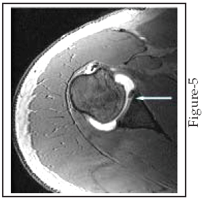
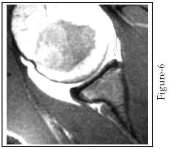
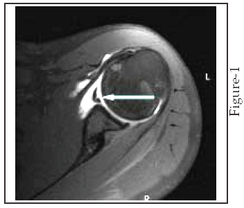
Fig-1: Bankart Lesion. An axial T1 weighted fat saturation spin echo MR arthrogram in 25 years male patient with history of shoulder dislocation. It shows avulsion and displacement of labroligamentous complex from the anteroinferior aspect of the glenoid, with complete distortion of the scapular periosteum, giving a typical appearance of fibrous Bankart lesion (arrow).
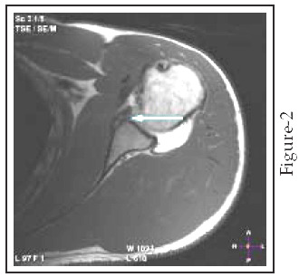
Fig-2: Bony Bankart Lesion. An axial T1 weighted spin echo MR arthrogram in a 31 years male patient with history of recurrent anterior shoulder dislocation .It shows fracture through the anteroinferior aspect of the bony glenoid together with an avulsed anteroinferior labrum (arrow) which is referred to as an osseous Bankart lesion.
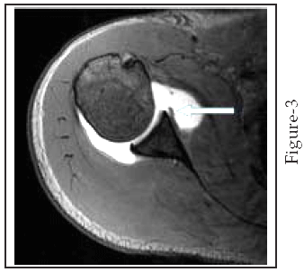
Fig-3: ALPSA Lesion. T1-weighted MR arthrogram of the left shoulder in a 24-year-old man with chronic shoulder instability. It demonstrates medial displacement of the labral ligamentous attachment (arrow) consistent with ALPSA lesion.
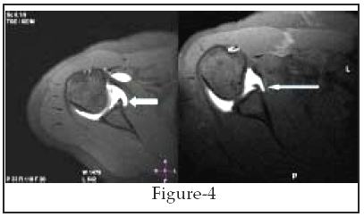
Fig-4: Perthes Lesion. An axial T1 weighted spin echo MR arthrogram with fat saturation shows an undisplaced avulsed anteroinferior labrum with medial stripping of an intact periosteum consistent with a Perthes lesion (arrow). This lesion is better visualized when stress is applied such as during abduction and external rotation ABER position.
Fig-5: GLAD Lesion. An axial 3D gradient echo shoulder MR arthrogram image in a 39-year-old man with chronic shoulder instability demonstrates a chondral lesion with an associated injury of the anteroinferior labroligamentous complex (arrow). The lesion was interpreted as a GLAD lesion at MR arthrography and arthroscopy.
Fig-6: An axial T1-weighted MR arthrogram for an 18 year male patient with shoulder instability. The glenoid labrum has a rounded and may be irregular contour (small arrow). This case was reported as Buford complex (normal variant) but it proved to be a Bankart lesion by arthroscopy.
DISCUSSION
Glenohumeral instability refers to symptomatic subluxation or dislocation of the humeral head in relation to the glenoid fossa. Anteroinferior instability is the most common type occurring in 95% of all patients. The remaining 5% of patients have posterior shoulder dislocation.
1-5,9 MR arthrography is increasingly recognized as the examination of choice in glenohumeral instability providing demarcation of complex anatomic structures of the joint and demonstration of subtle abnormalities, along with excellent delineation of associated intra-articular lesions.9,11,14-19 MR arthrography extends the capabilities of conventional MR imaging because contrast solution distends the joint capsule, outlines intraarticular structures, and leaks into abnormal areas. MR arthrography is possible in any joint in which conventional arthrography is performed.4,7,10,11,13 One of the major advantages of MR arthrography is visualization of the labral-ligamentous complex, which consists of the glenoid labrum in combination with the superior, middle, and inferior glenohumeral ligaments. The glenohumeral ligaments reinforce the joint capsule and function as a unit with the glenoid labrum, which anchors the ligaments to the glenoid rim.10,11,17,19-22 Normal glenohumeral ligaments have to be distinguished from torn labra. The ligaments can be followed away from the glenoid rim until they merge with the distended capsule. On the other hand, torn labral fragments can be followed back to the glenoid rim on sequential MR arthrographic images.In 1923, Arthur Bankart described the lesion of traumatic shoulder dislocation in the British Medical Journal, as follows: "The essential lesion is the detachment of the capsule from the fibro-cartilaginous glenoid ligament" and defined an "avulsed fibrous and fibrocartilaginous fragment" from the glenoid margin as the essential lesion of recurrent shoulder dislocation.
10 The avulsion of the labral-ligamentous complex from the anteroinferior aspect of the glenoid is termed a Bankart lesion or an osseous Bankart lesion if it is accompanied by a glenoid bony fracture. Several variants (Bankart variant lesions) including the following: Anterior Labral Periosteal Sleeve Avulsion (ALPSA) lesion, Perthes lesion and Glenolabral Articular Disruption (GLAD) lesion have been described. MR is essential for diagnosis of these variants.1,3,6,9,11,19 The Bankart lesion can be diagnosed if contrast medium is interposed between the glenoid and the detached labroligamentous complex. Loss of the triangular shape and increased signal intensity of the anteroinferior labrum are additional criteria for Bankart lesions, but only complete detachment of the labroligamentous complex can be assessed as a Bankart lesion.1,3-6,9,11,13 The presence of an associated adjacent glenoid rim fracture is referred to as an osseous Bankart lesion. A Hill-Sachs impaction deformity or fracture involving the posterosuperior humeral head is a frequently associated finding.The ALPSA lesion is characterized by the torn anteroinferior labrum which is displaced inferomedially by the inferior glenohumeral ligament and rolling up like a sleeve.
1,6,9,11,21,22 It differs from the classic Bankart lesion because the avulsed anterior labroligamentous structure is displaced medially along with an intact anterior scapular periosteum. Healing by fibrosis may occur in an abnormal anatomical location leaving a deformed and redundant labrum.1,10,13Criteria for a Perthes lesion include a nondisplaced tear of the anteroinferior labrum that is attached by a linear structure with decreased signal intensity believed to be the intact medial periosteum. As a result of the redundant intact periosteum, recurrent anterior instability may occur as the humeral head moves into this lax portion of the joint.
13,17,20Criteria for the glenolabral articular disruption (GLAD) lesion include a superficial tear of the anteroinferior labrum with an adjacent glenoid articular cartilage injury. The pattern of chondral injury can range from a cartilaginous flap tear to a depressed osteochondral injury of the articular cartilage and the underlying bone. This lesion results from impaction of the humeral head against the glenoid rather than from dislocation and is usually a stable lesion because of the non-displaced labrum and intact inferior glenohumeral ligament.
1,3-6,9 If lesions do not meet the MR criteria for Bankart, ALPSA, Perthes, or GLAD lesions, they are categorized as nonclassifiable at MR image analysis.To our knowledge, the frequencies of Bankart and Bankart variant lesions in a larger series of patients with acute and chronic anterior instability, excluding atraumatic and multidirectional instability have not yet been published.
1,3, 4,11,13-17 In this study, which included patients with acute and chronic instability, a considerable number of nonclassifiable lesions were found at arthroscopy 7/28, all of these lesions occurred in patients with a long-standing history of chronic instability with scar tissue formation and advanced degenerative changes in the labroligamentous complex. The classic Bankart lesion was the most frequent type of classifiable lesion15/28, followed by ALPSA 3/28 and Perthes lesions 2/28. Only one GLAD lesion was found in this series. With a sensitivity of 92.8% and a specificity of 100%, MR arthrography is highly effective in the detection of labroligamentous lesions. In this regard, the results are comparable with those of previous investigations on the diagnostic accuracy of MR arthrography. The study results compare well with the results of studies by Chandnani et al14 Palmer et al15 Palmer and Caslowitz16 and Tirman et al17 who reported sensitivities between 89% and 96% for the detection of anterior labral tears.14-17 In this study, imaging features of nonclassifiable lesions reflected the changes caused by repetitive injuries with labral tears, resynovialization, and scar tissue formation. Imaging features were a swollen inferior glenohumeral ligament complex with increased signal intensity without differentiation of the anterior labrum.Limitations of the study: The present study had several limitations. First, although arthroscopy was the best reference standard available for this study, it is an operator-dependent method, and arthroscopic findings cannot be reproduced in a retrospective analysis. In addition, findings at arthroscopy could have been biased by the availability of MR reports. Second, the limited number of patients used at this study. The reason for this is that the MR arthrogram has recently been introduced in the hospital where this study was conducted. Third, no assessment of interobserver variation; since all MR arthrograms were analyzed by one radiologist (the author) as only one musculoskeletal radiologist is currently working in the institute. And lastly the number of ALPSA, Perthes and GLAD lesions was too small to allow further statistical analysis.
CONCLUSIONS
MR arthrography is an effective method for detection and classification of anteroinferior labroligamentous lesions in patients with acute and chronic shoulder instability. In a substantial number of patients with a long-standing history of instability, classification of lesions was impossible at arthroscopy because of scar tissue formation and advanced degenerative changes in the labroligamentous complex.
ACKNOWLEDGMENT
The author would like to thank Professor Essam Dahabreh, Consultant orthopedic Surgeon Farah Center for Orthopedic, King Hussein Medical Center, Jordan, for his help in conducting this study.