|
|
||||
|
Published by : PROFESSIONAL MEDICAL PUBLICATIONS |
||||
|
ISSN 1681-715X |
||||
|
||||
|
- |
||||
|
ORIGINAL ARTICLE |
||||
|
- |
||||
|
Volume 25 |
January - March 2009 |
Number 1 |
||
|
|
||||
|
|
||||
|
|
||||
|
Published by : PROFESSIONAL MEDICAL PUBLICATIONS |
||||
|
ISSN 1681-715X |
||||
|
||||
|
- |
||||
|
ORIGINAL ARTICLE |
||||
|
- |
||||
|
Volume 25 |
January - March 2009 |
Number 1 |
||
|
|
||||
|
|
||||
Complications of laparoscopic cholecystectomy
at Isra University Hospital, HyderabadWaseem Memon1, Tariq Wahab Khanzada2,
Abdul Samad3, M. Hussain Laghari4ABSTRACT
Objective: The main objective was to determine the complications of laparoscopic cholecystectomy (LC) and its causes at Isra University Hospital, Hyderabad.
Methodology: This was a retrospective study carried out from July 2005 to June 2007. Data of all patients undergoing laparoscopic cholecystectomy during the study period and fulfilling the selection criteria was collected and analyzed retrospectively.
Results: A total of 216 patients underwent laparoscopic cholecystectomy with an overwhelming majority of females. The overall complication rate was 5%. The complications included bleeding (4/216, 1.8%) from cystic artery and gall bladder bed, port site infection (4/216, 1.8%), bile duct injury (2/216, 0.9%) and colonic injury (1/216, 0.4%). The common causes of these complications were accidental injury to cystic artery, gross spillage of infected bile and erroneous clipping of common bile duct.
Conclusions: Bleeding and Port site infections were the commonest complications followed by common bile duct and colonic injuries. The commonest cause of bleeding was cystic artery injury whereas the commonest cause of port site infection was gross spillage of infected bile.
KEY WORDS: Laparoscopic cholecystectomy, Gall stones, Complications.
Pak J Med Sci January - March 2009 Vol. 25 No. 1 69-73
How to cite this article:
Memon W, Khanzada TW, Samad A, Laghari MH. Complications of laparoscopic cholecystectomy at Isra University Hospital, Hyderabad. Pak J Med Sci 2009;25(1):69-73
1. Dr. Waseem Memon FCPS,
Assistant Professor Surgery,
2. Dr. Tariq Wahab Khanzada FCPS, FRCS,
Assistant Professor Surgery,
3. Dr. Abdul Samad FCPS,
Associate Professor Surgery,
4. Prof. M. Hussain Laghari FRCS,
Professor of Surgery,
1-4: Isra University Hospital,
Hyderabad, Pakistan.
Correspondence
Dr. Waseem Memon,
Assistant Prof. Surgery, Isra University Hospital,
P.O. Box: No. 313, Hala Road,
Hyderabad, Sindh - Pakistan.
E-mail: dr_waseem1973@hotmail.com
* Received for Publication: May 30, 2008
* Revision Received: November 20, 2008
* Revision Accepted: November 22, 2008
INTRODUCTION
Gall stones are major health problem worldwide. Phillip Mouret performed the first video-assisted laparoscopic cholecystectomy in Lyon, France.
1 Laparoscopic Cholecystectomy (LC) has caught the imagination of surgical community and we have moved from a position of skepticism to the point where the instrument makers are unable to keep pace with the surgical demand.2 Laparoscopic cholecystectomy has been generally accepted as the gold standard treatment for symptomatic gallstones disease. Several studies have shown the efficacy and safety of the procedure as well as the advantages such as reduced hospital stay, earlier recovery, less intra-abdominal adhesions and a better cosmetic outcome.3 LC can also be performed safely as a day care procedure.4 Unfortunately, this minimally invasive technique is associated with a higher incidence of complications.5,6 Formerly limited to uncomplicated cholelithiasis horizon of indications has progressively extended and at present, very few patients require the conventional ‘open approach’. The operation usually requires general anesthesia and is subject to same risks and complications as an open cholecystectomy, in addition to some complications specific to laparoscopic procedure like vascular or visceral injuries, bleeding, common bile duct (CBD) injury etc. The overall frequency of major complications is less than 5%.7 However, the incidence of complications is strongly related to the experience and recently a decrease in complication rate has been reported.8,9The main aim of this study was to determine the complications of LC and their causes at Isra University Hospital, Hyderabad.
METHODOLOGY
Medical records of all patients who underwent LC at Isra University Hospital, Hyderabad from July 2005 to June 2007 were reviewed retrospectively. Data recorded included demographic information, past medical history, indication for operation, duration of operation, operative findings and reason for conversion, peroperative complications and postoperative complications. Patients having history of jaundice, common bile duct dilatation (>8 mm in diameter on ultrasound), choledocholithiasis, pancreatitis, bleeding disorders, positive hepatitis B and hepatitis C viruses, sepsis, or malignancy were excluded from the study.
Preoperative work up included a complete blood count, blood urea, blood sugar, electrolytes, liver function tests, hepatitis profile, X-ray chest and ultrasound of abdomen. All patients were properly assessed by the anaesthetists preoperatively. All patients scheduled for elective cholecystectomy were admitted one day prior to surgery. Informed consent was taken and patients were fully informed about the nature of procedure, possible risks and complications and possibility of conversion to open procedure depending upon the operative findings. The operation was performed with standard four port technique, using carbon dioxide for peritoneal cavity insufflation. The Veress technique was used to create pneumoperitoneum. Cystic artery and cystic duct were skeletinized and clamped with metallic clips separately. The gall bladder was then dissected from its liver bed and removed through the epigastric port. Irrigation and suction was done in cases of bleeding and bile leakage from gall bladder perforation. Drain was placed in subhepatic space. After decompression of pneumoperitoneum abdominal closure was done with vicryl 2/0 for a rectus sheath and fine prolene suture for skin closure. Conversion to open procedure was carried out either due to complication or operative difficulty. Antibiotic prophylaxis was ensured with three intravenous first generation cephalosporin. Resected gall bladder specimens were sent for histopathological examination. Postoperative analgesia was achieved with intramuscular diclofenac sodium 75mg twice a day. All patients had oral liquids in the evening after operation and were encouraged to proceed with food, provided there was no nausea and vomiting. Any peroperative complications and post operative complications were noted. Drain was removed after 24 hours of operation, if there was no significant collection. Patients were discharged on next day if there was no problem. Skin sutures were removed on 8
th postoperative day. Follow up examination in surgical OPD was performed at one week, one month and three months intervals. The complications found during the follow up period were investigated and managed accordingly.The collected data was analyzed with especial reference to the frequencies of the complications and their causes.
RESULTS
This study included 216 patients comprising of 183 women and 33 men with female to male ratio of 5.5:1. The mean age was about 35 years with range of 20-70 years. Indications for Laproscopic Cholecystectomy (LC) included acute cholecystitis 42 (20%) and symptomatic gallstones 174 (80%). The overall complication rate was 5% whereas the mortality rate was 0%. Bleeding was the most frequent complication observed in 4 (1.8%) patients undergoing LC. Bleeding resulted from accidental injury to cystic artery in three patients and from gall bladder bed during removal of gall bladder in one patient. The bleeding from gall bladder bed was controlled by electrocautery laparoscopically and cystic artery injury in one patient was controlled by clipping of cystic artery laparoscopically. In remaining two patients with cystic artery injury, bleeding could not be controlled laparoscopically and required conversion to open procedure to control bleeding from damaged cystic artery. Four (1.8%) patients developed surgical port site infection. Out of these one patient had wound infection of the umbilical port and two had wound infection of the epigastric port. The patient having umbilical port infection was obese and two patients having epigastric port infection, had gross spillage of infected bile during extraction of gall bladder. All these patients were managed by antibiotics and local dressings. The fourth patient with port site infection had umbilical stitch sinus which was managed with opening of the wound, removal of subcutaneous stitch and appropriate antibiotics. Two patients (0.9%) had CBD injury and in both cases, the CBD was clipped as it was erroneously believed to be the cystic duct. This mistake went unrecognized intraoperatively and it was only suspected after three days when patients started to develop obstructive jaundice. Patients were re-operated, clips were removed, CBD exploration was done and T tube was placed in the CBD. In both of these patients, the post operative recovery was smooth and uneventful. After two weeks, T-tube cholangiogram was done showing free flow of contrast in duodenum with normal CBD caliber and then the T-tube was removed. One patient had colonic injury which was identified during the procedure requiring conversion to open method and the injured colonic wall was repaired primarily.
DISCUSSION
Complications from LC fall into two categories. Those directly resulting from the laparoscopic intervention like injury from Veress needle, trocar injuries etc and those associated with the operation itself like bile duct injury etc. The rates of complications in LC were much higher during the initial era of laparoscopy, technical limitation being the main reason. Complications can be seen during the creation of pneumoperitoneum especially with Veress needle. The incidence of major visceral or vascular injury is rare; but literature does report such injuries. Deziel et al
3 reported 13 cases of aortic injuries in a study of about 77604 operations. These insertional complications can be further minimized by using open technique for creating pneumoperitoneum.The incidence of CBD injury is strongly related to experience and a decrease in CBD injury has been reported.
9,10 Despite the knowledge on mechanism of injury and many reports which stress the value of preventive measures such as intraopreative cholangiography, the reported incidence still varies between 0-1%.11-13 In this study, the CBD injury was observed in two (0.9%) patients. In both of these patients, CBD was clipped and problem was identified postoperatively. Patients were explored again, clips were removed and T – tube was placed after exploration of CBD. These injuries can be prevented by adequate surgical experience, careful dissection and proper case selection. However, as surgeon obtains more experience, the frequency of this complication should decrease.The visceral injury can occur during introduction of Veress needle or trocar injuries as well as over judicious dissection of adhesions. Visceral damage may be evident peroperatively or remain unrecognized during operation and later manifest as peritonitis, abscesses or sepsis. In this study, one patient had colonic injury which was identified peroperatively and the primary repair was done. The overall incidence of serious visceral injuries during LC is reported to be 0-5% in the published literature.
3,7,9Bleeding is a frequently encountered and dangerous complication of laparoscopic cholecystectomy. The bleeding may occur during Veress needle insertion, dissection of gall bladder, slippage of clip from cystic artery or injury to cystic duct. In our study, four patients had bleeding. Only two patients needed conversion to open procedure as bleeding was not controlled laparoscopically. Minor bleeding can be controlled by pressure and application of suture or diathermy. Factors contributing to operative site bleeding may include inadequate exposure, acute inflammation, portal hypertension, adhesion, coagulopathy and rough technique.
8,9,14,15 Local study16 has reported bleeding in about 3.18% of the patients whereas another study by Usal et al17 reported major vessel injury (aorta, portal vein & inferior vena cava) in about 0.11% of the patients.Wound infection, usually involving the umbilical cannulation site through which the gall bladder is extracted, occurs in 0.3% -1% of cases.
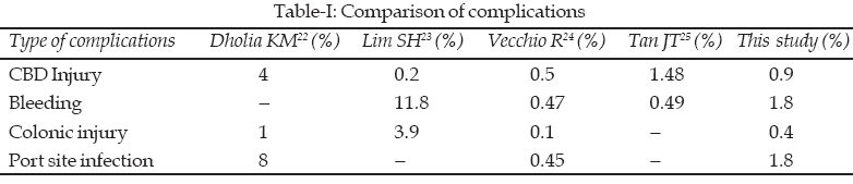
18-20 In this study, four patients (1.8%) developed surgical port site infections, which were managed by simple dressings and oral antibiotics. One of these patients had umbilical stitch sinus which was managed by opening of the wound, removing subcutaneous stitch and appropriate antibiotics. Wound infection was more common in obese patients. The hospital infection committee was informed about all these cases and necessary actions were taken. The reported incidence of surgical port site infection varies between 0.5 to 1%.18-21 The comparison of complications observed in this study with those observed in literature is mentioned in Table-I.CONCLUSION
The overall complication rate was 5% and these include bleeding and port site infection as the commonest complications followed by CBD and colonic injuries. The commonest cause of bleeding was cystic artery injury. The causes of port site infection were gross spillage of infected bile, obesity and umbilical stitch sinus.REFERENCES
1. Shamim M, Dahri MM, Memon AS. Complications of laparoscopic cholecystectomy. Pak J Surg 2006;22(2):70-7.
2. Gadacz TR. Update on laparoscopic cholecystectomy, including a clinical pathway. Surg Clin North Am 2000;80:1127.
3. Deziel DJ, Millikan KW, Economou SG, Doolas A, Ko ST, Airan MC. Complications of laparoscopic cholecystectomy: A national survey of 4292 hospitals & an analysis of 77604 cases. Am J Surg 1993;165:9-14.
4. Keulemans YCA, Eshuis J, Haesde H, Wit de LT, Gouma DJ. Laparoscopic cholecystectomy: Day care versus clinical observation. Ann Surg 1998;228:734-40.
5. Go PMNYH, Schol FPG, Gouma DJ. Laparoscopic cholecystectomy in the Netherland. Br J Surg 1993;80:1180-3.
6. McMohan AJ, Fullarton G, Baxter JN, O’Dwyer PJ. Bile duct injury and bile leakage in laparoscopic cholecystectomy. J Am Coll Surg 1995;180:101-25.
7. Jones DB, Soper NJ. Complications of laparoscopic cholecystectomy. Ann Rev Med 1996;47:31.
8. Schol FPG, Go PMNYH, Gouma DJ. Risk factors for bile duct injury in laparoscopic Cholecystectomy: analysis of 49 cases. Br J Surg 1998;81:1786-8.
9. Nair RJ, Dunn DC, Fowler S, McCloy RF. Progress with cholecystectomy: improving results in England & Wales. Br J Surg 1997;84:1396-8.
10. Richardson MC, Bell J, Fullarton JM. Incidence and nature of bile duct injury following laparoscopic cholecystectomy; An audit of 5913 cases. West of Scotland Laparoscopic cholecystectomy Audit Group. Br J Surg 1996;83:1356-60.
11. McMohan AJ, Fullarton G, Baxter JN, O’Dwyer PJ. Bile duct injury and bile leakage in laparoscopic cholecystectomy. Br J Surg 1995;82:307-13.
12. Strasberg SM, Heartl M, Soper NJ. An analysis of the problem of biliary injury during laparoscopic cholecystectomy. J Am Coll Surg 1995;180:101-25.
13. MacFayden BV, Vecchio R, Richrdo AF, Mathis CR. Bile duct injury after laparoscopic cholecystectomy. The United State experience. Surg Endosc 1998;12:315-21.
14. Soper NJ, Dunnegan DL. Laparoscopic cholecystectomy: experience of a single surgeon. World J Surg 1993;17:16-20.
15. Deveney KE. The early experience with laparoscopic cholecystectomy. An analysis of complications. Arch Surg 1993;128:627-32.
16. Arain Gm, Hasan A, Randhawa MH, Malik SA. Laparoscopic cholecystectomy and its complications. A study of 1100 cases. Pak J Gastroenterol 1998;12:1-2.
17. Usal H, Sayad P, Hayek N, Hallak A, Huie F, Ferzli G. Major vascular injuries during laparoscopic cholecystectomy. An institutional review of experience with 2589 procedures and literature review. Surg Endosc 1998;12(7):960-2.
18. Soper NJ, Stockmann PT, Dunnegan DL, Ashley SW. Laparoscopic cholecystectomy. The new gold standard? Arch Surg 1992;127:917-21.
19. Stoker ME, Vose J, O’Mara B, Maini BS. Laparoscopic cholecystectomy. A clinical and financial analysis of 280 operations. Arch Surg 1992;127:589-94.
20. Wittgen CM, Andrus JP, Andrus CH, Kaminski DL. Cholecystectomy. Which procedure is best for the high risk patients? Surg Endosc 1993;7:395-9.
21. Dunn D, Fowler S, Nair R. Laparoscopic cholecystectomy in England and Wales: Results of an audit by Royal College of Surgeons, England. Ann R Coll Surg Engl 1994;76:269.
22. Dholia KM, Memon AA, Sheikh MS. Laparoscopic cholecystectomy: Experience of 100 cases at a teaching hospital of Sindh. J Liaquat Univ Med Health Sci 2005;4(3):105-8.
23. Lim SH, Saleh I, Poh BK. Laparoscopic Cholecystectomy: An audit of our training programme. ANZ J Surg 2005;75(4):231-3.
24. Vecchio R, Macfadyen BV, Latteri S. Laparoscopic cholecystectomy: Analysis of 114,005 cases of United States series. Int Surg 1998;83:215-9.
25. Tan JT, Suyapto DR, Neo EL, Leong PS. Prospective audit of laparoscopic cholecystectomy at a secondary referral centre in South Australia. ANZ J Surg 2006;76(5):335-8.
HOME | SEARCH | CURRENT ISSUE | PAST ISSUES
Professional
Medical Publications
Room No. 522, 5th Floor, Panorama Centre
Building No. 2, P.O. Box 8766, Saddar, Karachi - Pakistan.
Phones : 5688791, 5689285 Fax : 5689860
pjms@pjms.com.pk