|
|
||||
|
Published by : PROFESSIONAL MEDICAL PUBLICATIONS |
||||
|
ISSN 1681-715X |
||||
|
||||
|
|
||||
|
- |
||||
|
ORIGINAL ARTICLE |
||||
|
- |
||||
|
Volume 25 |
January - March 2009 |
Number 1 |
||
|
|
||||
|
||||
|
|
||||
|
Published by : PROFESSIONAL MEDICAL PUBLICATIONS |
||||
|
ISSN 1681-715X |
||||
|
||||
|
|
||||
|
- |
||||
|
ORIGINAL ARTICLE |
||||
|
- |
||||
|
Volume 25 |
January - March 2009 |
Number 1 |
||
|
|
||||
|
||||
Prevalence of Hepatitis-C virus (HCV) among
Thalassemia Patients in Khuzestan Province, Southwest IranMehri Ghafourian Boroujerdnia1, Mohammad Ali Assareh Zadegan2,
Khoda Morad Zandian3, Morteza Haghirizadeh Rodan4ABSTRACT
Objective: The aim of this study was to determine the prevalence of HCV infection among thalassemia patients in Khuzestan province, southwest Iran.
Methodology: A retrospective cross-sectional study was conducted on 206 thalassemia patients referred to the Research Center of Thalassemia and Hemoglobinopathy (RCTH) of Ahvaz Shafa Hospital during March 2006 to April 2007. Demographic data were obtained from the patient files at the hospital. Serum specimens were tested with anti-HCV assays and a nested-PCR technique to assess HCV infection.
Results: Out of 206 patients, 97 (47.1%) and 109 (52.9%) were male and female, respectively, with a mean �SD age of 16.4�6.42 years. The overall prevalence rate of anti-HCV was 28.1% (58/206, 95% CI: 22.4-34.6). Forty six of anti-HCV positive patients (46/58, 79.3%) were also HCV RNA positive. HCV�positive patients were significantly older from HCV-negative ones (p<0.001). In addition, the results indicate that higher prevalence of anti-HCV or HCV RNA were significantly associated with longer duration of transfusion (p<0.003 and p<0.001, respectively).
Conclusion: Although it seems blood donor screening project reduced HCV infection, using more accurate technique is necessary in order to find viral infection and treat thalassemia patients with HCV infection more carefully.
KEY WORDS: Prevalence, Thalassemia, Hepatitis C, Khuzestan.
Pak J Med Sci January - March 2009 Vol. 25 No. 1 113-117
How to cite this article:
Ghafourian Boroujerdnia M, Assareh Zadegan MA, Zandian KM, Haghirizadeh Rodan M. Prevalence of Hepatitis C virus (HCV) among Thalassemia Patients in Khuzestan Province, Southwest Iran. Pak J Med Sci 2009;25(1):113-117.
1. Mehri Ghafourian Boroujerdnia,
Infectious and Tropical Diseases Research Center,
Hemoglobinopathy & Thalassemia Research Center
of Ahwaz Shafa Hospital,
Ahwaz Joundishapour University of Medical Sciences,
Ahwaz - Iran.
2. Mohammad Ali Assareh Zadegan,
3. Morteza Haghirizadeh,
2-3: Department of Immunology, Faculty of Medicine,
Ahwaz Joundishapour University of Medical Sciences,
Ahwaz, Islamic Republic of Iran.
4. Khoda Morad Zandian
4: Hemoglobinopathy & Thalassemia
Research Center of Ahwaz Shafa Hospital,
Shafa Hospital, Ahwaz, Iran.
Correspondence:
Mehri Ghafourian Boroujerdnia,
Infectious and Tropical Diseases Research Center,
Hemoglobinopathy & Thalassemia Research Center,
of Ahwaz Shafa Hospital,
Ahwaz Joundishapour University of Medical Sciences,
Ahwaz - Iran.
E-mail: Mehri_Ghafourian@yahoo.com
* Received for Publication: May 30, 2008
* Revision Received: December 17, 2008
* Revision Accepted: December 20, 2008
INTRODUCTION
Hepatitis C virus (HCV) is the most common cause of post-transfusion hepatitis (PTH) and end-stage liver disease in many countries. Regular blood transfusion in patients with hereditary hemolytic anemia, particularly thalassemia, has improved their overall survival, but carries a definite risk of acquisition of blood-borne virus infections, especially viral hepatitis.
1,2 Moreover, with respect to marked liver iron overload, which is often inevitable in patients on regular blood transfusion, HCV infection have been shown to have a potentiating effect on hepatic fibrogenesis in thalassemic patients.3 Khuzestan province is located in the southwest of Iran, a tropical area with an approximate population of 4.5 million (Census 2004). Khuzestan Province shares a land, river, and sea border with Iraq and Arabian countries along Persian Gulf. Khuzestan has suffered the heaviest damage of all Iranian provinces during a 28-year period including the Iran-Iraq War (1980-1988), the Gulf War (1990-1991), and the 18-year crisis in Iraq (1990-2008).4 Moreover, thalassemia is an important health problem throughout Iran, particularly in Khuzestan province. Therefore, the aim of the present study was to identify the prevalence of HCV infection in thalassemia patients in Khuzestan province.METHODOLOGY
Patients: This cross-sectional study was performed from March 2006 to April 2007 in Khuzestan Province on thalassemia patients attending the Research Center of Thalassemia and Hemoglobinopathies (RCTH) of Ahvaz Shafa Hospital in Khuzestan province. Whole blood specimens were collected from a total of 206 patients, after obtaining an informed consent. Serum samples were separated from the whole blood, aliquotted and stored at -70� C. Demographic data, such as age, duration and number of blood transfusions were obtained from patient records.
Laboratory assays: All sera were screened using anti-HCV assays with third-generation commercial ELISA microplate kits (DIA.PRO, Italy) according to the manufacturer�s instructions. The samples were considered positive when the sample absorbance/cut-off (SA/C) ratio was higher to 1.1 and negative when the SA/C ratio was <0.9 (values given by the manufacturer). Positive samples were confirmed using nested-RT PCR for HCV.
Two hundred microliters of serum was used for HCV-RNA extraction using high pure viral nucleic acid kits (Roche, Germany) according to the manufacturer�s instructions. HCV-RNA was immediately transcribed into cDNA using random primers (Fermentas, Lithuania). cDNA was targeted by a nested-PCR with specific primers for the conserved sequences in the 5` non-coding region (5`-NCR) of HCV.
5-7Statistical analysis
Prevalence and the corresponding 95% confidence interval (95% CI) were calculated with SPSS software version 13.0 (SPSS Inc., Chicago, IL, USA). Data comparisons were performed using the Chi-square, Fisher�s exact test and Student�s t-test. The differences were considered significant if p<0.05.
RESULTS
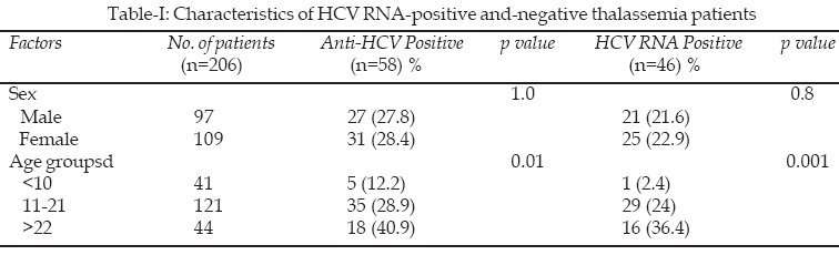
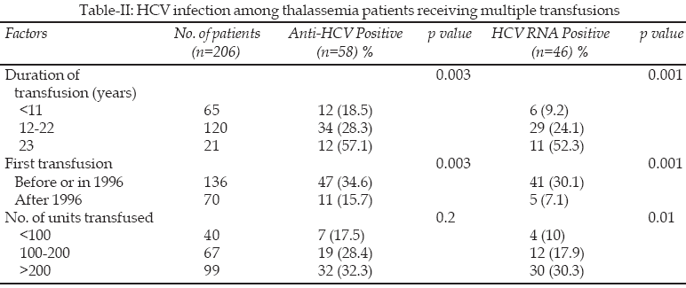
Two hundred six thalassemia patients were tested. There were 97 (47.1%) males and 109 (52.9%) females (Table-I); their mean age (�SD) was 16.4�6.42 years (range 2-34 years). Fifty eight of the 206 patients were anti-HCV positive by ELISA corresponding to a 28.1% (58/206, 95% CI: 22.4-34.6). The mean age of the fifty eight anti-HCV positive patients was higher than that of negative patients (18.98�6.65 vs. 15.47�6.06, p<0.001). No significant statistical difference in anti-HCV seropositivity was observed between males and females (p=1.0). The prevalence of anti-HCV seropositivity (47/136, 34.6%) was significantly higher (p<0.003) among patients who had started to receive transfusions before 1996 when serological screening for anti-HCV antibody had been in introduced to blood banks in Iran, than among patients (11/70, 15.7%) who had started to receive transfusions after 1996 (Table-II). The results indicate the incidence of anti-HCV was significantly associated with longer duration of transfusion (p<0.003).
The prevalence of HCV RNA was 22.3% (46/206, 95% CI: 17.1-28.5). Among the fifty eight anti-HCV positive patients, forty six individuals were positive for HCV RNA. Mean age was higher among HCV RNA-positive patients than among HCV
RNA-negative patients (20.17�6.32 vs. 15.39�6.05, p<0.001). The prevalence of HCV RNA was higher among females (22.9%) than among males (21.6%), although the difference was not significant (p = 0.8) (Table-I). There was a significant difference in the prevalence of HCV RNA (p< 0.001) between subjects who had started to receive transfusions before 1996 (30.1%) and those who had started to receive them after 1996(7.1%). The prevalence of HCV RNA was higher among the patients who had received more than 100 units of blood components (91.3%) than among those who had received less than 100 units (8.7%) (p<0.01) (Table-II).
DISCUSSION
There are more than 25,000 patients with thalassemia major in Iran.
8 It is an important health problem throughout Iran particularly in Khuzestan province. This study showed that the prevalence of anti-HCV in thalassemia patients is 28.1%. A recent report from northern Iran showed a 63.8% prevalence of HCV antibody in thalassemics compared to 0.5% in blood donors. In this report confirmatory immunoblotting test was employed using HCV-positive cases, which showed that 92.6% of samples were positive.9 Karimi et al.,9 from Shiraz, southern Iran, reported 73 of 466 thalassemic children with a history of multiple transfusions (15.7%) positive for anti-HCV. Results from another study on Iranian thalassemic patients, revealed 24.2% of them were anti-HCV positive.1 Previous single-center studies on Iranian thalassemic patients revealed a wide range of 16 - 64% for prevalence of HCV infection.8-10This is the first study to report on the prevalence of HCV infection among thalassemia patients in a war stricken area, Khuzestan province, in Iran. The prevalence of HCV infection in thalassemia patients ranges 33 to 67.3% in some neighboring Arabic countries,
11-13 which share a sea boarder with Khuzestan province. Therefore, comparing our result with these countries, the frequency of HCV infection in our thalassemia population is relatively low.The countries with a higher HCV prevalence in general population had a higher prevalence rate among thalassemia patients, too. For instance, a study in Egypt reported 75% of HCV prevalence among thalassemia patients, considering the fact that the prevalence in their blood donor population was 14.5%.
14 However, in India with a low HCV prevalence among blood donors (1.78%), the prevalence in thalassemics was reported relatively low (25.5%).15 It should be noted that the Iran-Iraq War of 1980-1988, has had a devastating impact on public health. Moreover, during a period of 18 years, due to poor security and living conditions, many Iraqi refugees have crossed over the Iraqi border to Iran, mainly to the southwestern regions.16 This geographical situation, mass immigration from Iraq, where a significantly higher prevalence of anti-HCV has been found among different populations13,17,18 and frequent travels between Khuzestan province to neighboring Arabic countries, all could affect prevalence of HCV in our region.When serologic tests for HCV became available, blood donor screening began to be performed in most countries. In Iran, mandatory anti-HCV screening was introduced to blood banks in 1996. The results of our study showed that anti- HCV seropositivity decreased significantly from 34.6% to 15.7%, after the implementation of screening blood components. It is important to consider that, in spite of the systematic screening of blood donors, testing blood donation recipients for HCV is still important. This finding demonstrates that more efforts should be made to improve blood transfusion safety. Serological tests are used worldwide for the screening of HCV infection. Some of the serological tests may have controversial results. The occurrence of HCV RNA detectable in patients with negative anti-HCV can be a consequence of immunosupression with decreased production of antibodies, or the window period of a recent infection.
19In this study, mean age �SD was significantly (p<0.001) higher in patients with positive HCV antibody (18.98 � 6.65) compared to negative subjects (15.47 � 6.06). The higher rate of HCV infection in older patients, reflecting more frequency of transfusion (p<0.01) and revealed the importance of providing safe blood to reduce the incidence of HCV infection in thalassemic population.
The results of our study showed HCV RNA-positive patients had a significantly longer duration and frequency of transfusion compared with HCV RNA-negative cases (p<0.001 and p<0.01).
However, these observations strongly indicated blood transfusion as the main risk factor for HCV infection acquisition among thalassemic patients, and confirmed the marked efficacy of donor screening in the prevention or viral transmission. The higher rate of HCV infection in older patients with thalassemia and in the subjects who had higher serum ferritin level- all reflecting transfusion of more units of blood- revealed the importance of providing safe blood to reduce the incidence of HCV infection in thalassemia patients.
Forty six of the anti-HCV positive patients (46/58) were also HCV RNA positive. The agreement between anti-HCV and HCV RNA detection was 79.3%, a finding similar to those reported for other populations, i.e., rates ranging from 65 to 86%.
20-22 Failure to detect HCV RNA in serum may be due to various factors such as inactivation of viral RNA during serum collection and storage, fluctuating viremia levels, resolved infection, or false-positive anti-HCV results.20,23In conclusion, although anti-HCV prevalence in thalassemia patients in Khuzestan province is lessr than those found in some other Iranian provinces and neighboring countries, they are still high. The fact that the risk of HCV infection diminished considerably after 1996 demonstrates the value of blood donor screening programs. However, simple measures such as enforced general asepsis rules, careful disinfection and equipment sterilization, routine testing of patients, serial determination of hepatic enzymes should be the common practice in dialysis centers in Iran.
ACKNOWLEDGEMENT
This study was supported by Infectious and Tropical Diseases Research Center and Hemoglobinopathies and Thalassemia Research Center of Ahvaz Shafa Hospital of Ahvaz, Joundishapur University of Medical Sciences.
REFERENCES
1. Alavian SM, Adibi P, Zali MR. Hepatitis C virus in Iran: Epidemiology of an emerging infection. Arch Iranian Med 2005;8(2):84-90.
2. Albertl A, Benvegnu L. Management of hepatitis C. J Hepatol 2003;38 Suppl 1:S104-18.
3. Ardalan FA, Osquei MRF, Toosi MN, Irvanloo G. Synergic effect of chronic hepatitis C infection and beta thalassemia major with marked hepatic iron overload on liver fibrosis: a retrospective cross-sectional study. BMC Gastroenterology 2004;4:17.
4. Assarehzadegan MA, shakerinejad G, Amini A, Rezaee SAR. Seroprevalence of hepatitis E virus in blood donors in Khuzestan Province, southwest Iran. Int J Infect Dis 2008;12(4):387-90.
5. Pham TN, MacParland SA Hepatitis C virus persistence after spontaneous or treatment-induced resolution of hepatitis C. J Virol 2004;78(11):5867-74.
6. Matar GM, Sharara HM, Abdelnour GE, Abdelnoor AM. Genotyping of Hepatitis C Virus Isolates from Lebanese Hemodialysis Patients by Reverse Transcription PCR and Restriction Fragment Length Polymorphism Analysis of 5' Noncoding Region. J Clin Microbiol 1996;34(10):2623-4.
7. Abdelnour GE, Matar GM, Sharara HM, Abdelnoor AM. Detection of Anti-Hepatitis C-Virus Antibodies and Hepatitis C-Virus RNA In Lebanese Hemodialysis Patients. Eur J Epidemiol 1997;13(8):863-7.
8. Mirmomem S, Alvian SM, Hajarizadeh B, Kafaee J, Yektaparast B, Zahedi MAJ et al. Epidemiology of Hepatitis B, Hepatitis C, and Human Immunodeficiency Virus Infections in Patients with Beta-thalassemia in Iran: A Multicenter Study. Arch Iranian Med 2006;9(4):319-23.
9. Ansar MM, Kooloobandi A. Prevalence of hepatitis C virus infection in thalassemia and hemodialysis patients in North Iran, Rasht. J Viral Hepat 2002;9(5):390-2.
10. Karimi M, Ghavanini AA. Seroprevalence of hepatitis B, hepatitis C and human immunodeficiency virus antibodies among multitransfused thalassemic children in Shiraz, Iran. J Pediatr Child Health 2001;37(6):564.
11. Al-Fuzae L, Aboolbacker KC, Al-Saleh Q. Beta thalassemia major in Kuwait. J Trop Pediatr 1998;44(5):311-2.
12. Al-Mahroos FT, Ebrahim A. Prevalence of hepatitis B, hepatitis C and human immune deficiency virus markers among patients with hereditary hemolytic anaemias. Ann Trop Paediatr 1995;15(2):121-8.
13. Al-Kubaisy WA, Al-Naib KT, Habib MA. Seroprevalence of hepatitis C virus specific antibodies among Iraqi children with thalassaemia. East Mediterr Health J 2006;12(1-2):204-10.
14. El-Gohary A, Hassan A, Nooman Z. High prevalence of hepatitis C virus among urban and rural population groups in Egypt. Acta Trop 1995;59(2):155-61.
15. Jaiswal SP, Chitnis DS, Naik G, Artwani KK, Pandit CS, Salgia P, et al. Prevalence of anti-HCV antibodies in central India. Indian J Med Res 1996;104:177-81.
16. Assarehzadegan MA, Shakerinejad G, Noroozkohnejad R, Amini A, Rezaee SAR. Prevalence of Hepatitis C and B Infection and HCV Genotypes among Hemodialysis patients in Khuzestan Province, southwest Iran. Saudi J Kidney Dis Transpl. In press.
17. Khattab OS. Prevalence and risk factors for hepatitis C virus infection in hemodialysis patients in an Iraqi renal transplant center. Saudi J Kidney Dis Transpl 2008;19(1):110-5.
18. Al-Kubaisy WA, Al-Naib KT, Habib MA. Prevalence of HCV/HIV co-infection among haemophilia patients in Baghdad. East Mediterr Health J 2006;12(3-4):264-9.
19. Abutaleb N. Notes on HCV-ELISA test results of the hemodialysis patients. Saudi J Kidney Dis Transpl 2008;19(5):817-9.
20. Alter MJ, Kruszon-Moran D, Nainan OV, McQuillan GM, Gao F, Moyer LA, et al. The prevalence of hepatitis C virus infection in the United States, 1988-1994. New Eng J Med 1999;341(8):556-62.
21. Dow BC, Follett EAC, Munro H, Buchanan I, Roy K, McOmish F, et al. Failure of 2nd- and 3rd-generation HCV ELISA and RIBA to detect HCV polymerase chain reaction-positive donations. Vox Sanguinis 1994;67(2):236-7.
22. Farma E, Boeri E, Bettini P, Repetto CM, McDermott J, Lillo FB , Varnier OE. Single-step PCR in molecular diagnosis of hepatitis C virus infection. J Clin Microbiol 1996;34(12):3171-4.
23. Lok ASF, Gunaratnam NI. Diagnosis of hepatitis C. Hepatology 1997;26(Suppl 1):48S-56S.
HOME | SEARCH | CURRENT ISSUE | PAST ISSUES
Professional
Medical Publications
Room No. 522, 5th Floor, Panorama Centre
Building No. 2, P.O. Box 8766, Saddar, Karachi - Pakistan.
Phones : 5688791, 5689285 Fax : 5689860
pjms@