|
|
||||
|
Published by : PROFESSIONAL MEDICAL PUBLICATIONS |
||||
|
ISSN 1681-715X |
||||
|
||||
|
- |
||||
|
ORIGINAL ARTICLE |
||||
|
- |
||||
|
Volume 22 |
July - September 2006 |
Number 3 |
||
|
|
||||
|
|
||||
|
|
||||
|
Published by : PROFESSIONAL MEDICAL PUBLICATIONS |
||||
|
ISSN 1681-715X |
||||
|
||||
|
- |
||||
|
ORIGINAL ARTICLE |
||||
|
- |
||||
|
Volume 22 |
July - September 2006 |
Number 3 |
||
|
|
||||
|
|
||||
Immuno Histochemistry of Cholesterol Cleft Granulomas
in Non-Specific Interstitial Pneumonia (NSIP)Abol Fazl Barkhordari1, RW Stoddart2, Sheena F. McClure3, John McClure4
ABSTRACT
Cholesterol cleft granulomas with clusters of giant cells were noted to be a common feature of Non-Specific Interstitial Pneumonia (NSIP). The giant cells commonly seen in granulomas are considered to be macrophage polykaryons formed by the fusion of alveolar macrophages attached to the same endocytic material. This study aimed to define the cell populations involved in the granulomas. The granulomas of 16 patients with cryptogenic fibrosing alveolitis (five cases with the histological features of NSIP, five with those of UIP and six cases of respiratory bronchiolitis) were examined histologically and the use of immunohistochemical markers. Granulomas were discrete, compact and present only in alveolar spaces. The granulomas contained central clefts surrounded by mononuclear and multinucleated giant cells, both of which were CD68 positive. The cells outside the granulomas and those lining the adjacent alveolar walls were AE1/AE3 and CAM5.2 positive and CD68 negative. Our observations indicate that cholesterol cleft granulomas occur with high frequency in NSIP, are present within alveolar spaces which are lined exclusively by type II pneumocytes and that external to this the interstitium is fibrotic with accumulations of mast cells. It is our speculation that these are linked in a pathogenetic mechanism related to the progression of NSIP. The results also suggest that the mononuclear and multinucleated cells of cholesterol cleft granulomas are derived from the macrophage-mononuclear cell lineage. The alveolar lining cells are type II pneumocytes which do not contribute to the granuloma cell population. The alveolar lining cells are type II pneumocytes which do not contribute to the granuloma cell population.
Key words: Cholesterol-cleft granuloma, Immuno Histochemistry, NSIP.
Pak J Med Sci July - September 2006 Vol. 22 No. 3 273-276
1. Dr. Abol Fazl Barkhordari
Department of Occupational Health,
Faculty of Health,
Shaheed Sadoughi University of Medical Sciences,
Yazd, Iran.2. Dr. R. W. Stoddart
3. Dr. Sheena F. McClure
4. Dr. John McClure
2-4: Department of the Laboratory Medicine
Academic Group, The Medical School,
University of Manchester, UKCorrespondence:
Dr. Abol Fazl Barkhordari
E-Mail: abfl340@yahoo.co.uk* Received for publication: April 29, 2005
* Accepted: March 12, 2006
INTRODUCTION
The pathological classification of Idiopathic Pulmonary Fibrosis (IPF) - also known as Cryptogenic Fibrosing Alveolitis (CFA) - has been a matter of difficulty and controversy for histopathologists.
1-5 A recent classification by Katzenstein and Myers6 includes Usual Interstitial Pneumonia (UIP), Desquamative Interstitial Pneumonia (DIP) / Respiratory Bronchiolitis Interstitial Lung Disease (RBILD), Acute Interstitial Pneumonia (AIP, Hamman-Rich disease) and Non-Specific Interstitial Pneumonia (NSIP). This last was defined by Katzenstein and Fiorelli7 as an idiopathic interstitial pneumonia with a pathological pattern distinct from UIP, DIP and AIP, although cases were first recognised because they could not be included in the first three categories.In a systematic evaluation of the histopathological features of a series of IPF we noted that half the cases of NSIP contained large numbers of discrete cholesterol cleft granulomas. Examination of the available literature revealed few references to these entities in NSIP. In two of the original series (n=64) of Katzenstein and Fiorelli,
7 loosely formed granulomas, one containing cholesterol clefts, were described. In 12 cases described by Cottin et al8 six had small, loosely formed granulomas consisting of a cluster of epithelioid cells, giant cells and cholesterol clefts. Finally, Katzenstein and Myers6 refer to rare, focal, poorly formed, non-necrotising granulomas. We were, therefore, prompted to examine the granulomas in our series in detail and to consider their pathogenesis and possible significance.MATERIALS AND METHODS
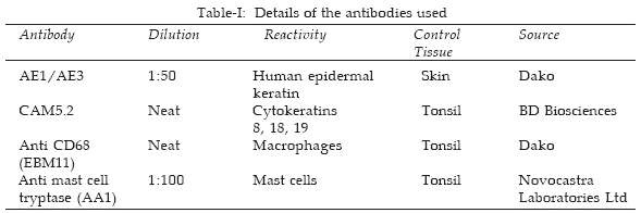
Nine formalin-fixed, paraffin-embedded blocks from six cases of Non-Specific Interstitial Pneumonia (NSIP) were obtained from the histopathology archive of the Wythenshawe Hospital Manchester. Paraffin-embedded sections (5�m), cut as near serially as possible, were dewaxed, blocked for endogenous peroxidase, rehydrated and immunostained with the monoclonal antibodies AE1/AE3, CAM5.2, anti-CD68 and anti-mast cell tryptase as detailed in Table-I. Sections were pre-treated with 0.03% (w/v) trypsin (type II, porcine, Sigma) stained directly with anti-CD68 and indirectly with AE1/AE3, CAM5.2 and anti-tryptase using the avidin-biotin method
9 with 3-3-diaminobenzidine tetrahy- drochloride as the substrate and Mayer�s haematoxylin as the counterstain.
RESULTS
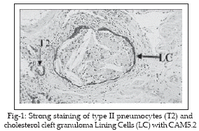
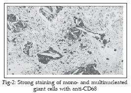
Half of the cases studied exhibited the presence of numerous dispersed cholesterol cleft granulomas (Figure-1). These were discrete, compact, uniform in size and present mostly in areas of alveolar wall thickening. The granulomas were universally present in alveolar spaces. They were never observed within alveolar walls nor in relationship to blood vessels. Typically the granulomas contained central clefts (mean length 52.6�m, range 18.0 109.3�m) surrounded by mono-and multinucleated cells which were CD68 positive, whereas the cells outside the granulomas and lining the alveolar walls were negative with this antibody (Figure-2). In contrast these latter cells were both AE1/AE3 and CAM5.2 positive (Figure-3). Elsewhere, in alveolar spaces not containing granulomas, there were occasional small populations of alveolar macrophages also showing CD68 positivity.
Anti-mast cell tryptase positive cells were present in the alveolar wall interstitium, frequently close to the lining cells, but never within the alveolar space nor in intimate relationship to the granulomas.
DISCUSSION
The most characteristic feature of NSIP is that the lesions are temporally uniform,
8 in distinction to the temporal heterogeneity of UIP. NSIP was originally defined by the exclusion of cases that could not be placed in the other well-defined categories of IPF. The presence of cholesterol cleft granulomas was a distinctive feature of half of our cases of NSIP. The granulomas were well defined with central clefts surrounded by macrophages and macrophage polykaryons. The granulomas were compact and confined to spaces lined clearly and separately from the cells of the granuloma, by a complete single layer of type II pneumocytes. Outside this, the interstitium was fibrotic and contained a population of mast cells demonstrable by anti-tryptase immunostaining. Immuno positive material had apparently diffused outside the cell boundaries suggesting tryptase release. Mast cells were never seen within the granulomas although an occasional cell was present in the type II pneumocyte lining and in the space between this lining and the cells of the granuloma.Granulomatous inflammation to cholesterol crystal clefts has been described in a number of pulmonary disorders including idiopathic cholesterol pneumonitis
12 and as a secondary phenomenon in bronchiectasis, chronic lung abscess and tuberculosis.13-15 The source of the cholesterol is likely to be pulmonary surfactant. In an electron microscopical study of lung tissue from heavy cigarette smokers, Corrin and Soliman16 found cholesterol crystal clefts in the cytoplasm of type II pneumocytes. Lipid extracts of bovine pulmonary surfactant contained 3% neutral lipid mainly as cholesterol and diacylglycerol and 97% phospholipid.17 The giant cells commonly seen in granulomas are considered to be macrophage polykaryons formed by the fusion of alveolar macrophages attached to the same endocytic material.18Whilst exogenous cytokines are influential in granuloma formation and activity, endogenous production of cytokines also occurs. Human non-caseating pulmonary tuberculous granulomas contain CD68 positive macrophage-like cells which produce mRNA for TNF-α, IFN-γ and IL-4 and these are likely to have functional significance.
19 Currently there are no available data on cytokine elaboration by pulmonary cholesterol cleft granulomas.This immuno histochemical study confirmed that the intra-alveolar cell populations in NSIP were composed predominantly of CD68 positive cells. It showed that the giant cells were also CD68 positive, but were cytokeratin negative. In contrast, the cells lining the alveolar spaces containing granulomas were cytokeratin positive (AE1/AE3 and CAM5.2), indicating that these were type II pneumocytes and showing that these cells had completely replaced type I cells at these loci. In addition, it is known that macrophages and macrophage polykaryons have distinctive glycoprofiles and that pneumocytes types I and II can be distinguished by their profiles.
20-22In summary, our observations indicate that cholesterol cleft granulomas occur with high frequency in NSIP, are present within alveolar spaces which are lined exclusively by type II pneumocytes and that external to this the interstitium is fibrotic with accumulations of mast cells. It is our speculation that these are linked in a pathogenetic mechanism related to the progression of NSIP. The results also suggest that the mononuclear and multinucleated cells of cholesterol cleft granulomas are derived from the macrophage-mononuclear cell lineage. The alveolar lining cells are type II pneumocytes which do not contribute to the granuloma cell population.
REFERENCES
1. Maheshwari U, Gupta D, Aggarwal AN, Jindal SK. Spectrum and diagnosis of idiopathic pulmonary fibrosis. Indian J Chest Dis Allied Sci 2004; 46: 23-6.
2. Selman M, Thannickal VJ, Pardo A, Zisman DA, Martinez FJ, Lynch JP. Idiopathic pulmonary fibrosis: pathogenesis and therapeutic approaches. Drugs 2004; 64: 405-30.
3. Williams K, Malarkey D, Cohn L, Patrick D, Dye J, Toews G. Identification of spontaneous feline idiopathic pulmonary fibrosis: morphology and ultrastructural evidence for a type II pneumocyte defect. Chest 2004; 125: 2278-88.
4. Flaherty KR, Travis WD, Colby TV, Toews GB, Kazerooni EA, Gross BH, et al. Histopathologic variability in usual and nonspecific interstitial pneumonias. Am J Respir Crit Care Med 2001; 164: 1722-7.
5. Takahashi H, Fujishima T, Koba H, Murakami S, Kurokawa K, Shibuya Y, et al. Serum surfactant proteins A and D as prognostic factors in idiopathic pulmonary fibrosis and their relationship to disease extent. Am J Respir Crit Care Med 2000; 162: 1109-14.
6. Katzenstein ALA, Myers JL. Idiopathic pulmonary fibrosis: clinical relevance of pathologic classification. Am J Respir Crit Care Med 1998; 157: 1301-15.
7. Katzenstein A-LA, Fiorelli RF. Nonspecific interstitial pneumonia/fibrosis: histological features and clinical significance. Am J Surg Pathol 1994; 18: 136-47.
8. Cottin V, Donsbeck AV, Revel D, Loire R, Cordier, JF. Non-specific interstitial pneumonia: individualization of a clinicopathologic entity in a series of 12 patients. Am J Respir Crit Care Med 1998; 158: 1286-93.
9. Guesdon JL, Ternynck T, Avrameas S. The use of avidin-biotin interaction in immunoenzymatic techniques. J Histochem Cytochem 1979; 27: 1131-9.
10. Waddell WR, Sniffen RC, Whytehead LL. Chronic pneumonitis: its clinical and pathological importance. J Thorac Surg 1949; 28: 134-44.
11. De Navasquez S, Haslewood GAD. Endogenous lipoid pneumonia with special reference to carcinoma of the lung. Thorax 1954; 9: 35-7.
12. Reid JD, Oliver AP, Cairney PC. Cholesterol pneumonitis. N Z Med J 1961; 60: 134-43.
13. Corrin B, Soliman SS. Cholesterol in the lungs of heavy cigarette smokers. Thorax 1978; 33: 565-8.
14. Yu S, Harding PG, Smith N, Possmayer F. Bovine pulmonary surfactant: chemical composition and physical properties. Lipids 1983; 18: 522-9.
15. Chambers TJ, Spector WG. Inflammatory giant cells. Immunobiology 1982; 161: 283-9.
16. Warfel AH, Hadden JW. Macrophage fusion factor elicited from BCG-sensitized lymphocytes. Am J Pathol 1978; 93: 753-70.
17. McNally AK, Anderson JM. Interleukin-4 induces foreign body giant cells from human monocytes/macrophages: differential lymphokine regulation of macrophage fusion leads to morphological variants of multinucleated giant cells. Am J Pathol 1995; 147: 1487-99.
18. DeFife KM, Jenney CR, McNally AK, Colton E, Anderson JM. Interleukin-13 induces human monocyte/macrophage fusion and macrophage mannose receptor expression. J Immunol 1997; 158: 3385-90.
19. Fenhalls G. In situ production of gamma interferon, interleukin-4 and tumor necrosis factor alpha mRNA in human lung tuberculous granulomas. Infect Immun 2000; 68: 2827-36.
20. Barkhordari A, McClure J, Stoddart RW, McClure SF. Cellular glycotypes in pulmonary sarcoidosis. J Pathol 1999; 189: 24A.
21. Barkhordari A, McClure J, McClure SF, Stoddart RW. Lectin binding profiles of pneumocytes in normal human lung. J Pathol 2000; 190: 54A.
22. Barkhordari A, Stoddart RW, McClure SF, McClure J. Lectin histochemistry of normal human lung. J Mol Histol 2004, 35: 147-56.
HOME | SEARCH | CURRENT ISSUE | PAST ISSUES
Professional
Medical Publications
Room No. 522, 5th Floor, Panorama Centre
Building No. 2, P.O. Box 8766, Saddar, Karachi - Pakistan.
Phones : 5688791, 5689285 Fax : 5689860
pjms@pjms.com.pk