|
|
||||
|
Published by : PROFESSIONAL MEDICAL PUBLICATIONS |
||||
|
ISSN 1681-715X |
||||
|
||||
|
- |
||||
|
ORIGINAL ARTICLE |
||||
|
- |
||||
|
Volume 22 |
July - September 2006 |
Number 3 |
||
|
|
||||
|
|
||||
|
|
||||
|
Published by : PROFESSIONAL MEDICAL PUBLICATIONS |
||||
|
ISSN 1681-715X |
||||
|
||||
|
- |
||||
|
ORIGINAL ARTICLE |
||||
|
- |
||||
|
Volume 22 |
July - September 2006 |
Number 3 |
||
|
|
||||
|
|
||||
Application of Polymerase Chain Reaction Technique
for Laboratory Diagnosis of Cutaneous TuberculosisAzar D. Khosravi1, Mohammad Omidian2
ABSTRACT
Objective: Evaluation of the role of polymerase chain reaction (PCR) for the detection of Mycobacterium tuberculosis (MTB) DNA as a diagnostic aid in cutaneous tuberculosis.
Study Design: Descriptive study.
Duration and place of study: TB reference laboratory, PHLS, Ahvaz, Iran from May 2001 to December 2001.
Patients & Methods: Thirty formalin-fixed, paraffin-embedded samples belonging to 28 patients were analyzed. Tissue sections were treated by lysis buffer containing proteinase K and DNA was extracted by using standard extraction kit. PCR amplification was performed using assay based on a repetitive sequence IS 6110 of MTB according to standard procedure.
Results: PCR was positive in six samples. Amongst them, two of the samples (7.1%) belonged to patients with chronic granulomatosis, which was previously confirmed histopathologically, were positive in entire applied tests, i.e. direct smear, culture and PCR. Using PCR technique, six out of the total specimens tested (21.4%), were positive for the presence of M. tuberculosis DNA. Statistically the difference between applied methods was significant (P<0.0016). Accounting histopathology as gold standard, the sensitivity of PCR in this study was determined as 75%.
Conclusions: Our study showed that from 8 cases of skin tuberculosis diagnosed by histopathology, 6 were positive by PCR technique, which shows the superiority of previous method to molecular technique. However, PCR assay has priority to conventional bacteriologic methods for detection of M. tuberculosis from cutaneous tuberculosis cases, and can be only used when the staining for acid fast bacilli is negative and there is a lack of growth on culture or when fresh material has not been collected for culture.
Key words: Cutaneous tuberculosis, PCR, M. tuberculosis, Culture.
Pak J Med Sci July - September 2006 Vol. 22 No. 3 291-294
1. Dr. Azar D. Khosravi PhD
Department of Microbiology,
School of Medicine, Infectious &
Tropical Diseases Research Center,
Ahvaz-Jundi Shapur University of Medical Sciences,
Ahvaz,
Iran.2. Dr. Mohammad Omidian MD
Dermatology Ward,
Imam Khomeini Hospital,
Ahvaz,
Iran.Correspondence:
Dr. Azar D. Khosravi
E-Mail: khosraviaz@yahoo.com* Received for Publication: July 22, 2005
* Revision Received: January 24, 2006
* Accepted: February 9, 2006
INTRODUCTION
Tuberculosis continues to be a health problem in both developed and developing countries. The development of resistance to anti-tuberculous drugs and the increase in diseases and conditions associated with immunodeficiency such as AIDS and chemotherapy has resulted in increasing number of tuberculosis case recently. As a result, the incidence of cutaneous tuberculosis has been increasing as well.
1,2Laboratory confirmation of cutaneous tuberculosis is often difficult using conventional techniques, which include the demonstration of acid fast bacilli (AFB) on histopathologic examination and culture. Moreover, in tuberculosis verrucosa cutis and lupus vulgaris which represent the pausibacillary forms of cutaneous tuberculosis, direct microscopy and tissue cultures lack either the specificity and/or sensitivity for the detection of Mycobacterium tuberculosis (MTB). In recent years, the polymerase chain reaction (PCR) has emerged as a promising tool in the diagnosis of various forms of cutaneous tuberculosis. Published data largely from case reports have indicated the usefulness of PCR in diagnosis of lupus vulgaris,
3,4 scrofuladerma5 and in the investigation of cases clinically suspected to be due to MTB infection.6 Based on there studies, which confirm the usefulness of PCR in the routine diagnosis for cutaneous tuberculosis, we report our experience in using PCR on archival skin biopsy specimens from patients suspected to have cutaneous tuberculosis upon clinico-pathologic correlation. Our results show that PCR can be very useful as a diagnostic aid for the detection of M. tuberculosis DNA in cases of cutaneous tuberculosis.MATEIALS AND METHODS
A total of 30 formalin-fixed, paraffin-embedded specimens from 28 patients were obtained from the histopatholgy files of the Dermatology ward from Imam Khomaini hospital, Ahvaz, Iran. The patients were diagnosed clinically and/or on biopsy as having cutaneous tuberculosis and in cases where it was being considered as a differential diagnosis. Of these samples, 4 were considered controls (2 positive and 2 negative controls). The positive controls belonged to patients who had a clinical history of mycobacterial infection and were confirmed by a positive smear for acid-fast bacilli (Ziehl-Neelsen staining) and positive culture for M. tuberculosis. The negative controls were from patients with dermatitis, which did not show any clinicopathological evidence of mycobacterial infection and were both Ziehl-Neelsen and culture negative for M. tuberculosis. The archival information regarding histopathology results and preliminary laboratory tests including acid fast staining and culture were provided along with the samples.
For tissue preparation and subsequent DNA extraction, two 5- �m sections were cut from each paraffin block, using ethanol swabbed scalpel blades for each section. Tissue sections were incubated overnight at 37
oC in lysis buffer containing proteinase K. DNA was extracted by using standard extraction kit (Cinnagen Co., Tehran, Iran) according to manufacturer�s instructions. As a negative extraction control, negative samples were processed in exactly the same manner as the positive controls and test samples. PCR amplification was performed using assay based on a repetitive sequence IS 6110 of MTB7 and by application of two oligonucleotide primers IS1 and IS2 as 5�- CCTGCGAGCGTAGGCGTCGG and 5�- CTCGTCCAGCGCCGCTTCGG for the detection of a 123-bp DNA fragment.8The reaction volume (25�l) composed of 50 mmol KCl, 10mmol Tris-HCl (pH 8.3), 1.5 mmol MgCl
2, 0.2 mmol of each deoxynucleotide triphosphate, 0.5�mol of each primer, 1 units of Taq polymerase (Cinnagen Co., Tehran, Iran),14�l of sterile distilled water and 5�l of processed sample. Amplification was performed with a four-stage protocol on a Techgene thermocycler using the program as below:An initial denaturation of 95
oC for 120s followed by 1 cycle of 95�C for 20s, 45�C for 360s and 72�C for 120s followed by 30 cycles of 95�C for 20s , 62�C for 60s and 72�C for 180s and final extension of 72�C for 10minutes. For analysis of the amplified products, they were loaded on a 2% agarose gel containing 0.5mg/ml ethidium bromide in electrophoresis and the products were visualized by UV transillumination.RESULTS
In this study, a total of 30 samples from 28 patients were analyzed, 75% of which were males (21 patients) and 25% were females (7 patients). The age distribution was between 19 and 70 years, but most of the patients belonged to age group 31-40 (14 patients).
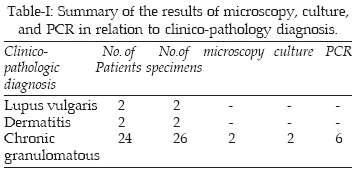
Histopathology examination of the sections was taken as the gold standard method, represented tissue changes compatible to lupus vulgaris, dermatitis and chronic granulomatosis. Eight of these were diagnosed as cutaneous tuberculosis and responded well to anti-TB drugs therapy. According to archival information provided, only two specimens were positive for AFB by direct microscopy. These specimens were positive by culture as well and were chosen as positive controls.
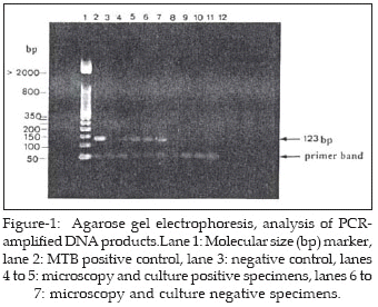
PCR gave positive results in 6 out of 8 specimens taken from patients with chronic granulomatosis. None of the specimens from patients diagnosed clinico-pathologically as lupus vulgaris or dermatitis were positive by direct microscopy, culture or PCR. PCR positive cases included two previously mentioned specimens with positive microscopy and culture (Figure-1).
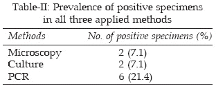
Of the PCR-positive cases, 5 belonged to male patients (23.8%) and one specimen was from a female patient (14.3%). A summary of the results is shown in Table-I. Based on the results obtained, 7.1% of the specimens were positive in both direct microscopy and culture. Using PCR technique, 21.4% of the total specimens tested, were positive for the presence of M. tuberculosis DNA (Table-II), while the difference between applied methods was significant (P<0.0016).
DISCUSSION
We have studied the usefulness of PCR in the detection of M. tuberculosis DNA using archival tissues from clinical specimens from patients diagnosed clinically and/ or on biopsy as having cutaneous tuberculosis and in cases where cutaneous tuberculosis was being considered as a differential diagnosis. From 30 analyzed specimens, two were positive by acid fast staining and culture. The majority were negative in direct microscopy and culture (data from archive), probably due to insufficient number of bacilli in pausibacillary forms of tuberculosis. Our findings were similar to studies showing the low positive results from direct smear and culture in cases of skin tuberculosis.
9,10Using PCR, 6 specimens were positive. Considering histopathology as gold standard, the sensitivity of PCR in this study was determined as 75%, which was in agreement with study of Tan and colleagues.
11 However according to the results, PCR was found to be more sensitive and specific as compared with direct microscopy and culture and the comparison was statistically significant. This confirms the superiority of PCR to traditional techniques as other investigators have shown previously.10,12,13Aside from the improvements to and the increasing use of PCR for the diagnosis of mycobacterial infections with fresh clinical specimens, there is still the need for the optimization of a sensitive and rapid PCR assay for the identification of mycobacteria from formalin fixed, paraffin embedded tissues. The effectiveness of the PCR method using archival material has been established in other studies.
12,14,15 The practical usefulness of this method is that patients need not to be subjected to repeated biopsies, which is important when there is no other lesions left for repeat biopsy. The PCR method also offers rapid diagnosis; the whole process takes few days, while conventional culture takes at least 2-4 weeks.We have not found the PCR method to be useful in the diagnosis of lupus vulgaris, which is largely based on presumptive clinical diagnosis supported by histopathologic features. In our study the sensitivity of PCR in detecting cutaneous tuberculosis compared to histopathology method was lower. It is in fact well known that the relatively frequent failure of the amplification of DNA from formalin fixed, paraffin embedded materials, can be due to the presence of inhibitors whose nature seems to be endogenous as well as induced by formalin fixation and by all other steps in tissue processing and deparaffinization.
16In conclusion, in the present study from 8 cases of skin tuberculosis diagnosed by histopathology, 6 were positive by PCR technique, which shows the superiority of previous method to molecular technique. However, PCR assay can be used for rapid detection of M. tuberculosis from cutaneous tuberculosis cases, particularly when the staining for acid fast bacilli is negative and there is a lack of growth on culture or when fresh material has not been collected for culture.
ACKNOWLEDGMENTS
The study was supported by a grant (No. 178) from Ahvaz Jundi Shapur University of Medical Sciences research affairs, Ahvaz, Iran. We are greatful to Mr. Bigan Barazandeh for his technical assistance.
REFERENCES
1. Chiewchanvit S, Mahanupab P, Walker PF. Cutaneous tuberculosis in three HIV-infected patients. J Med Assoc Thai 2000; 83: 1550-4.
2. Hamada M, Urabe K, Moroi Y, Koga T, Takeishi M, Fujita M, et al. A case of multifocal lupus vulgaris that preceded pulmonary tuberculosis in an immune compromised patient. J Dermatol 2004; 31: 124-8.
3. Serfling U, Penneys NS, Leonardi CL. Identification of Mycobacterium tuberculosis DNA in a case of lupus vulgaris. J Am Acad Dermatol 1993; 28: 318-22.
4. Steidl M, Neubert U, Volkenandt M, Chatelain R, Degitz K. Lupus vulgaris confirmed by polymerase chain reaction. Br J Dermatol 1993; 129: 314-8.
5. Taniguchi S, Chanoki M, Hamda T. Scrofuloderma: the DNA analysis of mycobacteria by the polymerase chain reaction. Arch Dermatol 1993; 129: 1618-9.
6. Penneys NS, Leonardi CL, Cook S. Identification of Mycobacterium tuberculosis DNA in five different types of cutaneous lesions by the polymerase chain reaction. Arch Dermatol 1993; 129: 1594-8.
7. Eisenach KD, Sifford MD, Cave MD, Bates JH. Detection of M. tuberculosis in sputum samples using a polymerase chain reaction. Am Rev Respir Dis 1991; 144: 1160-3.
8. Eisenach KD, Cave MD, Bates JH, Crawford JT. Polymerase chain reaction amplification of a repetitive DNA sequence specific for Mycobacterium tuberculosis. J Infect Dis 1989; 161: 977-81.
9. Bhutto AM, Solangi A, Khaskhely NM, Arakaki H, Nona M. Clinical and epidemiological observations of cutaneous tuberculosis in Larkana, Pakistan. Int J Dermatol 2002; 41: 159-65.
10. Barbagallo J, Tager P, Ingleton R, Hirsch RJ, Weinberg C. Cutaneous tuberculosis: diagnosis and treatment. Am J Clin Dermatol 2002; 3: 319-28.
11. Tan SH, Tan HH, Sun YJ, Goh CL. Clinical utility of polymerase chain reaction in the detection of Mycobacterium tuberculosis in different types of cutaneous tuberculosis and tuberculids. Ann Acad Med Singapore 2001; 30: 3-10.
12. Marchetti G, Gori A, Catozzi L, et al. Evaluation of PCR in detection of Mycobacterium tuberculosis from formalin-fixed, paraffin-embedded tissues: comparison of four amplification assays. J Clin Microbiol 1998; 36: 1512-7.
13. Hsiao PF, Tzen CY, Chen HC, Su HY. Polymerase chain reaction based detection of Mycobacterium tuberculosis in tissues showing granulomatous inflammation without demonstrable acid-fast bacilli. Int J Dermatol 2003; 42: 281-6.
14. Degitz K, Steidl M, Neubert U, Plewig G, Volkenandt M. Detection of mycobacterial DNA in paraffin- embedded specimens of lupus vulgaris by polymerase chain reaction. Arch Dermatol Res 1993; 285: 168-70.
15. Rish JA, Eisenach KD, Cave MD, Reddy MV, Gangadharam PRJ, Bates JH. Polymerase chain reaction of Mycobacterium tuberculosis in formalin-fixed tissue. Am J Respir Crit Care Med 1996; 153: 1419-23.
16. An SF, Fleming KA. Removal of inhibitor(s) of the polymerase chain reaction from formalin fixed, paraffin wax embedded tissues. J Clin Pathol 1991; 44:924-7.
HOME | SEARCH | CURRENT ISSUE | PAST ISSUES
Professional
Medical Publications
Room No. 522, 5th Floor, Panorama Centre
Building No. 2, P.O. Box 8766, Saddar, Karachi - Pakistan.
Phones : 5688791, 5689285 Fax : 5689860
pjms@pjms.com.pk