|
|
||||
|
Published by : PROFESSIONAL MEDICAL PUBLICATIONS |
||||
|
ISSN 1681-715X |
||||
|
||||
|
- |
||||
|
ORIGINAL ARTICLE- |
||||
|
- |
||||
|
Volume 22 |
July - September 2006 |
Number 3 |
||
|
|
||||
|
|
||||
|
|
||||
|
Published by : PROFESSIONAL MEDICAL PUBLICATIONS |
||||
|
ISSN 1681-715X |
||||
|
||||
|
- |
||||
|
ORIGINAL ARTICLE- |
||||
|
- |
||||
|
Volume 22 |
July - September 2006 |
Number 3 |
||
|
|
||||
|
|
||||
Oral Actinomyces Strain isolates in Patients suffering
from Progressive Periodontitis and Dentoalveolar AbscessSaeed Eshraghi1
ABSTRACT
Objective: The purpose of this study was to isolate and characterize of the Gram-positive anaerobic pleomorphic bacilli in particular Actinomyces strains from subgingival plaque and periapical abscesses specimens.
Patients and Methods: One hundred twenty six subgingival plaque samples from 100 patients with progressive periodontitis and 45 pus samples from 32 patients with dentoalveolar abscesses were included in this study. Sample collection criteria were contained deep pocket (over 3 mm), no recent antibacterial therapy and lack of systemic infection. The paper points specimens collected were transferred and cultured using appropriate media.
Results: The only strain of A. viscosus was obtained from a patient with progressive periodontitis with pocket depth of 6 mm and two strains of A. naeslundii with pocket depth of 4 mm. The peak incidence number of progressive periodontitis (35%) was in the third decade (31-40) and the lowest incidence (10%) was in the first decade (<20). Forty patients complained of bleeding of teeth and gingival disease with the lower incidence of (42.5%) in female. Of the 32 patients with dentoalveolar abscesses, the peak incidence of the dentoalveolar abscesses (25%) was seen in the group aged 31-35, while the low incidence (6.3%) was in the group aged 16-20 years. The causes of the progression of dentoalveolar abscesses were found as nineteen patients (59.4%) with dental carries, seven patients (21.9%) with a dental extraction and six patients (18.8%) with uncompleted endodontics treatment.
Conclusion: The present research indicates that most patients have neglected dental care and poor oral hygiene, suffer from the gingival disease and bleeding gum. The Columbia blood-agar with 10�L/mL cephadroxyl is recommended for the isolation of Actinomyces species, at 37�C for 5-7 days in anaerobic conditions. To obtain a higher recovery of these microorganisms, the Columbia blood-agar without antibiotic, in candle jars is recommended.
Key words: Actinomyces, subgingival plaque, Dentoalveolar abscesses, Gingivitis, Periodontal pocket.
Pak J Med Sci July - September 2006 Vol. 22 No. 3 238-243
1. Saeed Eshraghi PhD
Associate Professor in Microbiology,
Department of Pathobiology,
School of Public Health &
Institute of Public Health Research,
Tehran University of Medical Sciences,
P.O. Box: 6446,
Tehran-14155,
Islamic Republic of Iran.Correspondence:
Saeed Eshraghi PhD
E-Mail: eshraghs@sina.tums.ac.ir
saeed_eshraghi@yahoo.com* Received for Publication: June 20, 2005
* Revision Received: November 1, 2005
* Accepted: December 25, 2005
INTRODUCTION
It has been estimated that about 500 species of bacteria inhabit the human oral cavity.
1-3 While majority of these organisms are commensals, some of them are likely to become opportunistic pathogens that can cause systemic disease.4 For example, oral bacteria have been implicated in bacterial endocarditis,5 aspiration pneumonia,6 osteomyelitis in children,7 preterm low birth weight,8 and coronary heart disease and cerebral infarction (or stroke).9,10 Consequently, it is important to know what microorganisms are present in the oral cavity for the diagnosis and rational treatment of systemic as well as oral diseases. The Gram-positive anaerobic pleomorphic bacilli (GPAPB), especially the genus Actinomyces, are part of the oral microflora and they are generally associated with cervical caries.9 The genus Actinomyces is generally associated with root caries, and is also found in non- active sites in periodontal disease and in pulpar infections.11 The development of dental plaque is an example of autogenic succession whereby microbial factors influence the pattern of the development of the microflora.12,13Dental plaque is a microbial biofilm with high species diversity, embedded in polymers of salivary and bacterial origin, found on the tooth surface. The formation of dental plaque can be divided arbitrarily into a number of distinct stages.
14-16 Different species of Actinomyces, Lactobacillus, Fusobacterium and Bacteroides can be isolated from the dental plaque.3,12,14 The periapical or dento-alveolar abscess is collection of pus in the pulp, or around the root of the tooth. The abscess can result from necrosis of the pulp, usually from the progression of dental caries and contain a diverse collection of bacteria.15The main objective of this descriptive study was to isolate and characterize of (GPAPB) in particular Actinomyces strains from subgingival plaque and periapical abscesses specimens. The minimum schema of biochemical tests for the identification and differentiation of the Actinomyces species from other (GPAPB), investigating the drug sensitivity and resistance of the isolated bacteria, and providing the doctors and patients with necessary advice were also investigated.
MATERIALS AND METHODS
Sampling criteria: The samples were collected based on the three criteria:
a) periodontal plaque with deep pocket (>3 mm) / submandibular chronic dento-alveolar abscesses,
b) no antibiotic therapy for a period of at least two weeks, and
c) lack of systemic or contagious infectious.Patient specimens:
1. Subgingival plaque samples of patients with progressive periodontal disease were obtained from the periodontics department of the school of dentistry at Tehran University of Medical Sciences. After removal of supragingival plaque, three paper points were inserted into a periodontal pocket for 20 second. The specimens were then dispensed into small, screw-capped bottles filled with 2 ml reduced transport fluid and sent to the laboratory for processing within 30 minutes.
2. The patients with odontogenic abscesses were recruited in the Department of Oral and Maxillofacial Surgery. Pus was aspirated after decontamination of the mucosa and transferred to Stuart�s medium. Te samples were plated onto solid culture media within 2 hour. Two culture media were assayed for strain activation: Brain Heart Infusion (BHI) and thioglycolate broth as a soft agar media.
Culture and identification of microorganisms: The specimens were mixed properly using vortex for 30 records, and appropriate amount of inoculum (50�l) cultivated in the following culture media: thioglycolate broth (TGB), Mueller Hinton agar (MHA), brain heart infusion agar (BHIA), and Columbia blood-agar base (Difco) supplemented with 5% defibrinated horse blood, 5 mg/l haemin and vitamin K. The medium used with and without cephadroxyl (10�L/mL). All plates were incubated anaerobically at 37�C for 5-7 days using anaerobic jar and anaerocult A and C (Merck Darmstadt, Germany). The duplicate plates were also incubated in anaerobic condition in a candle jar for 5-7 days at 37�C. Identification of the bacteria species was based on colony morphology, cell morphology and biochemistry. The biochemical test plates were incubated in a candle jar for 14 days at 37�C. The results were recorded every 24 hour.
Antibiotic susceptibility test: Antibiotic susceptibilities were determined by inoculating of the bacteria onto the Mueller Hinton agar plates supplemented with the appropriate antibiotic discs. The plates were also incubated anaerobically at 37�C for 4 days. The minimal inhibitory concentration (MIC) was defined as the lowest concentration of the antibiotic inhibiting visible growth. Actinomyces naeslundii ATCC No. 12104 and Actinomyces viscosus ATCC No. 15984 served as control strains. The following antimicrobial agents were studied: penicillin G, amikacin, ampicillin, erythromycin, tetracycline, gentamycin, cephalothin and chloramphenicol.
RESULTS
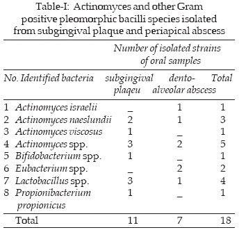
A total of 18 Gram positive pleomorphic bacterial were isolated from the specimens. The isolates included Actinomyces israelii, A. naeslundii, A. viscosus, A. spp, Bifidobacterium spp., Eubacterium spp., Lactobacillus spp., and Propionibacterium propionicus. Of the 171 cases analyzed, 100 presented with anaerobic bacteria growth in the plaque samples and 32 with periapical samples, while 39 cases did not yield appropriate bacterial growth. The highest recovery of Actinomyces (6 strains) was obtained from the patients with progressive periodontal disease and 4 strains from the patients with dento-alveolar abscesses. Apart from the Actinomyces species, one strain of Bifidobacterium, two strains of Eubacterium, 4 strains of Lactobacillus and one strains of Propionibacterium propionicus were also isolated (Table-I). The only strain of A. viscosus was obtained from a patient suffering progressive periodontitis with a pocket of 6 mm depth and two strains of A. naeslundii in a pocket of 4 mm depth. The rest of the Actinomyces (3 strains) from this site, did not show a typical colony characteristic that would allow their differentiation.
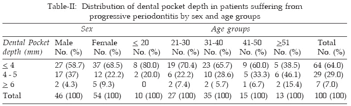
The peak incidence of progressive periodontal diseases (35%) was in the third decade (31-40) and the lowest (10%) in the first decade (<20) Table-II. Among the above patients, forty cases complained of gum bleeding and gingival disease with lower incidence of (42.5%) in female against the male (57.5%). No significant relationship between age, sex and the depth of periodontal pocket was observed.
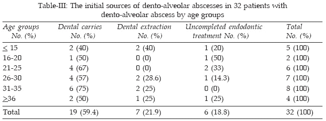
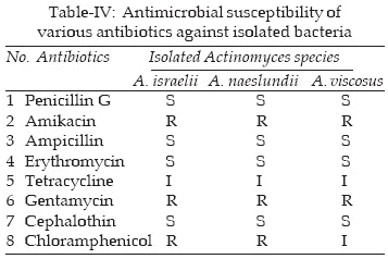
Of the 32 patients with dento-alveolar abscesses one strain of A. israelii and one strain of A.naeslundii were identified and 2 strains did not show a typical colony characteristic that would allow their differentiation (Table-I). The highest incidence of the dento-alveolar abscesses was seen in the group aged 31-35 years with (25%) and the lowest incidence in the group aged 16-20 years with (6.3%). The causes of the dento-alveolar abscesses were studied and the results are shown in Table-III. The influence of various antibiotics against Actinomyces strains isolated from subgingival plaque and dento-alveolar abscess are shown in Table-IV. The results suggest that the Actinomyces spp. may contribute to the etiology of the dento- alveolar abscesses.
DISCUSSION
Culture procedures have traditionally been used in the assessment of the micro organism associated with various infectious diseases, including progressive periodontitis and submandibular chronic dento-alveolar abscesses.
17-19 The culturing methods have a reasonable degree of agreement in terms of the identification of oral microorganisms compared with that of the molecular and serological techniques e.g. DNA-DNA hybridizing techniques.20 The major advantage of the culture procedure is its ability to enable the detection of unexpectedly viable cells (molecular procedures enable the detection of only target microbial species.21 Many molecular techniques assist in the identification of cultivable microorganisms.20 In the present study by using the culture procedures 1 strain of A. viscosus and 2 strains of A. naeslundii were isolated from the subgingival plaque sample of the patients with progressive periodontitis, and 1 strain of Actinomyces israelii and 1 strain of A. naeslundii were isolated from the pus sample of the patients with dentoalveolar abscess while, A. naeslundii was the most frequently isolated bacteria. The presence of other Gram positive non-spore forming pleomorphic bacteria was also evaluated in the current study. These microorganisms were found in 5 of 100 subgingival plaque sample (Bifidobacterium spp., Lactobacillus spp. and Propionibacterium propionicus) and 3 of 32 periapical abscess specimens (Eubacterium spp. and Lactobacillus spp). Theses findings are similar to those reported by other researchers.20,22,23 Our results indicate that the oral bacteria of subgingival plaque sample from patients with progressive periodontitis and pus sample from patients with dentoalveolar abscesses have polymicrobial characteristics and are predominated by anaerobic Gram negative bacilli. The role of anaerobic bacteria in progressive periodontitis and dentoalveolar abscesses cannot be overstated; in fact, these bacteria were isolated from the low percentage of above infection sites in this study. The Actinomyces strains isolates have shown to be highly sensitive to penicillin G, ampicillin, erythromycin and cephalothin with routine qualitative susceptibility criteria and resistant to amikacin, gentamycin and chloramphenicol. The bacterial strains that were determined to be unsusceptible to certain antibiotics according to the present criteria might actually be clinically susceptible to those antibiotics because they may be affected by other factors such as infection site or dosage.24In this study the patients with untreated gingivitis and progressive periodontitis, complained of gum bleeding and gingival irritation. The gingival disease with these patients probably was due to several factors including inadequate oral hygiene, pure dental care, accustomed to use the traditionally dental treatment, lack of periodical dental observation (economical disability), inadequate oral home care, smoking and lack of economical dental services.
Generally gums separate from the teeth, forming pockets (spaces between the teeth and gums) that become infected.
1,23 As the disease progresses, the pockets deepen and more gum tissue and bone are destroyed. Often, this destructive process has very mild symptoms. Eventually, teeth can become loose and may have to be removed.12,16,22-24CONCLUSION
Most patients have neglected dental care and poor oral hygiene, suffer from the gingival disease and bleeding of gum. These findings revealed that the patients have lack of periodical dental check up due to the economic reasons, inadequate oral home care, smoking and late call to a dentist etc. For recovery of Actinomyces species: use of the blood-agar with 10�L/mL cephadroxyl for the isolation of Actinomyces species, at 37�C for 5-7 days in anaerobic conditions in recommended. In this situation the antibiotic inhibits the Gram- negative microflora. To obtain a higher recovery of these microorganisms, use the Columbia blood-agar without antibiotic, at 37�C for 5-7 days incubation in candle jar.
REFERENCE
1. Moore WEC, Moore LVH. The bacteria of periodontal diseases. Periodontology 2000. 1994; 5: 66-77.
2. Socransky, SS, Haffajee AD. Evidence of bacterial etiology: a historical perspective. Periodontology 2000. 1994; 5: 7-25.
3. Wilson, MJ, Weightman AJ, Wade WG. Applications of molecular ecology in the characterization of uncultured microorganisms associated with human disease. Rev Med Microbiol 1997; 8: 91-101.
4. Curi MM, Dib LL, Kowalski LP, Landman G, Mangini C. Opportunistic actinomycosis in osteoradionecrosis of the jaws in patients affected by head and neck cancer: Incidence and clinical significance. Oral Oncology 2000; 36: 294-9.
5. Berbari, EF, Cockerill FR, Steckelberg JM. Infective endocarditis due to unusual or fastidious microorganisms. Mayo Clin Proc 1997; 72: 532-42.
6. Scannapieco FA. Role of oral bacteria in respiratory infection. J Periodontol 1999; 70: 793-802.
7. Dodman, T, Robson J, Pincus D. Kingella kingae infections in children. J Paediatr Child Health 2000; 36: 87-90.
8. Offenbacher S, Jared HL, O�Reilly PG, Wells SR, Salvi GE, Lawrence HP, et al. Potential pathogenic mechanisms of periodontitis associated pregnancy complications. Ann Periodontol 1998; 3: 233-50.
9. Beck J, Garcia R, Heiss G, Vokonas PS, Offenbacher S. Periodontal disease and cardiovascular disease. J Periodontol 1996; 67: 1123-37.
10. Trevisan Wu-TM, Genco RJ, Dorn JP, Falkner KL, Sempos CT. Periodontal disease and risk of cerebrovascular disease: the first national health and nutrition examination survey and its follow-up study. Arch Intern Med 2000; 160: 2749-55.
11. Schiphach P, Osterwalder V, Guggenheim B. Human root caries: Microbiota in Plaque covering sound, carious and arrested carious root surfaces. Caries Res 1995; 29: 382-95.
12. Liljemark WF, Bloomquist CG, Bandt. CL, Pihlstrom. BL, Hinrichs JE, Wolff-LF. Comparison of the distribution of Actinomyces in dental plaque on inserted enamel and natural tooth surfaces in periodontal health and disease. Oral Microbiol Immunol 1993; 8(1): 5-1.
13. Van Houte J, Jordan HV, Laraway R, Kent R, Soparkar PM, De Paola PF. Association of the microbial flora of dental plaque and saliva with human root surface caries. J Dent Res 1990; 69: 1463-8.
14. Fure S, Romaniec M, Emilson CG, Krasse B. Proportions of Streptococcus mutant, Lactobacilli and Actinomyces spp. in root surface plaque. Scand J Dent 1987; 95: 119-23.
15. Marsh P, Martin MV. Oral Microbiology, 4th edition MPG Books Ltd, Bodmin, Cornwall 2000; Chap 8, 3: pp 23-24 and 127-32. Oxford.
16. Mombelli A, Nyman S, Bragger U, Wennstrom J. Clinical and microbiological changes associated with an altered subgingival environment induced by periodontal pocket reduction. J Clin Periodontol 1995; 22: 780-7.
17. Cugini MA, Haffajee AD, Smith C, Kent RL Jr. Socransky SS. The effect of scaling and root planning on the clinical and microbiological parameters of periodontal diseases, 12 month results. J Clin Periodontol 2000; 27: 30-6.
18. Siqueira JF Jr. Aetiology of root-canal treatment failure. why well-treated teeth can fail. Int Endodontic J 2001; 34: 1-10.
19. de Annan SG, Valladares RR, de Cardenas. Effect of the Conditions of Incubation in the Recovery of Oral Actinomyces. Anaerobe 1999; 5: 101-4.
20. de Sousa ELR, de Almeida G BPF. Bacteriological study of root canals associated with periapical abscesses. Oral surg Oral med Oral Path 2003; 96: 332-9.
21. Papapanou PN, Madianos PN, Dahlen G, Sandros J. "Checker-board" versus culture: a comparison between two methods for identification of subgingival microbiota. Eur J Oral Sci 1997; 105: 389-96.
22. Chavez de Paz LE, Dahlen G, Molander A, Moller A, Bergenholtz G. Bacteria recovered from teeth with apical periodontitis after antimicrobial endodontic treatment. Int Endodontic J 2003; 36: 500-8.
23. Eick S, Pfister W, Straube E. Antimicrobial susceptibility of anaerobic and capnophilic bacteria isolated from odontogenic abscesses and rapidly progressive periodontitis. Int J Antimicrobial Agents 1999; 12: 41-6.
24. Kuriyama T, Karasawa T, Nakagawa K, Saiki Y, Yamamoto E, Nakamura S. Bacteriologic features and antimicrobial susceptibility in isolates from orofacial odontogenic infections. Oral Surg Oral Med Oral Pathol Oral Radiol Endod 2000; 90: 600-8.
HOME | SEARCH | CURRENT ISSUE | PAST ISSUES
Professional
Medical Publications
Room No. 522, 5th Floor, Panorama Centre
Building No. 2, P.O. Box 8766, Saddar, Karachi - Pakistan.
Phones : 5688791, 5689285 Fax : 5689860
pjms@pjms.com.pk