|
|
||||
|
Published by : PROFESSIONAL MEDICAL PUBLICATIONS |
||||
|
ISSN 1681-715X |
||||
|
||||
|
- |
||||
|
ORIGINAL ARTICLE |
||||
|
- |
||||
|
Volume 22 |
July - September 2006 |
Number 3 |
||
|
|
||||
|
|
||||
|
|
||||
|
Published by : PROFESSIONAL MEDICAL PUBLICATIONS |
||||
|
ISSN 1681-715X |
||||
|
||||
|
- |
||||
|
ORIGINAL ARTICLE |
||||
|
- |
||||
|
Volume 22 |
July - September 2006 |
Number 3 |
||
|
|
||||
|
|
||||
A Study of Esophageal Strictures after
Surgical repair of Esophageal AtresiaMehran Peyvasteh1, Shahnam Askarpour 2, Mohammad Hossein Sarmast Shoushtari3
ABSTRACT
Objective: The aim of this study was to find out the prevalence of esophageal stricture (ES) following surgical repair of esophageal atresia.
Patients and Methods: This retrospective study was carried out in two referral hospital (Bahrami & Children Medical Centre) from April 1999 till March 2000. Data was collected from patient’s file and follow up clinics. Seventy four patients with esophageal atresia were operated during this period. Twenty one patients who died and two patients with long gap esophageal atresia were excluded from the study. Hence fifty one patients were included in this study. End to end anastomosis was done in 45 and end to side anastomosis in 6 patients. Forty five patients had extrapleural thoracotomy while 6 patients had intrapleural thoracotomy. Single layer and double layer anastomosis were done in 36 and 25 patients respectively. Vicryl was used for repair in 47 babies and silk in 4 patients. Standard post operative care was provided to all patients.
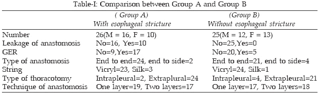
Results: Of 51 patients who survived 28 (54.8%) were male 23 (45.2%) female. Anastomotic leak was seen in 10 (19.6%) babies. 26 patients (M=16, F=10) developed ES. 22 (43.1%) patients had radiologically proven gastro-esophageal reflux. Incidence of stricture formation was significantly higher in babies who developed anastomosis leak after repair (p<0.05).
Conclusions: GER and leakage from the anastomosis site were significantly higher in the group A. The risk of stricture formation is a potential threat for up to a year in patients without G.E.R and up to 18 months in patients with GE Reflux. Leakage of anastomosis and GE Reflux play a major role in post operative strictures after esophageal atresia repair so treatment and prevention of these complications are very important.
Key words: Esophageal atresia, Anastomosis, Stricture.
Pak J Med Sci July - September 2006 Vol. 22 No. 3 269-272
1. Dr. Mehran Peyvasteh,
Pediatric Surgeon2. Dr. Shahnam Askarpour,
Pediatric Surgeon3. Dr. Mohammad Hossein Sarmast Shoushtari,
General Surgeon1-3: Jundishapour University of Medical Sciences,
Faculty of Medicine,
Department of Pediatric Surgery,
Ahwaz,
Islamic Republic of Iran.Correspondences:
Dr. Shahnam Askarpour,
Department of Surgery,
Imam Khomeini Hospital,
Azadegan Avenue
Ahwaz, IRAN.
E-Mail: shahnam_askarpour@yahoo.com* Received for Publication: August 27, 2005
* Revision Received: February 4, 2006
* Revision Accepted: June 17, 2006
INTRODUCTION
Esophageal stricture (ES) is the most common complication after esophageal atresia repair; other include anastomotic leak, recurrent tracheo-esophageal fistulas, GE reflux and esophageal motility disturbances.
1 A recent study showed the 52% babies developed ES after repair.2 Different factors such as ischemia at the anastomotic site, tension on the anastomosis,3 suture material,4 and GER play an important role in the development of complication.1,7 ES may remain asymptomatic or present with vomiting and dysphagia. Respiratory symptoms include stridor, dyspnoea and chocking during feeds. Aspirations pneumonia and other pulmonary complications can precipitate the baby’s condition. Diagnosis may be made by contrast studies and endoscopy.8 Repeated dilatation are necessary in nearly 40% of patients who develop ES.5,6 Patients who do not respond to the dilatation program or have long length of the stricture and in those complicated by GE Reflux, surgical intervention may be necessary. In case of GE reflux, funduplication may also be required.9-12 Regular follow-up is essential for operated cases of esophageal atresia for early detection of complications.1,8 We conducted this study with a view to find out the prevalence of ES following surgical treatment of esophageal atresia.PATIENTS AND METHODS
This retrospective study was carried out in Bahrami Children Hospital and Children Medical Centre from April 1999 to March 2000. Of 74 patients with OA who were operated 21 died, and two having type A esophageal were not included in this study. Hence fifty one patients with type C esophageal atresia were included in the study. At the time of operation surgical details such as layers of anastomosis, type of suture and thoracic exploration technique were recorded. After stabilization surgery was performed thorough right thoracotomy. Extrapleural approach was attempted in all patients but if pleura was opened it was converted to transpleural thoracotomy. Extrapleural thoracotomy was possible in 45 patients while six had intrapleural thoracotomy. Ligation of distal TE fistula and end to end anastomosis were done in 45 patients and end to side in 6 patients without division of distal fistula after ligation. Single layer and two layer anastomosis were done in 36 and 25 patients respectively. Polyglycolate (Vicryl) sutures were used in 47 and silk in 4 patients. Patients were divided in two groups. Patients who developed ES after surgery were placed in "Group A" while those who did not develop strictures were placed in group B. Patients were followed up for two years and dataó recorded in patient’s files. Data was analyzed by SPSS software. Kolmogrov-Smirov, Mann- Whitney, Gehan, and Chi-square tests were used for data analysis.
RESULTS
Gestational ages of patients were between 35-39 weeks with the mean of 37.1 weeks. Out of 51 patients, 26 patients (M=16, F=10) developed ES (Group A) and in 25 patients (M=12, F=13) stricture was not present (Group B). Ten patients in group A (38.46%) developed a leakage from anastomosis versus none in group B (P= 0.001). Seventeen patients (65.4%) in group A had GER versus 5 (20%) patients in group B (P=0.001). There was no significant difference between group A and B for the type of thoracotomy, anastomotic technique, and type of suture used (Table-I). 15 (57.7%) patients who developed ES presented between 0-6 months after surgery, 9 (34.9%) presented 6-12 months after surgery and 2 (7.7%) presented 12-18 months postoperatively. There was no new case of ES after 18 months. ES resolved after 1-3 dilatations in eleven patients (42.3%) whereas nine patients (34.6%) needed surgical revision. Fundoplication was done in six patients (23.1%) because of failure of medical therapy and non-responding anastomosis stricture with GER. There was no significant difference between patients with GER and with out GER for possible time of stricture developing (Gehan test, p=0.518)
DISCUSSION
Esophageal stricture is a common complication of esophageal atresia repair. Anastomosis techniques, repair under tension, leakage of anastomosis and GE Reflux may cause stricture
1 after a successful repair. Out of fifty one patients in our study twenty six (51%) developed stricture as compared to 30-40% reported in other studies.5,6 In a recent study of 50 patients ES developed in 40% cases after EA repair, which was directly related to tension at anastomosis site and gap between the two segments(p=0.03).3 Of the 26 patients who developed ES, 61.5% were male and 38.5% were female, but in patients without ES 48% were male and 52% were female. It had no statistical meaning (x2 =43%, p = 0.331). We compared gestational age of patients with ES and those without it. Mann-Whitney test was used for this distribution (value=286.5, p=0.447) and it was not significant. Of 51 patients ten (19.5%) developed leakage of anastomosis, but in other studies it was 8%.2 Tension at anastomosis site and technical problem were implicated for its causation. All patients with anastomotic leak developed stricture (P= 0.001, x2=11.961). Some studies showed protective affect of pleural or pericardial flap against leakage.13,14 Use of silk string may also be considered responsible for causation of ES.1-8 In patients without esophageal stricture (25 cases) silk was used in one (4%) and vicryl were used in twenty four (86%) patients. (X2=1.002 p = 0.32). Recent studies showed least incidence of stricture formation after use of Polydioxanone sodium sutures in contrast with silk which had the highest incidence of stricture formation.4 In our study silk was used in few patients thus a conclusive decision can not be made on its possible side effects. Patients who developed ES 73.1% had a single layer anastomosis and 26.9% had two layer anastomosis compared with patients without stricture (one layer=68%, two layer=32). This had no statistically significance (p = 0.764- x2 = 0.158). In a recent study anastomotic techniques were compared showing an improved survival with end to side anastomosis.15 It also showed that end to side anastomosis had less incidence of stricture formation and G.E Reflux.15 We did not find any direct correlation between type of suture used and anastomotic technique with subsequent stricture formation. In our study 7.5% patients with intrapleural thoracotomy and 92.3% with extra pleural thoracotomy developed stricture (x2=0.847- p= 0.41). Enguin SA et al mentioned that with extra pleural thoracotomy even after an anastomotic leak, 95% patients cured spontaneously, but may produce stricture.3 The reported incidence of GE Reflux is between 40-70%.3,6-11,12,16 The main reasons for GER are shortage of intra abdominal esophagus, stretch at anastomosis site, disturbance of esophageal motor function, manipulation during surgery, and esophageal motility problems.16-18 In our study 6(23.1%) patients who had GE Reflux and ES needed fundoplication. In some studies 45-75% of infants needed anti-reflux surgery, because medical treatment had failed or they had persistent anastomosis stricture, or distal ES.6,11,12 We also measured the probable time of ES formation after repair of EA. For compression Gehan static were used. (Gehan static = 0.4-7-p=0.518). Statistical review showed that GE reflux after 6 month was 0.64, after 12 months 0.66, and 1 after 18 month. This study shows that leakage of anastomosis and GE Reflux have a major role in post operative strictures after esophageal atresia repair so prompt treatment and prevention of these complications are very important.REFERENCES
1. Harman CM, Coran AG: Congenital Anomalies of the esophagus. In: O’Neill. JA, Rowe MI, Grosfeld JL, Fonkalsrad EW, Coran AG. Pediat Surg. St Louis, Mosby 1998; 941-61.
2. Konkin DE, O Nali WA, Webber EM, Blair GK: Outcomes esophageal atresia and tracheoesophageal fistula J Pediat Surg 2003; 38(12): 1726-9.
3. Michaud L, Guiber D, Sfeir R, Rakza T, Bajja H, Bonnevalle M, et al. Anastomotic stenosis after surgical treatment of esophageal atresia, frequency, risk factor and effectiveness of esophageal dilatations. Arch Pedia 2001; 8(3):268-74.
4. Mustafawi AR, Juouni WK, William.S. Esphageal stricture post repair of esophageal atresia. Saudi Med J 2003; 24(5 suppl): 539.
5. Enguin SA, Grosfeld JL, West KW, Rescorla FJ, Schere L. Analysis of morbidity and mortality in 227 cases of esophageal atresia and, or tracheoesophageal fistula over two decades. Arc Surg 1995; 130:502.
6. Spitz L, Keily E, Brerton. RJ, Drake D. Management of esophageal atresia. World J Surg 1993; 17:296.
7. Spitz L, Hitchcock R. Esophageal atresia and tracheooesophageal fistula. In: Freeman NV et al, Surgery of the newborn. New York Churchill livingstone1994.
8. Filston HC, Shorter NA. Esophageal atresia and tracheoesophageal malformations. (Eds) Ashcraft KW, Murphy JP, Sharp RJ, Sigalet DL, Snyder CL. Philadelphia WB. Saunders Company 2000; 348-70.
9. Corbally MT, Muftab M, Guiney EJ. Nissen funduplication for gastro- esophageal reflux in repaired tracheoesophgeal fistula. Eur J Pediat Surg 1992; 2:332.
10. Lindahl H, Rintala R, Louhimol. Failure of the Nissen fundoplication to control gastro -esophageal reflux in esophageal atresia patients. J Pediat Surg 1989; 24:985.
11. Manning PB, Morgan RA, Coran AG, Wesley GR, Polley T, Behrendt DM, et al: Fifty years, experience with esophageal atresia and tracheoesophageal fistula beginning with Cameron Haights first operation in 1935. Ann Surg 1989; 204:449.
12. Wheatley MJ, Coran AG, Wesley JR. Efficacy of the Nissen fundoplication in the management of gastro esophageal reflux following esophageal atresia repair. J Pediat Surg 1993; 28:53-5.
13. Chavin K, Field G, Chandler J, Tagge E, Othersennn HB, et al. Save the Child’s esophagus: Management of major disruption after repair of esophageal atresia, J Pediate Surg 1996; 31:48.
14. Wheatley MJ, Coran AG. Pericardial flap interposition for the definitive management of recurrent tracheoesophageal fistula. J Pediat Surg 1992; 27:1122.
15. Touloukian RJ, Seashore JH, Thirty-five year institutional experience with end to side repair for esophageal atresia. Arch Surg 2004; 139(4): 371-4 Discussion 374.
16. Tovuar JA, Diezpadro JA, Murica J, Prieto G, Molina M, Polaneo I, et al: Ambulatory 24-hour manometric and PH metric evidence of permanent impairment of clearance capacity in patient with esophageal atresia. J Pediat Surg 1995; 30:1224.
17. Jolly SG, Johnson SG, Roberts CC, Herbst JJ, Matlak ME, Mccombs A, et al: Patterns of gastro esophageal reflux in children following repair of esophageal atresia and distal tracheoesphageal fistula. J Pediat Surg 1980; 15:857.
18. Ashcraft KW, Goodwin F, Amoury RA, Holder TM, et al: Early recognition and aggressive treatment of gastro esophageal reflux following repair of esophageal atresia. J Pediat Surg 1977; 12:317.
HOME | SEARCH | CURRENT ISSUE | PAST ISSUES
Professional
Medical Publications
Room No. 522, 5th Floor, Panorama Centre
Building No. 2, P.O. Box 8766, Saddar, Karachi - Pakistan.
Phones : 5688791, 5689285 Fax : 5689860
pjms@pjms.com.pk