|
|
||||
|
Published by : PROFESSIONAL MEDICAL PUBLICATIONS |
||||
|
ISSN 1681-715X |
||||
|
||||
|
- |
||||
|
ORIGINAL ARTICLE |
||||
|
- |
||||
|
Volume 22 |
October - December 2006 |
Number 4 |
||
|
|
||||
|
|
||||
|
|
||||
|
Published by : PROFESSIONAL MEDICAL PUBLICATIONS |
||||
|
ISSN 1681-715X |
||||
|
||||
|
- |
||||
|
ORIGINAL ARTICLE |
||||
|
- |
||||
|
Volume 22 |
October - December 2006 |
Number 4 |
||
|
|
||||
|
|
||||
Isolation of Brucella melitensis and Brucella abortus
from brucellosis patients by conventional culture
method and polymerase chain reaction technique
Azar D. Khosravi1, Effat Abassi2, Seyed Mohammad Alavi3
ABSTRACT
Objective: Isolation of B. melitensis and B. abortus from brucellosis patients by application of conventional culture and PCR methods and comparison of sensitivity of both techniques in diagnosis of human brucellosis.
Duration and place of study: Infectious diseases Ward, Ahwaz University’s hospitals over a period of 9 months (September 2003 to May 2004).
Patients & Methods: A total of 30 peripheral blood samples were collected from patients with brucellosis referred to Infectious Diseases Unit of Ahwaz University’s hospitals. The samples were inoculated into Castaneda medium and after 7-30 days, the isolated colonies were identified by inoculation into brucella agar containing thionine and fuschin and by performance of biochemical tests. DNA was extracted from colonies and serum samples and examined by PCR involving specific primers for B. melitensis and B. abortus based on IS 711 in the brucella chromosome.
Results: We identified 8 isolates of B. melitensis using culture and biochemical methods. When PCR technique was applied to serum samples, 28 cases were positive for B. melitensis and we were able to isolate more cases compared to culture method and the difference was significant (P<0.05). The sensitivities of culture and PCR in this study were determined as 26.6% and 93.3% respectively. Neither of examined cultures or sera subjected to PCR were positive for B. abortus.
Conclusions: The results of present study showed that PCR assay is a rapid and sensitive technique for diagnosis of brucellosis compared to culture method. However it is more valuable when coupled with conventional methods.
KEY WORDS: B. melitensis, B. abortus, Blood Culture, PCR.
Pak J Med Sci October - December 2006 Vol. 22 No. 4 396-400
1. Dr. Azar D. Khosravi
2. Mrs. Effat Abassi
1-2: Department of Microbiology,
School of Medicine & Infectious &
Tropical Diseases Research Center,
Ahwaz Jondi Shapur University of Medical Sciences,
Ahwaz, Iran.
3. Dr. Seyed Mohammad Alavi
Infectious Diseases Ward, Razi Hospital,
Infectious and Tropical Diseases Research Center,
Ahwaz Jondi Shapur University of Medical Sciences,
Ahwaz, Iran.
Correspondence:
Dr. Azar Khosravi
E-Mail: khosraviaz@yahoo.com
* Received for Publication: September 30, 2005
*
Revision Received: April 18, 2006* Revision Accepted: April 25, 2006
INTRODUCTION
Brucellosis is a zoonosis that produces severe morbidity in humans, and, is still of both health and economic significance in many developing countries.1 The disease exists worldwide, especially in the Mediterranean basin, the Middle East, India, and Central and South America.2 Half a million new cases are reported worldwide each year, but according to the World Health Organization, these numbers greatly underestimate the true incidence of this human disease.3 The disease is produced by several different species of brucellae, of which four, namely, Brucella abortus, B. suis, B. canis and especially B. melitensis are able to cause human infections.4
The clinical picture of brucellosis is very nonspecific and may, moreover, show great variability.5 Its diagnosis, therefore, requires microbiological confirmation. Peripheral blood is the clinical sample most commonly used for isolation of brucella spp. with acute forms produced by B. melitensis. The number of positive results from blood culture is usually high, i.e. 70 to 80% of samples,6 however in patients with long illness and focal complications, the percentage of positive samples rarely exceeds 30 to 50%.7 Furthermore, culture of blood is time-consuming and requires prolonged incubation. It also possess considerable risk to laboratory personnel because brucella spp. are class III pathogens and need expert handling.8
The serological tests that are used for the diagnosis of brucellosis, despite that they are easy to perform, lack the required specificity, especially in the endemic area of the disease, in patients with a suspected relapse and in patients with a recent history of brucellosis.9 Moreover, most serological tests can produce cross-reaction with other bacteria.10
Amplification of DNA by polymerase chain reaction (PCR) has been in use for decades to diagnose several infectious diseases caused by fastidious or slowly growing bacteria. Previous studies have detected the small amounts of brucella DNA in pure cultures of human and animal samples by means of PCR.11-13
In this study, we investigated the potential role of the PCR assay in detection of B. melitensis and B. abortus from patients suspect to brucellosis using serum samples, and compared its sensitivity with conventional diagnostic method.
patients AND METHODS
Sampling:
This was a prospective cross-sectional study conducted in Ahwaz, Iran, over a period of 9 months (September 2003 to May 2004). A total of 30 peripheral blood samples were obtained from patients suspect to brucellosis referred to Infectious Diseases units of Ahwaz University’s hospitals. The samples were taken before adequate antibiotic treatment was begun. Informed consent was taken from patients or their families in the presence of the research assistants. Beside, permission was obtained from human ethics committee at the university, during the approval of the proposal. The patients were 15 men and 15 women, and their age ranged from 18 to 65 years with a mean of 35.7.The diagnosis of brucellosis was established by the presence of a compatible clinical picture including undulant fever, night sweat and low back pain together with the demonstration of specific antibodies at significant titres. This was considered to be a Wright’s seroagglutination test titer of >1:80 and 2ME test of >1:40.
Culture and biochemical tests: Ten milliliters of peripheral blood were taken from each patient and divided into identical parts. One part was collected in EDTA and the serum was separated from the second part, were aliquoted and stored at –20oC until processing. The first part of the blood with anticoagulant was inoculated into Castaneda biphasic blood culture medium (Kushafar Giti, Tehran, Iran) containing both a solid and a liquid phase,14 and incubated at 37oC in the presence of 10% CO2. After 7-30 days, colonies grown in the solid phase, were identified by inoculation into brucella agar (PatanTeb, Tehran, Iran) containing Thionine and Fuschin and by performance of biochemical tests.15
DNA extraction: DNA was extracted from colonies grown on solid media by simple boiling method. In short, few colonies were removed and suspended in 500µl of sterile double distilled water and kept in boiling water bath for 10 min. After centrifugation at 12000 g for 3 min, 5µl of supernatant was used for the PCR. DNA was extracted from serum samples by using DNA extraction and purification kit (Cinnagen, Tehran, Iran) according to manufacturer’s instruction.
PCR: Oligonucleotide primers specific for IS711 B. melitensis and B. abortus which were used in this study, were: 5’-AAA TCG CGT CCT TGC TGG TCT GA and 5’-TGCCGA TCA CTT AAG GGC CTT CAT for B. melitensis and 5’- GAC GAA CGG AAT TTT TCC AAT CCC and 5’-TGCCGA TCA CTT AAG GGC CTT CAT for B. abortus.16 PCR assay was performed in a final volume of 25 µl mixture containing 50 mmol KCl, 10mmol Tris-HCl (pH 8.3), 1.5 mmol MgCl2, 0.2 mmol of each deoxynucleotide triphosphate, , 0.5 µmol of each primer, 1.25 unit Taq polymerase and 5µl template (Cinnagen, Tehran, Iran).
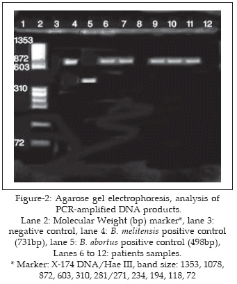
The amplifications were carried out in a Techgene thermocycler, with the following steps: initial denaturation at 95oC for 3 min followed by 35 cycles of 95oC for 2 min, 55oC for 2 min and 72oC for 2 min with a final extension at 72oC for 4 min.17 The products were analyzed by electrophoresis through a 2% (w/vol) agarose gel, after which the gel was stained with 0.5 mg/ml ethidium bromide, and DNA fragments were visualized by UV transilluminator. Positive and negative controls of PCR were included in each experiment. Negative control, containing all the reagents but lacking template DNA were processed exactly as has been described to monitor for contamination with brucella DNA. All were negative in all experiments. Positive controls with 100ng of genomic DNA isolated from a suspension of B. melitensis and B. abortus were also included. All PCR reactions were carried out in duplicate. A strain of staphylococci from our department was used as a control for the DNA extraction and contamination; its PCR was negative in all the experiments carried out.
Data were analyzed with the help of SPSS software, using Chi square and t-test.
RESULTS
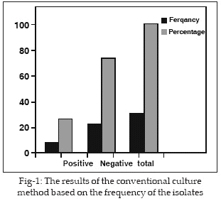
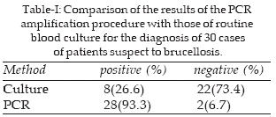
For eight patients (26.6%), the diagnosis of brucellosis was established by isolating the pathogen in blood cultures. All the strains isolated were identified as B. melitensis (Figure-1). When PCR technique was applied to serum samples, 28 cases (93.3%), were positive and gave 731bp B. melitensis-specific bands (Figure-2). All the culture positive samples were confirmed by PCR of pure culture and relevant serum samples. Neither of examined cultures or sera subjected to PCR were positive for B. abortus. The sensitivity of the PCR in this study was 93.3%. Table-I, shows the diagnostic results of PCR compared with those of conventional culture methods for the 30 cases of brucellosis studied. The difference was significant (P<0.05).
DISCUSSION
Our results showed that the sensitivity of the PCR assay using serum samples was far superior (93.3%), to that of conventional culture, which was positive for only 26.6% of cases. This very good sensitivity, confirms that the PCR assay could be a useful tool for the diagnosis of human brucellosis as other investigators showed by using whole blood18-20 or serum samples.21
The explanation for the low yield of conventional culture in present study, appears to be related more to the low number of pathogen in the blood sample and use of different antibiotic treatments for various diagnostic suspicion in other clinical sectors, before referring to Infectious Diseases Units, than to the technical difficulty of isolation brucella spp. from clinical samples. The high degree of sensitivity of the PCR assay even for patients who had previously received nonspecific antibiotic therapy, seems to be related to the high degree of detection capacity of the technique. As it was estimated in other studies, the detection capacity is about 10 to 100fg of DNA.20 Moreover, PCR is able to amplify intra-macrophage pathogens or pathogens which are damaged or nonviable as a result of previous treatment and which would be impossible to isolate in conventional culture.
In most of the PCR works on brucellosis, whole- blood was used. In this study, we used serum samples instead of whole- blood for PCR assay. There is only one study in which serum is the preferred clinical specimen for diagnosis of human brucellosis.21 In their study, the diagnostic sensitivities for serum and whole-blood assay were 94% and 61% respectively. The use of serum instead of whole-blood samples, offers several advantages for nucleic acid amplification methods. Inhibition by anticoagulants, haemoglobin, human DNA, or any other substance present in whole-blood but not in serum, is circumvented. Besides, red blood cell lysis, washings by centrifugation, and measurement and adjustment of isolated DNA concentrations are not required.21 Regarding the origin of pathogen nucleic acids in serum samples, most probably, they are released in the circulation as breakdown products during bacteremia, as several studies have documented the presence of circulation pathogen DNA in serum samples.22,23
Finally, in addition to the high yield of the PCR assay for the diagnosis of human brucellosis according to present study, and focal complications in such patients as previously reported,20 other important aspects are:
1) PCR is fast, providing results in 24 hour, which is much less than the time required for conventional methods to rescue a fastidious microorganism such as brucella spp.,
2) the technique almost completely obviates the necessity for direct handling of the pathogen, thus drastically reducing the risk of infection of laboratory personnel, and
3) the sample can be stored at –20oC until processing, thus enabling it to be collected by any physician and processed immediately, or else stored and safely sent to another laboratory if necessary.
In conclusion, the high degrees of sensitivity of PCR assay, together with its speed, versatility in sample handling, and risk reduction for laboratory personnel, make this technique a very useful tool for the diagnosis of brucellosis, compared with conventional culture method.
ACKNOWLEDGEMENTS
This study was supported by funds (Grant No. 418) of research affairs, Ahwaz Jundi Shapur University of Medical sciences, Ahwaz, Iran. We thank the personnel of Infectious Diseases Ward and Razi hospital’s central laboratory for their collaboration.
REFERENCES
1. Matyas Z, Fujikura T. Brucellosis as a world problem. Dev Bio Stand 1984; 56:3-20.
2. Corbel MJ. Brucellosis: an overview. Emerg Infect Dis 1997; 3:313-21.
3. World Health Organization: Brucellosis. Fact Sheet No. 173, 1997. WHO. Geneva, Switzerland.
4. Verger JM, Grimont F, Grimont PAD, Grayon M. Taxonomy of the genus brucella. Ann Inst Pasteur/Microbiol 1987; 138: 235-8.
5. Colmenero JD, Reguera JM, Martos F, Sanchez-Mora D, Delgado M, Causse M, et al. Complications associated with Brucella melitensis infection: a study of 530 cases. Medicine 1996; 75:195-211.
6. Rufi G, Pujol M, Gudiol F. Characteristics of and risk factors for relapse of brucellosis in humans. Clin Infect Dis 1995; 20:1241-9.
7. Kiel FW, Khan NJ. Analysis of 506 consecutive positive serologic tests for brucellosis in Saudi Arabia. J Clin Microbiol 1987; 25:1384-7.
8. Yagupsky P. Detection of Brucella melitensis by BACTEC NR660 blood culture system. J Clin Microbiol 1994; 32:1899-1901.
9. Ariza J, Pellicer T, Pallares R, Foz A, Gudiol F. Specific antibody profile in human brucellosis. Clin Infect Dis 1992; 14:131-40.
10. Young EJ. Serologic diagnosis of human brucellosis: analysis of 214 cases by agglutination tests and review of the literature. Rev Infect Dis 1991; 13:359-72.
11. Leal-Klevezas D, Martinez-Vazquez IO, Lopez-Merino A, Martinez-Soriano JP. Single-step PCR for detection of brucella spp. from blood and milk of infected animals. J Clin Microbiol 1995; 33: 3087-90.
12. Matar GM, Khneisser IA, Abdelnoor AM. Rapid laboratory confirmation of human brucellosis by PCR analysis of a target sequence on the 31-kilodalton brucella antigen DNA. J Clin Microbiol 1996; 34:47-8.
13. Romero C, Gamazo C, Pardo M, Lopez-Goni I. Specific detection of brucella DNA by PCR. J Clin Microbiol 1995; 33:615-7.
14. Yagupsky P. Detection of brucellae in blood cultures. J Clin Microbiol 1999; 37:3437-42.
15. Koneman EW, Allen SD, Janda WM, Schreckenberger PC, Winn WC. Brucella species. In: diagnostic Microbiology, 5th ed. Lippincott, Philadelphia, USA. 1997; pp.:431-6.
16. Bricker BJ, Halling SM. Differentiation of Brucella abortus bv.1,2, and 4, Brucella melitensis, Brucella ovis and Brucella suis bv.1 by PCR. J Clin Microbiol 1994; 32:2660-26.
17. Leyla G. Comparison of polymerase chain reaction and bacteriological culture for the diagnosis of sheep brucellosis using aborted fetus samples. Veter Microbiol 2003; 93:53-61.
18. Queipo-Ortuno MI, Morata P, Ocon P, Manchado P, Dios Colmenero J. Rapid diagnosis of human brucellosis by peripheral blood PCR assay. J Clin Microbiol 1997; 35:2927-30.
19. Casanas MC, Queipo-Ortuno MI, Rodriguez-Torres A, Orduna A, Colmenero JD. Specificity of a polymerase chain reaction assay of a target sequence on the 31-kilodalton brucella antigen DNA used to diagnose human brucellosis. Eur J Clin Microbiol Infect Dis 2001; 20:127-31.
20. Morata P, Queipo-Ortuno MI, Reguera JM, Miralles F, Lopez-Gonzalez JJ, Colmenero JD. Diagnostic yield of a PCR assay in focal complications of brucellosis. J Clin Microbiol 2001; 39:3743-6.
21. Zerva L, Bourantas K, Mitka S, Kansouzidou A, Legakis NJ. Serum is the preferred clinical specimen for diagnosis of human brucellosis by PCR. J Clin Microbiol 2001; 39:1661-4.
22. Bougnoux ME, Dupont C, Mateo J, Saulnier P, Faivre V, Payen D, Nicolas-Chanoine MH. Serum is more suitable than whole blood for diagnosis of systemic candidiasis by nested PCR. J Clin Microbiol 1999; 37:925-30.
23. Murdoch DR, Walford EJ, Jennings LC, Light GJ, Schousboe MI, Chereshsky S, et al. Use of polymerase chain reaction to detect legionella DNA in urine and seum samples from patients with pneumonia. Clin Infect Dis 1996; 23:475-80.
HOME | SEARCH | CURRENT ISSUE | PAST ISSUES
Professional
Medical Publications
Room No. 522, 5th Floor, Panorama Centre
Building No. 2, P.O. Box 8766, Saddar, Karachi - Pakistan.
Phones : 5688791, 5689285 Fax : 5689860
pjms@pjms.com.pk