|
|
||||
|
Published by : PROFESSIONAL MEDICAL PUBLICATIONS |
||||
|
ISSN 1681-715X |
||||
|
||||
|
- |
||||
|
Short Communication |
||||
|
- |
||||
|
Volume 22 |
October - December 2006 |
Number 4 |
||
|
|
||||
|
|
||||
|
|
||||
|
Published by : PROFESSIONAL MEDICAL PUBLICATIONS |
||||
|
ISSN 1681-715X |
||||
|
||||
|
- |
||||
|
Short Communication |
||||
|
- |
||||
|
Volume 22 |
October - December 2006 |
Number 4 |
||
|
|
||||
|
|
||||
Mycological Studies in
15 Cases of OtomycosisAli Zarei Mahmoudabadi1
Abstract
Otomycosis is a subacute or acute superficial mycotic infection of the outer ear canal that is caused by opportunistic fungi. The infection is usually unilateral and characterized by inflammation, pruritus, scaling and severe discomfort such as suppuration and pain. In this study 15 patients (11 female and 4 male) with symptomatic otomycosis were confirmed by direct microscopy and cultures. The most common fungal pathogens were Aspergillus niger (8 cases) followed by A. flavus (2 cases), A. fumigatus, Penicillium Spp., Candida albicans, C. parapsilosis and Rhizopus Spp each 1 case.
Key words: Otomycosis, Aspergillus niger, Aspergillus, Penicillium, Candida, Rhizopus.
Pak J Med Sci October - December 2006 Vol. 22 No. 4 486-488
1. Dr. Ali Zarei Mahmoudabadi, BSc. MSc, PhD,
Department of Medical Mycoparasitology,
Jundishapour University of Medical Sciences,
Ahwaz, Iran.Correspondence:
Dr. Ali Zarei Mahmoudabadi
Email: zarei40@hotmail.com* Received for Publication: November 25, 2005
* Accepted: May 30, 2006
Introduction
Otomycosis is an acute, subacute or chronic fungal infection of the pinna, the external auditory meatus and the ear canal.
1 However the disease may occur in the middle ear in case of perforated tympanic membrane.2 Infection is caused by some species of the saprophytic fungi, such as moulds and yeasts; especially Aspergillus niger.3,4 Other etiologic agents include: A. flavus, A. fumigatus, Allescheria boydii, Scopulariopsis, Penicillium, Rhizopus, Absidia and Candida Spp.4-6 In addition, otomycosis is a secondary infection deals to predisposing factors such as bacterial otitis externa corticosteroids therapy and swimming.2 The presenting symptoms include: scaling, pain, pruritus and erythematous, etc. Wax formation is also prominent. Otomycosis can occur in both temperate and tropical environment.3 The prevalence of disease is greatest in hot, humid and in dusty areas. In this study, fungal agents, predisposing factors and sex distribution for otomycosis were investigated.Patients and methods
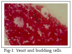
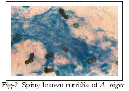
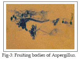
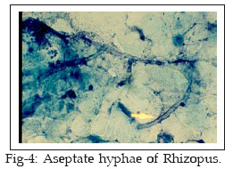
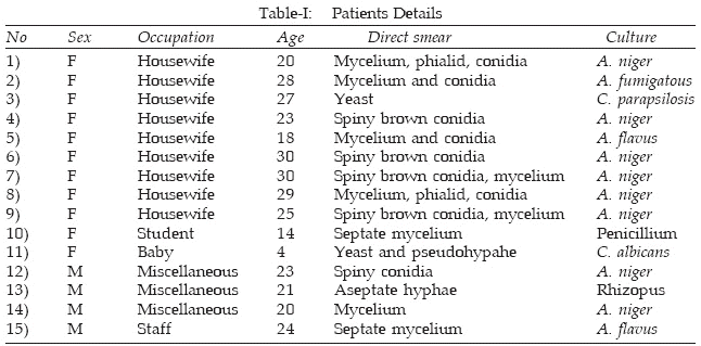
Fifteen patients attending at the Jahad and Razi Laboratories supposed to be suffering from disease were involved in this investigation. It included 11 female (73.3%) and 4 males (26.7%). The ages of the patients ranged between 4-30 years with a mean of 17 years. All patients had one or more of the aural symptoms (itching, otalgia, hearing loss). Secretion and pus were collected from the ear by two sterile cotton wool swabs. One swab was used for direct microscopy and other for culture examination. Direct examination of the samples was carried out by staining the smears with methylene blue and Gram techniques. Otomycosis was confirmed by the presence of aseptate mycelium, septate mycelium, Aspergillus conidia, fruiting bodies, yeast and pseudohyphae (Figs 1-4). The presence of fungal elements in stained smears was re-confirmed by fungal culture fungal colonies. Any kind of clinical materials, especially liquid samples (swabs, pus) should be examined as quickly as possible. Swabs did not require processing and were directly used for culture. Swabs were rolled and inoculated over the surface of Sabouraud�s Dextrose Agar with chloramphenicol (SC). Cultures were incubated at laboratory ambient (25-27�C) for 2-3 weeks, aerobically. Fungal isolates (moulds) were identified on the basis of colonial morphology and slide cultures. Yeast colonies, also detected by germ tube test, production of chlamydoconidia on corn meal agar and API 20 C AUX system.
Results
This report includes 15 patients with otomycosis. Mycelial elements, branching mycelium, fruiting heads and brown spiny conidia were observed in 12 samples. In the direct smears of 8 patients and growth besides in the culture medium showed A. niger. In direct smears of 4 patients septate branching mycelium conidia were seen and isolated species from the culture medium were as follow A. flavus (2 cases), A. fumigatus (1), Penicillium Spp.(1). The direct smears of two patients showed yeast cells, budding cells and pseudohyphae while in the culture medium C. albicans and C. parapsilosis had grown respectively. Broad aseptate mycelia were also seen in direct smear and yielded Rhizopus Spp. in culture. (Table-I)
Discussion
Otomycosis occur more commonly in female (especially housewife) than male and our finding confirmed the results other researcher�s have reported.
5,7 Out of 15 patients, 11 were female and 4 male. Otomycosis usually occurs most frequently in adults, and less in children.3,7 In our study, we found that otomycosis was more common in young men which is similar to the findings of the other researchers.2,3,5 Nine patients were housewife; two patients one each were student and baby. Four cases (male) had miscellaneous occupations.Infectious mould agents which are present in environment including: A. niger, A. flavus, A. fumigatus, Penicillium, Scopulariopsis, Rhizopus, Mucor, etc. A. niger has been reported as the most common causes of otomycosis. In two studies on otomycosis in Babol and north-western area of Iran, A. niger was major cause of cases.
7,8 Ozcan et al.2 and Hurst9 reported A. niger as a major etiologic agent of otomycosis in Turkey and Australia, respectively. However in Kaur et al.3 study A. fumigatus was reported as major agent, followed by A. niger. Other species of Aspergillus that have been associated with otomycosis are A. flavus.5 Also Penicillium Spp. has been reported by Pavelenko.10 Other fungi that have been associated with otomycosis are C. albicans and C. parapsilosis.5 In this study A. niger was the most common isolate, followed by A. flavus.Otomycosis is a secondary infection of the ear and predisposing factors are responsible for the invasion of fungi. Secondary bacterial infection was one of the most common predisposing factors in the history of our patients followed previous antibiotic therapy for one to four months duration and lastly swimming was the causative factor. These factors may differ from region to region. Our patients were admitted in the spring and summer, when it is hot and humid in Ahwaz. In three patients an impairment of hearing (deafness) were observed too. All patients were refereed for treatment to physicians.
conclusion
In this study we found that otomycosis is common in female than male and A. niger is the major etiologic agent in Ahwaz.
References
1 Anaissie EJ, McGinnis MR, Pfaller MA. Clinical Mycology, Philadelphia, Elsevier Sciences 2003, p. 464.
2 Ozcan KM, Ozcan M, Karaarslan A, Karaarslan F. Otomycosis in Turkey: predisposing factors, etiology and therapy. J Laryngol Otol 2003; 117: 39-42.
3 Kaur R, Mittal N, Kakkar M, Aggarwal AK, Mathur MD. Otomycosis: a clinicomycologic study. E N T J 2000; 79: 606-9.
4 Miertusova S, Simaljakova M. Yeasts and fungi isolated at the mycology laboratory of the First Dermatovenerology Clinic of the Medical Faculty Hospital of Comenius University in Bratislava (1995-2000). Epidemiol Microbiol Imunol 2003; 52:76-80.
5 Pradhan B, Tuladhar NR, Amatya RM. Prevalence of otomycosis in outpatient department of otolaryngology in Tribhuvan University Teaching Hospital, Kathmandu, Nepal. Ann Otol Rhinol Laryngol 2003; 112: 384-7.
6 Roland P S. Chronic external otitis. E N T J 2001; 80:12-6.
7 Ghiacei S. Survey of Otomycosis in north-western area of Iran. Med J Mashhad Uni Med Sci 2001; 43: 85-7.
8 Sephidgar A, Kyakajouri K, Meyrzaei M, Sharifi F. Fungal infection of external ear in otomycosis. J Babol Med Sci 2001; 13: 25-9.
9 Hurst WB. Outcome of 22 cases of perforated tympanic membrane caused by otomycosis. J Laryngol Otol 2001; 115: 879-80.
10 Pavelenko SA. Otomycosis in the Kuznetsk region and organization of medical services for this group of population. Vestin Otorinolarigol 1990; 4: 70-4.
HOME | SEARCH | CURRENT ISSUE | PAST ISSUES
Professional
Medical Publications
Room No. 522, 5th Floor, Panorama Centre
Building No. 2, P.O. Box 8766, Saddar, Karachi - Pakistan.
Phones : 5688791, 5689285 Fax : 5689860
pjms@pjms.com.pk