|
|
||||
|
Published by : PROFESSIONAL MEDICAL PUBLICATIONS |
||||
|
ISSN 1681-715X |
||||
|
||||
|
- |
||||
|
ORIGINAL ARTICLE |
||||
|
- |
||||
|
Volume 24 |
October - December 2008 (Part-I) |
Number 5 |
||
|
|
||||
|
|
||||
|
|
||||
|
Published by : PROFESSIONAL MEDICAL PUBLICATIONS |
||||
|
ISSN 1681-715X |
||||
|
||||
|
- |
||||
|
ORIGINAL ARTICLE |
||||
|
- |
||||
|
Volume 24 |
October - December 2008 (Part-I) |
Number 5 |
||
|
|
||||
|
|
||||
Antioxidants, Oxidative Stress status and
Waist/Hip ratio in normolipidaemic
AMI patientsArun Kumar1, Ramiah Sivakanesan2, Susil Gunasekera3
ABSTRACT
Objectives: The main objectives were to compare antioxidants and lipid peroxidation markers in normolipidaemic AMI patients to age/sex-matched controls.
Methodology: The study was conducted in Hindustan Institute of Medical Sciences and Research, Sharda Hospital, India on 165 AMI patients, (123 males and 42 females) admitted in ICCU, Sharda Hospital with age/sex-matched 165 controls (123 males and 42 females). Apart lipid profile measurement, the activities of superoxide dismutase (SOD), glutathione peroxidase (GPx), and catalase were determined. Oxidative stress markers, malondialdehyde (MDA) and conjugated diene (CD) were also determined.
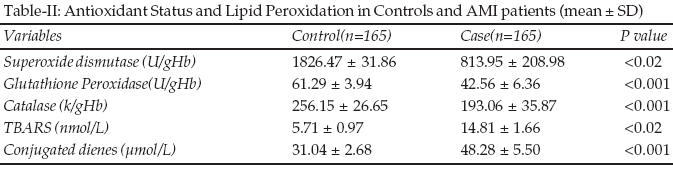
Results: The mean SOD activity in patients were 813.9 (U/gHb) compared to 1826.5 (U/gHb) in controls which was significantly lower (p<0.02). The mean GPx activity in patients was 42.6 (U/gHb) significantly lower than controls (p<0.001), 61.3 (U/gHb). Significant changes (p<0.001) were observed in mean catalase activities in patients, 193.06 (k/gHb) compared to control 256.2 (k/gHb).The mean MDA levels were significantly higher in patients (p<0.02) showing 14.81 (nmol/L), compared to control, 5.71 (nmol/L). Similarly conjugated diene levels in patients were higher, 48.28 (�mol/L) than controls 31.0 (�mol/L) (p<0.001).
Conclusions: Myocardial Infarction is a multifactorial disease in which normolipidaemic subjects could also be a victim even though the risk is higher in dyslipidemic subjects. Apart from lipid profile measurement in health check up package, inclusion of antioxidants studies should also be incorporated as the current studies highlight the facts with special attention to higher waist/ hip ratio subjects.
KEY WORDS: Antioxidants, Malondialdehyde, Conjugated diene, AMI, Waist / Hip ratio, Normolipidemia.
Pak J Med Sci October - December 2008 (Part-I) Vol. 24 No. 5 689-693
How to cite this article:
Kumar A, Sivakanesan R, Gunasekera S. Antioxidants, Oxidative Stress status waist/hip ratio in normolipidaemic AMI patients. Pak J Med Sci 2008;24(5):689-93.
1. Arun Kumar,
Research Scholar,
Department of Biochemistry,
Faculty of Medicine,
University of Peradeniya,
Sri Lanka.
2. Prof. Ramiah Sivakanesan
Head, Department of Biochemistry,
Faculty of Medicine,
University of Peradeniya,
Sri Lanka.
3. Dr. Susil Gunasekera,
Senior Lecturer,
Department of Biochemistry,
Faculty of Medicine, University of Peradeniya,
Sri Lanka.Correspondence
Dr. Arun Kumar,
Assistant Professor,
Phullari Campus,
Department of Biochemistry,
Manipal College of Medical Sciences,
Pokhara,
Nepal.
Email: arun732003@gmail.com
* Received for Publication: October 22, 2008
* Revision Received: February 13, 2008
* 2nd Revision Received: August 25, 2008
* Final Revision Accepted: August 26, 2008
INTRODUCTION
Cardiovascular disease would be the leading cause of mortality and morbidity in the world by the year 2015
1 and people from Indian subcontinent are at higher risk. It is a multifactorial disease accompanied by factors like hereditary, hyperlipidemia, obesity, hypertension, environmental factors and life style variables like stress, smoking, alcohol consumption, etc.2 Fatty diet aggravates coronary artery disease (CAD) and dyslipidemia add additive risk especially in Asians. Elevated low-density lipoprotein cholesterol (LDL) stands important due to its oxidative properties and atherogenic potential however, some patients of CAD are normolipidaemic.3 Under oxidative stress apart from circulating LDL molecules other lipoproteins also become a victim to structural changes induced due to oxidation including malondialdehyde and conjugated diene formation. Literature survey reveal the risk of myocardial infarction is aggravated in dyslipidemia.4As we came across patients of AMI with normolipidaemia, the current study was planned to evaluate the balance of lipid of antioxidants and lipid peroxidation in these patients, moreover, extensive literature search revealed not much studies was conducted in this area.
METHODOLOGY
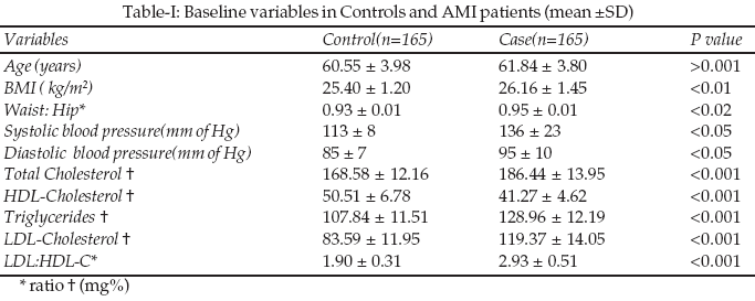
Setting Design and Patients: The study was conducted in Hindustan Institute of Medical Sciences and Research, Sharda Hospital, India. The study comprised of 165 normolipidemic AMI patients, male to female ratio was 3:1, with ages ranged from 48-69 years, mean � SD (61.8 � 3.8 y). One hundred sixty five age-sex matched subjects, with similar ratio of male: female as in patients were recruited as controls. Their ages ranged from 48-69 years, mean � SD (60.55 � 3.98 y). The AMI cases were diagnosed as per diagnostic criteria: chest pain lasting for >3 hours, ECG changes (ST elevation of 2 mm or more in at least two leads), increased creatine phosphokinase (CPK-MB) and aspartate aminotransferase enzyme. Informed consent was obtained from patients and controls recruited for the study and the study was approved by the ethical committee of the Institution.
Exclusion Criteria: Patients with diabetes mellitus, hyperlipidemia, renal insufficiency, current and past smokers, hepatic disease or taking lipid lowering drugs or antioxidant vitamin supplements.
Inclusion Criteria: Normolipidemic AMI patients as per the NCEP ATP -111 guidelines.
Criteria for Normolipidemics: Normal lipid profile was defined if LDL was <160mg/dl, HDL >35mg/dl, Total cholesterol (TC) <200 mg/dl and Triglycerides (TG) were <150mg/dl.
5 Blood (10 ml) was collected after overnight fasting in different containers.1. EDTA vial: 5.0 ml of blood was taken. Red cells were washed 3-4 times with ice-cold normal saline and used for estimation of glutathione peroxidase, superoxide dismutase and catalase.
2. Plain vial: Remaining blood was taken and serum was separated by centrifugation for five minutes at 5000 rpm and was used for determination of lipid profile, malondialdehye and conjugated dienes.
Total cholesterol, triglycerides, and HDL-cholesterol were estimated by enzymatic methods using the kits obtained from Randox Laboratories Limited, Crumlin, UK. Plasma LDL-cholesterol was determined from the values of total cholesterol and HDL-cholesterol using the following formulae:
LDL-c = TC - TG � HDL-c (mg/dl)
5
All chemicals of analytical grade were obtained from Sigma chemicals, New Delhi, India.
Glutathione Peroxidase (GPx): The glutathione peroxidase activity in red cells was determined by the procedure of Paglia and Valentine.
6 Briefly, the oxidized glutathione produced during GPx enzyme reaction was immediately reduced by NADPH and glutathione reductase. Therefore, the rate of NADPH consumption was monitored as a measure of formation of oxidized glutathione. Results were expressed as units of GPx per gram of hemoglobin.Superoxide dismutase (SOD): Superoxide dismutase enzyme activity in red cells was measured by SOD assay kit using rate of inhibition of 2-(4-indophenyl)-(4-Nitrophenol)-5-phenyltetrazolium chloride (I.N.T) reduction method, modified method of Sun et al
7 using Ransod assay kit SD 125, Randox Lab. One unit of SOD activity was defined as the amount of protein that inhibits the rate of I.N.T. reduction by 50%. Enzyme activity was expressed as Unit per gram hemoglobin (U/gHb). Hemoglobin was measured by Drabkin�s method.Catalase: Catalase activity in red cells was measured spectrophotometrically as described by Beutler.
8 One Unit of enzyme activity was expressed as micromole hydrogen peroxide decomposed per min per gram hemoglobin.MDA Method: MDA levels in serum were estimated by thiobarbituric acid (TBA) reaction.
9 Using 40% trichloroacetic acid, proteins were precipitated from 0.5 ml serum, and precipitated proteins were incubated with TBA reagent in a boiling water bath for one hour. After bringing down to room temperature, the colored complex formed was measured using spectrophotometer at 532 nm.1, 1, 2, 3-tetraethoxypropane (1 nmol/l) was used as a standard for MDA estimation. The concentration was expressed in terms of (nmol/l).Conjugated dienes (CD): CD levels in serum were measured by Recknagel and Glende method
10 with little modification. Briefly, the principle of the assay is based on with the rearrangement of double bonds in polyunsaturated fatty acids leading to the formation of CD, which absorb light at 233 nm. The oxidation index of the lipid sample at 233 nm and 215 nm is computed which reflect the diene content and the extent of peroxidation. The lipid peroxidation (LP) products measured in serum were treated with antioxidant butylated hydroxytoluene (BHT) twice, immediately after obtaining and before adding the test reagents to suppress artefactual changes during handling and assay procedures. The first stage of LP consists of the molecular rearrangement of the double bonds in polyunsaturated fatty acids residues of lipids, which leads to CD formation and conversion of CD in hydroperoxide. Serum was chosen to avoid possible influences of substances required for plasma preparation. Serum sample (150 �l) and (150�l) of 0.9% NaCl (reagent blank contains only isotonic saline) were incubated at 37�C for 25 minutes. 0.25% BHT (150�l) was added and the lipids were extracted by heptane/isopropanol (1:1). Then samples were acidified by 5mol/L HCl and extracted by cold heptane (1600�l). After centrifugation for five minutes at 3000 rpm the absorbance of heptane fraction were measured spectrophotometrically at absorbance maximum between 220nm and 250nm. The amount of hydroperoxides produced was calculated using Molar Coefficient of 2.52 X 10⁴ m-1.Statistical Analysis: The data from patients and controls were compared using Student�s �t�-test. Values were expressed as mean � standard deviation (SD). Microsoft Excel for windows 2000 was used for statistical analysis. �P� value of less than 0.05 was considered to indicate statistical significance.
RESULTS
The baseline variables, antioxidant status and lipid peroxidation in AMI patients compared to control are shown in Table-I&II. Body mass index (BMI), waist / hip ratio, blood pressure, and lipid profile parameters were significantly higher in AMI patients (Table-I).
The antioxidant enzymes and lipid peroxidation are shown in Table-II. Serum SOD, GPx and catalase were significantly lower in AMI patients compared to control. The MDA and CD levels were significantly higher in patients compared with controls (Table-II).
DISCUSSION
Atherosclerosis is the root cause of AMI. Antioxidants play an important role by inhibiting LDL oxidation further minimizing the progression of atherosclerosis. It is a chronic inflammatory condition followed by acute clinical event, by plaque rupture leading to thrombosis. There fore inflammation occupies a central place, in all phases of atherosclerosis before emerging as a clinical event like AMI.
11 Involvement of oxygen free radicals in inflammation, ischemia and reperfusion damage have been reported earlier.12 Demonstration of elevated serum lipid peroxides, justifies the claim.Significant rise in MDA (p<0.02) and CD levels (p<0.001) in the current study is suggestive of increased oxidative stress in AMI patients. The current study concurs with the work of Dubois Rande et al,
13 Mc Murray14 and Senthil et al15 reported similar findings.OFRs are generated in early phase of MI and due to involvement of GPx to reduce hydrogen peroxide radicals, their level decreases. The current study observed similar pattern of reduction in GPx activity as reported by Kharb,
16 Simic17 and Blaustein et al.18 Perfect negative correlation was established between GPx and MDA levels in AMI patients (r = -0.62). This suggests GPx being antioxidant gets succumbed to peroxidised products during oxidative damage. Ondregjickova et al.,19 demonstrated increased glutathione disulfide with concomitant decrease in GPx in myocardium during coronary occlusion. Their study confirms the abnormal balance between the oxidative and protective mechanisms in MI patients which is further supported by the findings of the current study.The SOD activity was significantly lower in patients (p<0.02) compared to controls in current study. Similar findings were reported by Kumar and Das.
20Even catalase activities were significantly lowered in AMI patients (p<0.001) compared to control. Senthil et al
21 reported the reduction of catalase in cardiogenic shock patients. The results of these studies are suggestive of increased utilization of free radical scavenging system to combat the toxic radicals. It could also be due to decreased catalase activities, the lipid peroxides are increased. Studies by Dusinovic et al,22 Pandey et al23 and Bhakuni et al24 on antioxidants defense system in MI patients, also observed similar findings. With the observations of all the past studies along with the current study findings indicates the asynchrony between oxidants and antioxidants in AMI patients.CONCLUSION
Myocardial Infarction is a multifactorial disease in which normolipidaemic subjects could also be a victim even though risk is higher in dyslipidemic subjects. Apart from lipid profile measurement in health check up package, inclusion of antioxidants studies should also be incorporated as the current studies highlights the facts with special attention to higher waist/ hip ratio subjects.
REFERENCES
1. Reddy KS. Cardiovascular disease in India. World Health Stat Q 1993;46:101-7.
2. Chopra V, Wasir H. Implications of lipoprotein abnormalities in Indian patients. J Assoc Physicians of India 1998;46:814-8.
3. Vasisht S, Narula J, Awtade A, Tandon R, Srivastava LM. Lipids and lipoproteins in normal controls and clinically documented coronary heart disease patients. Ann Natl Acad Med Sci (India) 1990;26:57-66.
4. Patil N, Chavan V, Karnik ND. Antioxidant Status in patients with Acute Myocardial Infarction. Ind J Clinical Biochemistry 2007;22:45-51.
5. Executive Summary of The Third Report of The National Cholesterol Education Program (NCEP) Expert panel on Detection, Evaluation and treatment of high Blood Cholesterol in Adults (Adult Treatment Panel III). Expert Panel of Detection, Evaluation and Treatment of High Blood Cholesterol in Adults. JAMA 2001;285(19):2486-97.
6. Paglia DE, Valentine WN. Studies on quantitative and qualitative characterization of erythrocyte glutathione peroxidase. J Lab Clin Med 1967;70:158-69.
7. Sun Y, Oberly LW, Li Y. A simple method for clinical assay of superoxide dismutase. Clin Chem 1988;34:497-500.
8. Beutler E. Red Cell Metabolism: A Manual of Biochemical Methods, 3rd edition. New York, Grune and Stratton, 1984;105.
9. Bernheim S, Bernheim MLC, Wilbur KM. The reaction between thiobarbituric acid and the oxidant product of certain lipids. J Biol Chem 1948;174:257-64.
10. Recknagel RO, Glende EA. Spectrophotometric detection of lipid conjugated dienes. Methods Enzymol 1984; 105:331-337.
11. Libby P. Vascular biology of atherosclerosis: Overview and state of art. Am J Cardiol 2003;91(suppl):3A-6A.
12. Goldhaber J, Weiss JN. Oxygen free radicals and cardiac reperfusion abnormalities. Hypertension 1992;20:118-127.
13. Dubois-Rande JL, Artigou JY, Darmon JY, Habbal R, Manuel C, Tayarani I, et al. Oxidative stress in patients with unstable angina. Eur Heart J 1994;15(2):179-83.
14. McMurray J. Evidence of oxidative stress in chronic heart failure in humans. Eur Heart J 1993;14(11):1493-7.
15. Senthil S, Veerappan RM, Ramakrishna Rao M, Pugalendi KV. Oxidative stress and antioxidants in patients with cardiogenic shock complicating acute myocardial infarction. Clin Chim Acta 2004;348:131-7.
16. Kharb S. Low blood glutathione levels in acute myocardial infarction. Ind J Med Sci 2003;57(8):335-7.
17. Simic D, Mimic-Oka J, Pljesa M, Milanovic D, Radojevic S, Ivanovic B. Time course of erythrocyte antioxidant activity in patients treated by thrombolysis for acute myocardial infarction. Jpn Heart J 2003;44:823-32.
18. Blaustein A, Dencke SM, Stolz RI. Myocardial glutathione impairs recovery after short periods of ischaemia. Circulation 1989; 80:1449-57.
19. Ondrejickova O, Horakova L, Juranek I, Ziegelhoeffer A. Effect of stobadine on lipid peroxidation in brain and heart after ischemia and reperfusion of the brain. Life Science 1999;65:1959-61.
20. Kumar KV, Das UN. Are free radicals involved in the pathobiology of human essential hypertension? Free Radic Res Commun 1993;19(1):59-66.
21. Senthil S, Veerappan RM, Ramakrishna RM, Pugalen-di KV. Oxidative stress and antioxidants in patients with cardiogenic shock complicating acute myocardial infarction. Clin Chim Acta 2004;348(1-2):131-7.
22. Dusinovic S, Mijalkovic D, Saicic ZS, Duric J, Zunic Z, Niketic V, et al. Antioxidative defense in human myocardial reperfusion injury. J Environ Pathol Toxicol Oncol 1998;17(3-4):281-4.
23. Pandey NR, Kaur G, Chandra M, Sanwal GG, Mishra MK. Enzymatic oxidant and antioxidants of human blood platelets in unstable angina and myocardial infarction. Int J Cardiol 2000;76(1):33-8.
24. Bhakuni P, Chandra M, Misra MK. Levels of free radical scavengers and antioxidants in post perfused patients of myocardial infarction. Current Science 2005;89(1):168-170.
HOME | SEARCH | CURRENT ISSUE | PAST ISSUES
Professional
Medical Publications
Room No. 522, 5th Floor, Panorama Centre
Building No. 2, P.O. Box 8766, Saddar, Karachi - Pakistan.
Phones : 5688791, 5689285 Fax : 5689860
pjms@pjms.com.pk