|
 |
|
Published
by : PROFESSIONAL MEDICAL PUBLICATIONS |
|
ISSN 1681-715X |
|
|
|
- |
|
ORIGINAL
ARTICLE |
|
- |
|
Volume 25 |
October - December 2009
(Part-I) |
Number
5 |
|
|
|
Incidence of head and neck birth defects in Iran:
A cross-sectional study from southwest region
Saki Nader1, Saki Ghasem2, Rahim
Fakher3, Nikakhlagh Sohayla4
ABSTRACT
Objective: The aims of the study were to assess the
prevalence of birth defects (BDs) in Ahwaz, Iran amongst the live births born
between 1 Oct 2006 and 31 Oct 2007.
Methodology: There were a total of 4176 live births
in Ahwaz during this study period. The interview included the Father and
mother’s date of birth, Number of gravidity, paternal smoking, maternal
delivery age and the occupation of the mother and father.
Results: Out of 4176 subjects, 1.43 per 1000 live
births presented with multiple anomalies. The overall occurrence of
malformation among working mothers were 1.67 and 7.42 for working fathers.
Parents in the occupational group ‘Commerce’ had the highest rates (3.83 per
1000 live births). The frequencies of BDs in smokers were 3.5 (per 1000 live
births). The gravidity profiles showed that all three types were very close to
each other. The occurrence rate of BDs increased in the maternal delivery age
of 18-35 years (5.74 per 1000 live birth).
Conclusion: The prevalence of BDs in Southwest of
Iran is comparable to those in other countries. Some of the birth defects are
not diagnosed at birth and may occur later in the life.
KEY WORDS:
Birth
defects (BDs), Gravidity, Paternal smoking, Maternal delivery age, Occupation
of mother and father.
Pak J Med Sci October - December 2009
(Part-I) Vol. 25 No. 5 770-775
How to cite this article:
Nader S, Ghasem S, Fakher R, Sohayla N. Incidence of head and neck birth
defects in Iran: A cross-sectional study from southwest region. Pak J Med Sci
2009;25(5):770-775.
1. Saki Nader,
2. Saki Ghasem,
Department of Anatomical Science,
Faculty of Medicine,
3. Rahim Fakher,
Physiology Research Center,
4. Nikakhlagh Sohayla,
1,4: Department of ENT,
Imam Khomeini Hospital,
Faculty of Medicine, Apadana Private Hospital,
Ahwaz, Khuzestan, Iran
1-4: Ahwaz Joundishapour University of Medical Science.
Correspondence
Ghasem Saki
Department of Anatomical Science,
Faculty of Medicine,
Ahwaz Joundishapour University of Medical Science,
Ahwaz – Iran.
E mail: ghasemsaki@yahoo.com
* Received for Publication: February 4, 2009
* Accepted for Publication: August 31,2009
INTRODUCTION
Birth defects constitute the leading cause of infant death
among all racial/ethnic groups.
1,2
A broad definition would include functional and metabolic disorders that,
although present, may not necessarily be recognizable at birth, but are
determined only when the child is older.3
There are two most commonly used classification systems: (A) the Birth defect
classification according to the International Classification of Diseases;4,5
and (B) the International Clearing-house for Birth Defects Monitoring System (ICHBD).6
Worldwide surveys have shown that the frequency of Birth defects varies
greatly from country to country. The frequency depends on the time of
observation after birth, the types of malformation included, and the
differences in reporting and statistical procedures.7
In a multiracial and heterogeneous population like that of Iran, it is
important to know the significance of various Birth defects and their possible
associated factors. It is vital to ascertain the epidemiology of Birth
defects, as a Birth defect can be a significant medical, psychological and
socio-economic burden on both the family and the community. Occupational and
environmental agents are the suspected causes for about 60% of birth defects
with unknown etiology.8
The existence of hazards in the workplace has raised concerns about the
potential of these substances for adverse reproductive effects. Historically,
studies assessing the role of occupational exposure as etiological agents for
birth defects focused on maternal exposures during pregnancy. The role of
paternal exposure received less attention despite animal evidence showing that
exposures of males to toxic agents may result in congenital malformations in
offspring.9-11
With increasing concern about male reproductive function in
the past decade, epidemiological studies are being published considering the
role of paternal exposures by evaluating paternal occupations and risk of
birth defects. Several studies have investigated whether parental cigarette
smoking increased the risk of having offspring with BDs.
12,13
In this observation the epidemiological evidence about the
relation between paternal and maternal occupations, paternal smoking, maternal
delivery age, gravidity with distribution of birth defects is summarized.
METHODOLOGY
Subjects:
The subjects included all live births born to Ahwazian parents between 18th
Oct 2006 and 31st
Oct 2007, who were registered at the Imam Khomeini and Apadana Hospitals.
Birth defects eligible to be included in the study were all live births with
the presence of one or more BD during the data collection period. The
inclusive criteria were based on the classification of cases according to the
International Clearing-house for Birth Defects Monitoring System (ICHBD).
Paternal and Maternal Interviews: The study was
approved by the Institutional Ethics Committee of Ahwaz Jondishapour
University of Medical Sciences (AJUMS).Written consents were taken from all
the participants of this study. Interviews were conducted with mothers and
fathers of study cases in Persian. Interviews were completed an average of two
months for cases after the date of delivery.
Data Collection: Information on live births and BD
cases obtained from the filled questionnaire and interview included the Father
and mother’s date of birth, Number of gravidity (Gravidity means number of
previous pregnancies. The gravidity was classified in three groups include; 1:
one previous pregnancy; 2: two previous pregnancies; 3: more than two previous
pregnancies), paternal smoking, maternal delivery age and the occupation of
the mother and father (if employed, the industrial sector involved).
Data processing and statistical analysis: Before being
added to the final database, all the raw data from the different sources were
checked and merged to produce a unique record number for each subject. The
entire database was then re-examined to identify and resolve any
discrepancies. New variables were created from the basic information in the
raw data. The final dataset comprised a comprehensive list of variables that
included an array of potential risk factors as well as socio-demographic
information. Statistical analyses were performed with standard contingency
tables using the SPSS program, version 15.0, on a personal computer.
RESULTS
During the one year period (2006–2007) covered by this
study, there were 31 cases with one or more congenital defects retrieved from
the records of a total of 4176 registered live newborns in Imam Khomeini and
Apadana Hospitals, Ahwaz. The overall rate of BDs was 7.42 per 1000 live
births. The occurrence of congenital defects (per 1000 live births) across the
period was 8.62 in 2006 and 7.02 in 2007—showing a downward trend from 2006 to
2007.
Among the live born with congenital defects in this study,
1.43 (per 1000 live births) had multiple anomalies. The commonest single BD
was ‘Cleft lip and Palate Cleft’, which accounted for 3.35 (per 1000 live
births) of all malformations (Table-I).

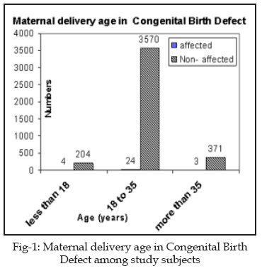
Maternal delivery age:
The mean maternal delivery age was 30.3
± 4.7 years (mean ± SD) for all live births, with 30.3 ± 4.7 years for
non-defects and 30.7 ± 5.0 years for defects. There was a significant increase
of distribution of the defect rates in maternal delivery age of 18-35 years
with a value of 5.74 per 1000 live birth (Fig-1).
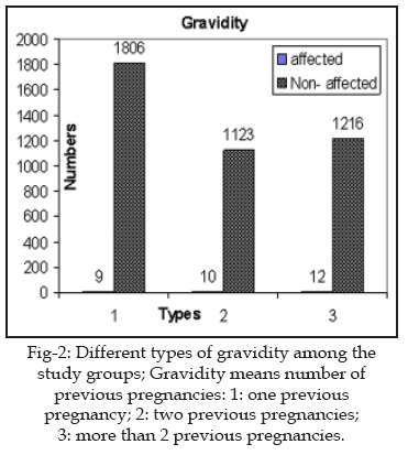
Gravidity:
The gravidity profiles showed that all three types were very close to each
other (Fig-2).
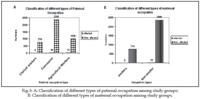
Distribution of BDs by parental and maternal occupation:
In total, 97.2% of mothers reported that they were not working; 2.8% held a
clerical occupation. In contrast to the large group of non-working mothers,
all the fathers were employed. The proportions of the fathers in other
occupations ranged from 18 to 24%. The overall malformation occurrences (per
1000 live births) of working mothers versus non-working mothers were 1.67
versus 5.74, and 7.42 for working fathers. In paternal occupational
classification: the lowest defect rate of 1.19 per 1000 was recorded for
‘Clerical workers’, while the highest rate of 3.83 per 1000 was found for
‘Commerce’ and 2.39 per 1000 was found for ‘Agricultural Workers’ (Figure 3 a
& b).
Paternal Smoking: Among all paternal groups who
participate in this study 1980 individuals have shown history of smoking and
the rest had no smoking history. The relevant frequency of BDs in smokers were
15 (3.5 per 1000 live births) and in non-smokers were 16 (3.8 per 1000 live
births), respectively (Fig-4).

DISCUSSION
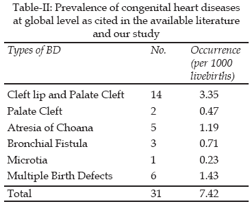
Based on our findings, the prevalence rate of BD was 0.74%
(7.42/1000). Comparison of international frequencies of BD that is available
in literature to our study described in Table-II.
14-24
Previous studies have suggested an association between these non-occupational
risk factors and adverse reproductive outcomes.7
Father’s drinking and coffee consumption also were not available.
These variables could be potential confounders, which may
over or underestimate the associations of fathers’ occupations and BDs. We
found the prevalence of overall BDs (7.42 per 1000) in our study to be fairly
close to that of other studies. According to the Finnish register of BDs, the
prevalence of BDs among newborns in Finland varied from 13 to 20 per 1000 in
1963–1980, and the mean for the period was 14 per 1000.5
Not many studies have reported national BD rates. Most published data are
based on hospital births over a period of time, and are not population based.
Dryden25
studied the rate of BDs in 10 000 babies born consecutively in a general
hospital in Papua New Guinea and reported an overall prevalence of congenital
defects of 11.6 per 1000. Riley et al.26
reported that 32 per 1000 babies had at least one malformation during a 13
year study period (1983–1995) in Victoria, Australia. However, the study
included 25 231 infants (born at 20 weeks or more) and 1566 terminations of
pregnancy before 20 weeks’ gestation. Between January 1992 and January 1995, a
total of 24 233 babies born consecutively in Corniche Hospital, which is the
only maternity hospital in Abu Dhabi, United Arab Emirates, were surveyed for
the presence of major congenital malformations. A total of 401 infants (16.6
per 1000) had a major defect.27
The time trend during our five year study indicated that, in the first four
years, the overall occurrence of BDs in live births decreased. Meanwhile,
increasingly sophisticated prenatal diagnostic procedures have been developed
to detect congenital defects in utero. The detection of serious congenital
anomalies earlier in pregnancy and with greater accuracy has led to a
corresponding increase in elective terminations of affected pregnancies.28
There were also considerable variations in the distribution of different
malformations.+
These results highlight the need to conduct a more detailed
study to identify the possible associations between parental occupations and
BDs. In summary, the prevalence of BDs in Ahwaz, Iran is comparable to those
in other countries. Comparison of international reports of most frequent type
of BDs that is available in literature to our study that shows Cleft lip and
Palate Cleft is most frequent type of BDs (3.35 per 1000), has been discussed
here. The prevalence of cleft lip and palate in different studies are, in the
United States 1.42 per 1000,30
Brazil 0.19 per 1000,31
Ireland 1.14 per 1000,32
1.03 per 1000 in Ireland,33
1.84 per 1000 in Lithuania,34
China 0.61 per 1000,35
Korea 1.81 per 1000,36
0.62 per 1000 in Oman,37
1.4 per 1000 in Japan,38
5.2 per 1000 in Tehran,39
and other Asian countries 1.22 per 1000.40
In conclusion, the reasons of inconsistency between our
results which represents congenital abnormalities ever investigated in Iran
and other studies are probably as bellow: Some of the birth defects are not
diagnosed at birth and may occur later in the life, therapeutic advances,
application of appropriate preconception care, and prenatal diagnosis program.
Also this inconsistency might be explained by diagnosis of both minor and
major BD in all systems.
REFERENCES
1. Lynberg MC, Khoury MJ. Contribution of birth defects to
infant mortality among racial/ethnic minority groups, United States, 1983.
MMWR CDC Surveill Summ 1990;39:1-12.
2. Ferencz C. Epidemiology of congenital heart disease: the
Baltimore–Washington infant study 1981–1989. In: Ferencz , Rubin JD, Loffredo
CA, Magree CA, eds. Perspective in Pediatric Cardiology,Vol.4.Mount
Kisco.NewYork: Futura Publishing Co., 1993;1-11.
3. Epstein CJ. Genetic disorders and birth defects. In:
Rudolph AM, Hoffman JIE, Rudolph CD, eds. Rudolph’s Pediatrics, 19th edn.
Appleton & Lange, 1991;265-9.
4. WHO. ICD-9. International Classification of Diseases,
9th revision. Geneva: WHO. (Year Missing)
5. WHO. ICD-10. International Classification of Diseases,
10th revision. Geneva: WHO. (Year Missing)
6. BPA. Classification of Diseases Vols 1 & 2, 2nd edn.
British Pediatric Association, 1987.
7. Taskinen HK. Effects of parental occupational exposures
on spontaneous abortion and congenital malformation. Scand JWork Environ
Health 1990;16:297-314.
8. Sever LE. Congenital malformations related to
occupational reproductive hazards [review]. Occup Med 1994;9:471-94.
9. Titenko-Holland N, Ahlborn T, Lowe X. Micronuclei and
developmental abnormalities in 4-day mouse embryos after paternal treatment
with acrylamide. Environ Mol Mutagen 1998;31:206-17.
10. Muller WU, Streffer C, Wojcik A. Radiation-induced
malformations after exposure of murine germ cells in various stages of
spermatogenesis.Mutat Res 1999;425:99-106.
11. Nagao T. Congenital defects in the offspring of male
mice treated with ethylnitrosourea. Mutat Res 1988;202:25-33.
12. Van Den Eeden S, Karagas MR, Daling J, Vaughan TL. A
case-control study of maternal smoking and congenital malformations. Paediatr
Perinatol Epidemiol 1990;4:147-55.
13. Shaw GM, Wasserman CR, Lammer EJ, O’Malley CD, Murray
JC, Basart AM, et al. Orofacial Clefts, Parental Cigarette Smoking, and
Transforming Growth Factor-Alpha Gene Variants. Am J Hum Genet 1996;58:551-61.
14. Al-Gazali LI, Dawodu AH, Sabarina T. The profile of
major congenital abnormalities in the United Arab Emirates populations. J Med
Genet 1995;32:743-7.
15. Sheikha SA. Epidemiology of congenital abnormalities in
Bahrain. East Mediterr Health J 1995;1:248-52.
16. Delport SD, Christianson AI, Van den B. Congenital
anomalies in black South African live born neonatal an urban academic
hospital. S Afr Med J 1995;85:111-15.
17. Hsich TT, Lo LM, Hsu JJ. Congenital malformation in
newborns. Analysis of 501 cases. Changgeg Yi Xue Za Zhi 1995;18:14-9.
18. Bittar Z. Major congenital malformation presenting in
the first 24 hours of life in 3865 conscecutive births in south of Beirut.
Incidence and pattern. J Med Libya 1998;46:250-60.
19. Hemmatoghlu O, Tiras MB, Gursoy R. The incidence of
congenital malformations in a Turkish.Int J Gyneocolo Obstet 1998;55:117-21.
20. Mkheidze MO. Birth defects monitoring (BDM) in St.
Petersburg; Reality or Rhetoric? Proceeding of the 10th international Congress
of Human Genetics; 2001; Vienna, Austria, P187.
21. Roya R, Gupta D, Ghosh SK. Spectrum of congenital
surgical malformations in newborns. JIMA 2002;100:321-25.
22. Shaw GM, Carmichael SL, Kaidarova Z, Harris JA.
Differential risks to males and females for congenital malformations among 2.5
million California births, 1989-1997. Birth Defects Res A Clin Mol Teratol.
2003;67(12):953-8.
23. Golalipour MJ, Ahmadpour-Kacho M, Vakili MA. Congenital
malformations at a referral hospital in Gorgan, Islamic Republic of Iran. East
Mediterr Health J 2005;11(4):707-15.
24. Sípek A, Gregor V, Horácek J. Birth defects in the
Czech Republic in the period 1994 - 2005—perinatology data. Ceska Gynekol
2007;72(2):103-9.
25. Dryden R. Birth defects recognized in 10,000 babies
born consecutively in Port Moresby General Hospital, Papua New Guinea. P N G
Med J 1997;40:4-13.
26. Riley MM, Halliday JL, Lumley JM. Congenital
malformations in Victoria, Australia, 1983–95: an overview of infant
characteristics. J Paediatr Child Health 1998;34:233-40.
27. Al Talabani J, Shubbar AI, Mustafa KE.Major congenital
malformations in United Arab Emirates (UAE): need for genetic counselling. Ann
Hum Genet 1998;62:411-18.
28. Limb CJ, Holmes LB. Anencephaly: changes in prenatal
detection and birth status, 1972 through 1990. Am J Obstet Gynecol
1994;170:1333-8.
29. Terry PB, Bissenden JG, Condie RG, Mathew PM. Ethnic
differences in congenital malformations. Arch Dis Child 1985;60:866-8.
30. Thomas PC. Multidiscilpinary care of the child born
with CL/P. ORL Head. Neck Nurs 2000;18:6-16.
31. De Castro Monterio Loffredo L, Freitas JA. Prevalence
of Oral Clefts from 1975-1994, Brazil. Rev Saued Publica 2001;35:571-5.
32. Newson AR, Mchammara CM. Cleft lip/ Palate in the west
of Ireland. Spec Care Dentist 2000;21:103-6.
33. Newson AR, Mchammara CM. Cleft lip/ Palate in the west
of Ireland. Spec Care Dentist 2000;21:103-6.
34. Vasiliauskas A., Utkus A., Matulevičienë A.,
Linkevičienë L., The incidence of cleft lip and/or palate among newborns in
Lithuania, 1993–1997. ACTA MEDICA LITUANICA. 2004;11(2):1-6.
35. Wangy LJ. Nonsyndromic Cleft lip with or without CP in
Chinese population. Hua Xi Yi Ke Dax Bao 2000;31:401-10.
36. Kim S, Kim WJ. Cleft palate / Lip incidence among the
live birth in the Republic of Korea. J Korean Med Sci 2002;18:6-16.
37. Rajab A., Thomas C. Oral clefts in the Sultanate of
Oman. Eur J Plast Surg 2001;24:230-3.
38. Natsume N, Kawai T, Kohama G, Teshima T, Kochi S,
Ohashi Y, et al. Incidence of cleft lip or palate in 303 738 Japanese babies
born between 1994 and 1995. British J Oral and Maxillofacial Sur
2000;38:605-7.
39. Jamalian A, Nayeri F, Babayan A. Incidence of cleft lip
and palate in Tehran. J Indian Soc Pedod Prevent Dent 2007;7:174-6.
40. Cooper ME, Ratay JS, Marazita ML. Asian oral-facial cleft birth
prevalence. Cleft Palate J 2006;43(5):580-9.
HOME
| SEARCH
| CURRENT
ISSUE | PAST
ISSUES
Professional
Medical Publications
Room No. 522, 5th Floor, Panorama Centre
Building No. 2, P.O. Box 8766, Saddar, Karachi - Pakistan.
Phones : 5688791, 5689285 Fax : 5689860
pjms@pjms.com.pk