|
 |
|
Published
by : PROFESSIONAL MEDICAL PUBLICATIONS |
|
ISSN 1681-715X |
|
|
|
|
|
- |
|
REVIEW ARTICLE |
|
- |
|
Volume 25 |
October - December 2009
(Part-I) |
Number 5 |
|
|
|
Is biopsy needed in every gastritis
found during endoscopy?
Muhammad Zubair1, Muhammad Ali Channa2,
Masoom Raza Mirza3, Lubna Habib4
ABSTRACT
Objective: To find out concordance between
endoscopic and histological diagnosis of gastritis in dyspeptic patients.
Methodology: All dyspeptic patients who underwent
upper gastrointestinal endoscopy with endoscopic diagnosis of gastritis and
available biopsy report from July 2006 to June 2008 in Hamdard Medical
University Hospital and different private hospitals of Karachi were included
in this study. The endoscopist formed a global impression on the presence or
otherwise of gastritis according to the Sydney System. Patients with ulcer,
growth and any other endoscopic diagnosis apart from gastritis were excluded.
With standard biopsy forceps, minimum of two gastric biopsies from inflamed
mucosa were taken for histological evidence of gastritis.
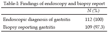
Results: Total 112 patients (44 males and 68
females) were included in the study with mean age of 41.95 years. Most common
symptom experienced by patients was epigastric discomfort in 103 (92%) of
patients. In all patients endoscopic diagnosis was gastritis. Biopsy reports
showed chronic gastritis in 109 (97.3%) patients.
Conclusion: There is good concordance of endoscopic
findings of gastritis with histological evidence of gastritis and thus biopsy
is not required in every case of gastritis.
KEYWORDS:
Endoscopic gastritis, Dyspepsia, Biopsy, Histology.
Pak J Med Sci October - December 2009
(Part-I) Vol. 25 No. 5 849-851
How to cite this article:
Zubair M, Channa MA, Mirza MR, Habib L. Is biopsy needed in every gastritis
found during endoscopy? Pak J Med Sci 2009;25(5):849-851.
1. Dr. Muhammad Zubair, FCPS, FRCS
Senior Registrar,
Surgical Unit IV,
Dow University of Health Sciences and
Civil Hospital, Karachi - Pakistan.
2. Dr. Muhammad. Ali Channa, FCPS
Senior Registrar,
3. Dr. Masoom Raza Mirza, FRCS
Associate Professor,
4. Dr. Lubna Habib, FCPS
Assistant Professor,
2-4: Hamdard College of Medicine & Dentistry,
Hamdard University Hospital,
Karachi - Pakistan.
Correspondence
Dr. Muhammad Zubair, FCPS, FRCS
Email: drmzubair@hotmail.com
* Received for Publication: April 6, 2009
* Revision Received: September 5, 2009
* Revisions Accepted: September 7, 2009
INTRODUCTION
Dyspepsia is a common condition that is reported from 19%
to 41% of the general population.
1
About one out of every four
subjects with dyspepsia consults his general practitioner and these accounts
for 1-4% of all consultations in primary care.2,3
Dyspepsia is defined as an upper gastrointestinal symptom complex
characterized by epigastric pain or discomfort and may include heartburn, acid
regurgitation, excessive burping/belching, abdominal bloating, feeling of
abnormal or slow digestion, early satiety or nausea.4
Management requires whether to
arrange initial investigation by endoscopy or barium X-ray before starting
therapy, to ‘test and eradicate’ Helicobacter pylori (H. pylori) infection or
to start empirical symptomatic therapy. In practice, empirical anti-secretory
treatment is commonly the first step unless the patient has new onset symptoms
and is older or has alarm features (vomiting, gastrointestinal bleeding,
abdominal mass, dysphagia, unexplained weight loss and anaemia), despite
concerns that this approach may miss clinically significant upper
gastrointestinal tract lesions.4,5
Most of the time patient is referred for upper GI endoscopy & it is a common
practice for end-oscopists to make judgements on the presence or absence of
gastritis on the basis of the endoscopic appearance of the gastric mucosa.6
The concept of "endoscopic gastritis" was given further acceptance by the
acknowledgement of its existence by the working party that formulated the
Sydney System of classifying gastritis.7
While doing endoscopy it is
customary to take biopsy of inflammed mucosa in order to confirm the
endoscopic findings & to test for H. pyl-ori infection. The practical role of
gastric biopsy in the management of gastritis is controversial.
Overall, endoscopic examinations are 2-dimentional and
performed in-situ whereas histological examination are pinpoint and performed
on in-vitro specimens, which is the reason for discrepancies in diagnosis.
8
Numerous studies have shown that there is low concordance between endoscopy
and histology with regard to the diagnosis of gastritis9,10
while some other studies have suggested that the concordance is good.11,12
In Pakistan like many developing countries facilities for
endoscopy are not so commonly available to our vast majority of patients. In
those cases where endoscopy is performed and gastritis was diagnosed, doing
routine biopsy is a costly procedure for most of our poor patients. Also there
are chances of infecting patients with hepatitis with this invasive procedure,
which causes a breach in mucosa as compared to simple visual endoscopy. The
objective of this study was to find out the concordance between endoscopic and
histological diagnosis of gastritis.
METHODOLOGY
All dyspeptic patients who underwent upper GI endoscopy
with endoscopic diagnosis of gastritis and available biopsy report were
included in this retrospective study from July 2006 to June 2008 in Hamdard
University Hospital and different private hospitals of Karachi. Endoscopy was
performed by the conventional method after six hours of fasting prior to the
procedure under throat anaesthesia with 4% xylocaine spray. Intravenous
sedation with midazolam was used selectively.
The endoscopist formed a global impression on the presence
or absence of gastritis according to the Sydney System (oedema, erythema,
friability, exudates, erosions, rugal hyperplasia and atrophy, visibility of
vascular pattern, intramural bleeding spots and course nodularity) in the
antrum and body of the stomach.
7
Patients with ulcer, growth and any other endoscopic diagnosis apart from
gastritis were excluded. With standard biopsy forceps, at least two gastric
biopsies from the inflammed mucosa of the antrum were taken for histological
evidence of gastritis.
RESULTS
Total 112 patients (44 males and 68 females) were included
in the study. Mean age of the patients was 41.95 years (Range 15-68). Mean
duration of symptoms was 2.1 years (Range: 0.3-20). The most common symptom
experienced was epigastric discomfort in 103 (92%) of patients. In all
patients the endoscopic diagnosis was chronic gastritis. Biopsy reports showed
chronic gastritis in 109 (97.3%) patients Table-I.
DISCUSSION
Upper gastrointestinal diseases are leading causes of
morbidity and mortality globally. Among diagnostic modalities, endoscopy holds
an important role.
It enables visualization, photography,
ultrasonography, and biopsies of suspicious lesions and also facilitates the
performance of therapeutic procedures such as sclerotherapy, polypectomy, and
gastrostomy.12.13
Our results showed there is good
concordance between endoscopic diagnosis of gastritis as compared with
histological evidence of chronic gastritis. This may suggest that in every
case of gastritis, biopsy should not be taken for confirmation, firstly
because of additional cost of biopsy to most of our poor patients and
secondly, chances of spread of hepatitis. Different studies have shown that
endoscopic instruments can be a good source of hepatitis C virus.14
A study to detect HCV genome on endoscopes and biopsy-forceps used in patients
with known chronic HCV infection showed that HCV genome is detected in 27% of
cases in the biopsy-suction channel after an endoscopic procedure performed on
chronic HCV infected patients. The biopsy-forceps are PCR positive in 6% of
cases.15
The infected gastric juice may play a role in the contamination of the
endoscopes. Another study reported 100% frequency of contamination of the
endoscopes by hepatitis C virus in 19 patients with chronic replicative
hepatitis C.14
The complete disinfection procedure
seems effective to eliminate HCV but cannot guarantee complete disinfection
all the time especially in our setup with overall poor quality control.
Endoscope reprocessing is a multi-stepped process that renders a contaminated
endoscope safe for reuse. Its steps include meticulous cleaning, complete
immersion in a liquid chemical sterilant (LCS) or disinfectant to achieve
high-level disinfection (or "liquid sterilization"), water rinsing, and proper
handling and storage.16
We encounter large number of unscreened hepatitis patient in our practice and
we can minimize the chances of infecting normal patient with this chronic
disease by selective use of biopsy.
Limitations of the study:
Only patient with endoscopic gastritis were included in the study. To find the
sensitivity and specificity of endoscopic gastritis, biopsy should be required
for patient with normal macroscopic changes. But in clinical practice we
didn’t take any biopsy of normal looking mucosa.
CONCLUSION
There is good concordance of endoscopic findings of
gastritis with histological evidence of gastritis. Biopsy is not required in
every case of gastritis. More work is required about biopsy findings in
dyspeptic patients with normal looking mucosa to generalize our findings.
REFERENCES
1. Arents NLA, Thijs JC, Kleibeuker JH. A rational approach
to uninvestigated dyspepsia in primary care: review of the literature.
Postgrad Med J 2002;78:707-16.
2. Penston JG, Pounder RE. A survey of dyspepsia in Great
Britain. Aliment Pharmacol Ther 1996;10:83-9.
3. Heikkinen M, Pikkarainen P, Takala J. General
practitioners’ approach to dyspepsia. Scand J Gastroenterol 1996;31:648-53.
4. Veldhuyzen van Zanten SJ, Flook N, Chiba N. An evidence-
based approach to the management of patients with uninvestigated dyspepsia in
the era of Helicobacter pylori. Can Med Assoc J 2000;162(12 Suppl.):S3–S20.
5. Chiba N, Bernard L, O’Brien BJ, Goeree R, Hunt RH. A
Canadian physician survey of dyspepsia management. Can J Gastroenterol
1998;12:83-90.
6. Thomson AB, Barkun AN, Armstrong D, Chiba N, White RJ,
Daniels S, et al. The prevalence of clinically significant endoscopic findings
in primary care patients with uninvestigated dyspepsia: the Canadian Adult
Dyspepsia Empiric Treatment - Prompt Endoscopy (CADET-PE) study. Aliment
Pharmacol Ther 2003;17(12):1481-91.
7. Tytgat GNJ. The Sydney System: Endoscopic division.
Endoscopic appearances in gastritis/duodenitis. J Gastroenterol Hepatol
1991;6:223-34.
8. Guindy AE, Ghoraba H. A study of the Concordance between
endoscopic gastritis and histological gastritis in nonulcer dyspeptic patients
with and without Helicobacter Pylori infection. Tanta Med Sci J
2007;2(2):67-82.
9. Kaur G, Raj SM. A study of the concordance between
endoscopic gastritis and histological gastritis in an area with a low
background prevalence of Helicobacter pylori infection. Singapore Med J
2002;43(2):090-2.
10. Atkins L, Benedict EB. Correlation of gross
gastroscopic findings with gastroscopic biopsy in gastritis. N Eng J Med
1956;254:641-4.
11. Fung WP, Papadimitriou JM, Matz LR. Endoscopic,
histological and ultrastructural correlations in chronic gastritis. Am J
Gastroenterol 1979;71(3):269-79.
12. Tytgat GNJ. Role of endoscopy and biopsy in the work up
of dyspepsia. Gut 2002;50(Suppl. 4):13-16.
13. Axon ATR, Bell GD, Jones RH, Quine MA, McCloy RF.
Guidelines on appropriate indications for upper gastrointestinal endoscopy.
BMJ 1995;310:853-6.
14. Deflandre J, Cajot O, Brixko C, Crine M, Labalue J,
Senterre JM. Risk of contamination by hepatitis C of endoscopes utilized in
gastroenterology hospital service. Rev Med Liege 2001;56(10):696-8.
15. Becheur H, Harzic M, Colardelle P, Deny P, Coste T,
Dubeaux B, et al. Hepatitis C virus contamination of endoscopes and biopsy
forceps. Gastroenterol Clin Biol 2000;24(10):906-10.
16. Muscarella LF. Inconsistencies in endoscope-reprocessing and
infection-control guidelines: The importance of endoscope drying. Am J
Gastroenterol 2006;101(9):2147-54.
HOME
| SEARCH
| CURRENT
ISSUE | PAST
ISSUES
Professional
Medical Publications
Room No. 522, 5th Floor, Panorama Centre
Building No. 2, P.O. Box 8766, Saddar, Karachi - Pakistan.
Phones : 5688791, 5689285 Fax : 5689860
pjms@