|
|
||||
|
Published by : PROFESSIONAL MEDICAL PUBLICATIONS |
||||
|
ISSN 1681-715X |
||||
|
||||
|
- |
||||
|
CASE REPORT |
||||
|
- |
||||
|
Volume 23 |
October - December 2007 (Part-II) |
Number 6 |
||
|
|
||||
|
|
||||
|
|
||||
|
Published by : PROFESSIONAL MEDICAL PUBLICATIONS |
||||
|
ISSN 1681-715X |
||||
|
||||
|
- |
||||
|
CASE REPORT |
||||
|
- |
||||
|
Volume 23 |
October - December 2007 (Part-II) |
Number 6 |
||
|
|
||||
|
|
||||
Testicular carcinoma misdiagnosed
as hydrocele: lesson to learn
Syed Abdullah Iqbal1, Fareya Usmani2, Muhammad Shamim3
ABSTRACT
About 99% of testicular tumors are malignant though they only constitute about 1-2% of malignant tumors in male. They are more readily accessible to examining fingers than a tumour of any other organ in the body, however too often they escape detection until it has metastasised. Worst error is to plunge a trocar and cannula into the enlarge testis or operating from scrotal approach in the belief that it is a hydrocele. Proper pre-operative assessment and diagnosis has the key role in the life expectancy of patient. Hydrocele and testicular tumor both have different operative approaches. For hydrocele scrotal approach and for tumor inguinal approach is recommended. For tumor, if scrotal approach has been adapted it may result in disaster. Two cases of testicular carcinoma initially diagnosed and operated as cases of hydrocele are reported.
KEY WORDS: Testicular carcinoma, Hydrocele, Misdiagnosis.
Pak J Med Sci October - December 2007 (Part-II) Vol. 23 No. 6 950-952
1. Syed Abdullah Iqbal, FCPS
Professor of Surgery,
2. Dr. Fareya Usmani, MBBS
Postgraduate Trainee,
3. Muhammad Shamim, FCPS
Assistant Professor
1-3: Department of Surgery,
Baqai Medical University,
Karachi - Pakistan.
Correspondence
Prof. Syed Abdullah Iqbal FCPS
Dept. of Surgery,
Baqai Medical University,
Karachi - Pakistan.
Email:
drsabdullah_iqbal@yahoo.com
* Received for Publication: January 22, 2007
* Revision Received: August 17, 2007
* Revision Accepted: October 25, 2007
INTRODUCTION
Testicular cancer accounts for about 1% of cancers in men, with increase in the incidence over some last decades.
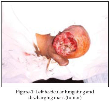
1 The peak incidence occurs in men aged 25–35 years. However, despite the rise in incidence, mortality from testicular cancer has fallen the overall cure rate is now over 90%.2 Maldescent predisposes to malignancy. Some studies suggest that a long treatment delay shortens survival.3,4 Proper pre-operative assessment and diagnosis has the key role in the life expectancy of patient. Hydrocele and testicular tumor both have different operative approaches. We are reporting two cases of testicular cancer that were first diagnosed and treated as hydrocele by scrotal approach but later when correct diagnosis was made they were treated by radical orchidectomy.Case-1: A 40-years-old male patient presented with a fungating and discharging mass on the left side of the scrotum. One year back patient noticed heaviness and swelling of the left testis. With the passage of time, it increased in size for which he went to some doctor who referred him to a surgeon where he was operated for hydrocele via scrotal approach. Intra-operatively testicular biopsy was also taken which turned out to be Embryonal cell carcinoma. He was at that time advised for surgery but he deferred. Postoperatively the swelling remained about the same size. Gradually the mass increased in size which was non tender and fungates with foul smelling greenish yellow prulent discharge. The swelling was associated with low grade fever, weight loss and Jaundice. On examination it revealed about 10x12cm fungating mass on the left side of scrotum (Fig-1) with greenish yellow discharge with enlarged left inguinal lymph nodes. He was planned for radical orchidectomy.
Investigations showed the Hemoglobin of 4.5g/dl, Serum LDH 809I.U/L (253-548), ß-HCG 65.77miu/ml (0.0-2.6), Serum α-feto protein 2481IU/ml (0.5 - 5.5). Ultrasound scan showed left sided inguinal lymphadenopathy. CT scan showed soft tissue density ill-defined 4x3x6cm scrotal mass in left inguinal region & scrotum. Multiple enlarged left inguinal lymphnodes were consistent with metastasis. Rest of the abdomen and pelvis was tumor free. Bone scan was normal. He was transfused 5 units of packed cell and his left sided orchidectomy was done via inguinal approach (Fig-2).

His immediate post operative recovery was un-eventful. Biopsy showed embryonal cell carcinoma & post operative α-feto proteins level was 1346I U/ml. His chemotherapy had to be started but within ten days he developed secondaries in high inguinal region which increased so rapidly with in a period of 05 days and acquired a size of 10cm x 5cm. Immediately he was operated and inguinal clearance was done. Tumor was found to be encroaching the walls of penile urethra. On second post operative day he was shifted to oncology department where he received 06 cycle of chemotherapy. On follow up after six months patient was healthy with good recovery.
Case-2: A 30-years-old male patient presented with Right Scrotal Swelling for the last two years which was gradually increasing in size. He went to a surgeon who diagnosed him as a case of primary hydrocele and operated for via scrotal approach. Intra-operatively he took right testicular, biopsy was taken with the suspicion of tumor, which turned out to be embryonal cell carcinoma. Patient came to us three weeks after the first operation with complaint of swelling of the right testis. On examination he was anemic, there was a non tender 4x5cm firm mass on the right testis which was not tranilluminable. Inguinal lymph nodes were not palpable, rest of the examination was unremarkable.
Investigations showed the hemoglobin 10g/dl, Serum ß-HCG 115.5miu/ml (0.0-2.6), Serum α-feto protein 3324 IU/ml (0.5-5.5). CT scan showed 4x5x5cm ill-defined mass in right testis. Rest of the testes and abdomen was normal and bone scan showed no metastasis. His right orchidectomy was done, post operative recovery was un-eventful. He was referred to oncology department where he received six cycles of chemotherapy and at six months follow up he is doing well.
DISCUSSION
Testicular tumor are more readily accessible to examining fingers than a tumor of any other organ in the body. Too often it escapes detection until it has metastasized. However the worst error is to push a trocar or cannula into the enlarged testis or operating from scrotal approach with the belief that it is a hydrocele, not applying the test of translucency as wall as not performing scrotal ultrasound. It has been reported that some patients consulted their GPs for testicular disease, sometimes long delays occurred because cancerous lumps were confused with cysts, or because their doctors did not recognize some of the less common symptoms of testicular cancer, such as sweating, swollen breasts, sensitive nipples, or backache.
5Accurate palpation of the testis may be obscured by the presence of a hydrocele.
6 In such cases where there is a suspicion or doubt, scrotal ultrasound7,8 and tumor markers9 are of great help. Accurate diagnosis pre-operatively can change the operative approach like instead of scrotal approach inguinal will be adapted which in turn has a great effect on prognosis of patient as there is less chances of metastasis and a high cure rate cannot be anticipated with equal frequency in those patients with high-volume non-seminomatous germ cell tumors of testicle.10 Therefore a patient which could be diagnosed preoperatively and operated via inguinal approach makes his prognosis good without inguinal metastasis.These two cases other a lesson which is sometimes forgotten that detailed clinical history and good physical examination is the most important in reaching correct diagnoses resulting in appropriate treatment If both these patients were properly examined and thoroughly investigated, prognosis would be better as compared to after second surgery of radical orchidectomy.
REFERENCES
1. Power D, Brown R, Brock C. Trends in testicular carcinoma in England and Wales, 1971-1999. Br J Urol Int 2001;87:361-5.
2. Dearnaley D, Huddart R, Horwich A. Managing testicular cancer. BMJ 2001;322:1583-8.
3. Wishnow K, Johnson D, Preston W. Prompt orchiectomy reduces morbidity and mortality from testicular carcinoma. Br J Urol 1990;65:629-33.
4. Hernes E, Harstad K, Fossa S. Changing incidence and delay of testicular cancer in southern Norway (1981-1992). Eur Urol 1996;30:349-57.
5. Chapple A, Sue Ziebland S, McPherson A. Qualitative study of men’s perceptions of why treatment delays occur in the UK for those with testicular cancer. British J General Practice 2003;54:25-32.
6. Roy CR, Peterson NE. Positive hydrocel cytology accompanying testis Seminoma. Urol 1992;39(3):292-3.
7. Powell TM, Tarter TH. Management of non palpable testicular masses. J Urol 2006;176(1):96-8.
8. Skvortsov SV; The use of tumoral markers in clinical practice. Voen Med Zh 2006;327(8):35-42.
9. Logothetis CJ. Emerging strategies in the treatment of testis cancer. Curr Opin Urol 1998;8(50):411-2.
10. Clarke NW. Management of testicular tumours. Surg Int 2007;73:163-8.
HOME | SEARCH | CURRENT ISSUE | PAST ISSUES
Professional
Medical Publications
Room No. 522, 5th Floor, Panorama Centre
Building No. 2, P.O. Box 8766, Saddar, Karachi - Pakistan.
Phones : 5688791, 5689285 Fax : 5689860
pjms@pjms.com.pk