|
|
||||
|
Published by : PROFESSIONAL MEDICAL PUBLICATIONS |
||||
|
ISSN 1681-715X |
||||
|
||||
|
- |
||||
|
ORIGINAL ARTICLE |
||||
|
- |
||||
|
Volume 22 |
April - June 2006 |
Number 2 |
||
|
|
||||
|
||||
|
|
||||
|
Published by : PROFESSIONAL MEDICAL PUBLICATIONS |
||||
|
ISSN 1681-715X |
||||
|
||||
|
- |
||||
|
ORIGINAL ARTICLE |
||||
|
- |
||||
|
Volume 22 |
April - June 2006 |
Number 2 |
||
|
|
||||
|
||||
Role of Toluidine Blue in Early
Detection of Oral Cancer
Imtiaz Ather Siddiqui1, M. Umer Farooq2,
Riaz Ahmed Siddiqui3, S.M. Tariq Rafi4
Abstract
Background: Oral cancer is one of the ten most common cancers in the world with marked geographic incidences in occurrence. The range of frequency of this dreadful cancer varies form negligible in Japan to 45% in Sir Lanka, Nepal, India and Pakistan when compared with the over all incidence of all bodily cancers. In Pakistan oral cancer constitute 10% of all malignant tumors standing second to bronchogenic carcinoma in males and breast carcinoma in females. The more early it is diagnosed the better is the prognosis.
Objective: This prospective study was planned to use Toluidine Blue as a diagnostic material for early diagnosis of oral cancer.
Patients and Methods: Patients with oral lesions having no clinical evidence of oral cancer, but presenting as non-healing oral ulcers more than 6 months or raised margins were selected. They belonged to both sexes and all age groups. Toluidine Blue (ORA SCAN) was used to stain the lesion and biopsy was taken from stained area, 30 patients which in some cases was more than one place. These patients were randomly selected.
Results: Histopathology revealed squamous cell carcinoma in twelve patients and dysplasea in five patients.
Conclusion: Tuluidine Blue can be used as early diagnostic aid in cancer of oral mucosa.
KEY WORDS: Oral Cancer, Tuloidine Blue, Diagnosis, early diagnosis, Ora scan, Tobacco chewing, Smokeless Tobacco, Oral lesion, Oral pathology.
Pak J Med Sci April - June 2006 Vol. 22 No. 2 184 - 187
1. Dr. Imtiaz Ather Siddiqui MBBS, DLO
Senior Registrar
2. Dr. Mohammed Umer Farooq DLO, FCPS
Assistant Professor
3. Dr. Riaz Ahmed Siddiqui MBBS, DLO
Registrar
4. Prof. S.M. Tariq Rafi DLO, FCPS
Professor of ENT
1-4: Department of ENT
Jinnah Postgraduate Medical Centre,
Rafiqui Shaheed Road,
Karachi-75510.
Correspondence:
Dr. Imtiaz Ather Siddiqui
E-Mail: siddykee@yahoo.com
* Received for Publication: July 30, 2005
Accepted: October 16, 2005
Introduction
Oral cancer is one of the most common cancers in the world with marked geographic incidences in occurrences.1 The range of frequency of this dreadful cancer varies from negligible in Japan to 45% in Sri Lanka, Nepal, India and Pakistan when compared with overall incidence of all body cancers.2 Oral cancer constituted 10% of all malignant tumors standing second only to Bronchogenic carcinoma in males and breast carcinoma in females. A study conducted along with oncologist showed that about 20% of all cancers seen at ENT and oncology department were oral malignancies.3,4
The importance of early diagnosis in oral cancer, as indeed in all bodily tumors can hardly be emphasized. In the former case however, the significance increases manifold as the overall prognosis of any form of treatment is directly linked with the stage at which treatment is offered. Suffice is to say that keeping in the view the most regrettable overall results of treatment of oral cancer, the chances of survival can be relatively improved if a tumor is diagnosed at an early T-Stage.
Caner experts agree that early diagnosis of oral carcinoma greatly increases the probability of cure with minimal impairment and deformity.5 The location and appearance of oral lesions does not always facilitate early detection. However appearance shows that most lesions are diagnosed only after becoming symptomatic.
Clinicians may fail to recognize patients at high risk of developing oral cancer and the appearance of early sequences carcinoma in the oral cavity can easily be overlooked.6 Despite good prospects of early diagnosis small developing lesions often go unrecognized by clinicians. 60% of lesions are well advanced by the time of discovery.
Until now, clinicians have relied on clinical judgment and non-specific visual evidence to decide when and from where the biopsy to be taken from suspected lesions. Toluidine blue 1% allows the clinician to more reliably evaluate high risk sites in the oral cavity. The routine use of Toluidine 1% on high risk patients will facilitate earlier detection and treatment.
Patients and Methods
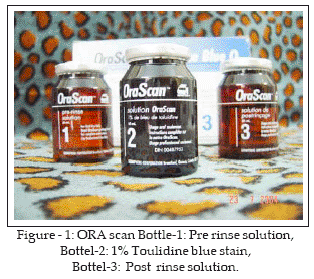
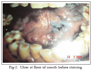
It is a descriptive case series study. A three component oral scan system was used in this study, one component is flavored 1% toluidine blue dye solution. The other two are pre and post rinse solutions consisting of flavored 1% acetic acid. (Figure-1) Toluidine blue is a metachromatic stain when used for histological analysis. Clinically one mechanism of action appears to be greater penetration and temporary retention of the dye in the intercellular spaces of rapidly dividing cancer cells in vivo. There is no evidence of chemical reaction with any component of the cell. Subjects with oral lesions having no clinical evidence of oral cancer presenting as non healing oral ulcers, for more than six months or raised margins were selected for the study. Traumatic lesions and oral inflammatory charges were excluded from the study to avoid false positive results. Subjects were selected from both sexes and all age groups.
Ora scan was used after a complete head and neck examination. Visual examination of whole oral cavity was performed before any instrumentation of soft tissues to detect any soft tissue injury. The observation and notation of the pre-staining appearance of soft tissues and teeth were completed before any structures were stained by ora scan. Instrumentation to the oral cavity can cause cuts which may retain some of the stain material and lead to false positive results.
Patient’s were provided clothing with a bib. As expectoration was required, patients were positioned near a sink. Following procedures were adopted for staining:
1. Patients oral cavity rinsed with approximately one half of the contents of the ora scan pre-rinse (Bottle No.1) for 20 seconds & expectorated. This solution removed excess saliva and provides a consistent oral environment.
2. Patient rinsed with water for 20 seconds and expectorated.
3. Patient rinsed and gargled with one half of the ora scan toluidine blue solution (bottle No. 2) for one minute and expectorated.
4. Patient then rinsed with ora scan post rinse solution (bottle No.3) for 20 seconds and expectorated. This step was repeated again with the remaining solution.
5. Patient again rinsed with water. This step was also repeated.
6. Observations were made using appropriate oral soft tissue examination techniques with retraction; well balanced lighting, and magnification if found necessary.
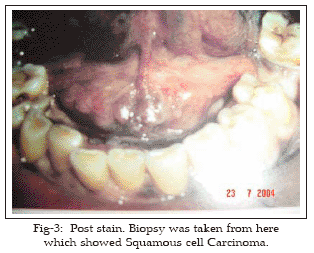
7. Location, size, morphology, color and surface characteristic of suspected lesions that have retained the blue color of the stain were recorded.
8. Any retention of stain, even an equivocal one, that can not be removed with the application of the one post rinse solution, was considered positive and biopsy was taken from there.
9. The areas which retained the stain but were not considered positive were regular papillar crevices on the dorsum of tongue, dental plaques, gingival margins around each tooth, diffuse stain of soft palate transferred from the retained stain on dorsum of tongue and ulceration lesions which are easily distinguished.
One hundred patients were included in the study and thirty out of them showed positive stain. Biopsy was taken from stained area.
Results
Histology revealed squamous cell carcinoma in twelve patients and dysplasia in five, rest were negative for squamous cell carcinoma or dysplasia. Those who showed positive stain but were negative on histopathology for squamous cell carcinoma or dysplasia were subjected to second evaluation of lesions after 14 days thereby allowing for the clinical healing of traumatic and non malignant inflammatory lesions. They all showed negative on this second evaluation. Hence they were false positive on first evaluation and zero percent on second evaluation.
Total number of patients 100, sixty were male and forty female. Minimum age 14 year and maximum age 67 years. Mean age 33, male: female ratio 3:2
No allergic or any other reaction to the stain material was noted. There was a prolonged stain of vermillion bonder, dorsum of tongue and dental plaque which usually disappeared in 4-6 hours.
Discussion
Toluidine blue was first used by Richart in 19637 to stain uterine cervical carcinoma in Situ. Unfortunately false positives have appeared high in early oral studies because of the conclusion of traumatic lesions and other oral inflammatory studies.
In 1980 Masberg Showed that false positives using applications for asymptomatic lesions could be reduced to 8.5% by a second evaluation of lesions in 10-14 days there by allowing for the clinical healing of traumatic and non malignant inflammatory lesions.10
Traumatic lesions and inflammatory changes in oral mucosa were among the exclusion criteria in our study design 13% stained false positives in first staining but they turned zero on second evaluation after 14 days.
A rinse protocol was developed that established the diagnosis of early asymptomatic carcinoma and carcinoma in Situ.11,12 Subsequent studies point to the favorable use of toluidine blue rinse as an effective adjunct to clinical diagnosis. This protocol improves patient outcome, and positively influences risk management.13-15
Cigarette smoking, smokeless tobacco chewing, Pan, Beetal nut chewing especially in combination are highest int his region of Southeast Asia. There are high risk factors in development of oral, upper aero digestive and lung squamous cell carcinoma. Toluidine blue is a useful diagnostic adjunct; this can be used as a screening rinse in high risk patients to encompass the entire oral mucosa after a clinical examination and as a guide to improve biopsy yields. Oral cancer examination and Toluidine blue screening are recommended for every patient with high risk factors for oral cancer. This should be scheduled at least once a year or more frequently according to the judgment of patient’s dentist or Ear, Nose, Throat surgeon.
References
1. Sankaranarayanan, Kerala. Oral cancer in India: An epidemiologic and clinical review. Oral Surg Oral Med Pathol 1990; 69: 325-30.
2. Pindborg JJ, Kiaer J, Gupta PC. Studies in oral Leukoplakia; prevalence or Leukoplakia among 10,000 persons in Lucknow, India, with special references to use or tobacco and betel nut. Bull WHO 1967; 37:109-16.
3. Malignant tumors-Report of a Multicenter Study PMRC-1982.
4. Zaidi SHM, Jafery N. A Cancer problem in Pakistan with special reference to oropharyngeal cancer. J Pak Med Assoc 1960; 16:44.
5. American Cancer Society: Cancer facts and figures – 1991.90-500 M-No. 5008.91 Atlanta, G.A.
6. Mashberg A, Samit: Early Detection, Diagnosis and Management or oral and oropoharyngeal cancer. A Cancer Journal for Clinicals 1989; (39 & 27): 67.
7. Richart, RM. A clinical staining test for in vivo delination of Dysplasia and Carcinoma in Situ. Am J Obst Gynecol 1963; 86:703.
8. Strong MS. Toluidine Blue in the management of carcinoma of the oral cavity. Arch Otolaryngol 1968; 87: 527-31.
9. Sheded DP, Hukill P, Bank, S. In vivo staining properties of Oral Cancer Arch Surg 1980; 46: 758-63.
10. Mashberg A. Re-evaluation of Toluidine Blue application as a diagnostic Adjunct UN the detection of Asymptomatic Oral Squamous Carcinoma, Cancer.1980; 46: 758-63.
11. Mashberg A. Tolonium (Tolonium Blue) Rinse- A Screening Method for Recognition of Squamous Carcinoma; J Pak Med Assoc 1981; 245: 2408-10.
12. Mash berg A. Final Evaluation of Tolonium Chloride Rinse-A Screening of High Risk Patients with Asymptomatic Squamous Cell Carcinoma. J Am Dent Assoc 1983; 106: 319-23.
13. Silverman S, Migliorati C, Barbosa J. Toulidine Blue staining in the Detection Oral Pre-cancerous and Malignant lesions. Oral Surg Oral Med Pathol 1984; 57:37.
14. Silverman, S., Oral Cancer. New York Am Cancer Society 1985; 37: 29-33.
15. Kaugars GE, Mehailescu WL. Toluidine Blue: Its significance and Application in Dentistry. General Dentistry 1989; 37: 404-7.
HOME | SEARCH | CURRENT ISSUE | PAST ISSUES
Professional
Medical Publications
Room No. 522, 5th Floor, Panorama Centre
Building No. 2, P.O. Box 8766, Saddar, Karachi - Pakistan.
Phones : 5688791, 5689285 Fax : 5689860
pjms@pjms.com.pk