|
|
||||
|
Published by : PROFESSIONAL MEDICAL PUBLICATIONS |
||||
|
ISSN 1681-715X |
||||
|
||||
|
- |
||||
|
CASE REPORT |
||||
|
- |
||||
|
Volume 24 |
April - June 2008 (Part-I) |
Number 2 |
||
|
|
||||
|
|
||||
|
|
||||
|
Published by : PROFESSIONAL MEDICAL PUBLICATIONS |
||||
|
ISSN 1681-715X |
||||
|
||||
|
- |
||||
|
CASE REPORT |
||||
|
- |
||||
|
Volume 24 |
April - June 2008 (Part-I) |
Number 2 |
||
|
|
||||
|
|
||||
Angiofibroma with oropharyngeal origin
Ali Eftekharian1, Fariba Samiei2, Mohammad Rakhshan3
SUMMARY
A case is described as orophayageal angiofibroma which is presented as a pink pear-like lesion hanging from the posterior tonsillar pillar of a young man. Pathologic and immunohistochemical analysis confirmed that it was an angiofibroma, with a rare site of origin.
KEY WORDS: Angiofibroma, Oropharynx, Tonsil.
Pak J Med Sci April - June 2008 (Part-I) Vol. 24 No. 2 319-320
1. Ali Eftekharian, MD
2. Fariba Samiei, MD
Departments of Otolaryngology
Head & Neck Surgery,
3. Mohammad Rakhshan, MD
Departments of Otolaryngology,
Head & Neck Surgery and Pathology,
1-3: Loghman Hospital,
Shahid Beheshti University of Medical Sciences,
Tehran – Iran.
Correspondence
Ali Eftekharian,
Department of Otolaryngology
Head & Neck Surgery,
Loghman Hospital,
Kamali st, South Kargar Ave.11457,
Tehran – Iran.
E-mail: alishko@gmail.com
* Received for Publication: August 31, 2007
* Revision Received: January 20, 2008
* Revision Accepted: January 22, 2008
INTRODUCTION
Angiofibromas are benign uncommon vascular neoplasms with a strong liking for the nasopharynx. Although it is the most common benign neoplasm of the nasopharynx, it accounts for less than 0.05% of all head & neck tumors.
1 Though angiofibromas extend beyond the nasopharynx commonly, they rarely originate outside the nasopharynx.2-4 We describe a case of angiofibroma which originated from the posterior tonsillar pillar in oropharynx.CASE REPORT
A 19-year-old young man presented with sensation of something in his throat. He had been aware of a mass in his throat for 6-7 years which had a slow growing pattern. He had no pain or other complaints other than sensation of something in his throat.
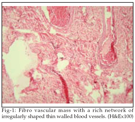
Examination revealed a pink pear-like mass which was hanging from his right posterior tonsillar pillar. The mass was resected under local anesthesia. It was a 3.4cm long pear-like mass which had originated from right posterior tonsillar pillar. Resection was accompanied with no complication or any significant hemorrarhage. Pathologic exam revealed fibro vascular mass with a rich network of irregularly shaped blood vessels. (Fig-1)
As its location was not a common site for angiofibroma, immunohistochemical analysis was done. Stromal cells appeared to be strongly immunoreactive to Vimentin and occasionally reactive to smooth muscle actin. These features suggest that the mass was an angiofibroma.
DISCUSSION
Angiofibromas rarely originate out side the nasopharynx.
2-4 In 2004, Windfuhr and Remmert reviewed the literature and compiled 65 cases of extranasopharyngeal angiofibroma in which four cases had oropharyngeal origin and the maxilla was the most commonly affected site (24.6%).4To this date we have found only five cases of oropharyngeal angiofibroma in the literature.
5-9 Angiofibromas are histologically composed of a proliferating vascular component set in a fibrous stroma. The former is characterized by blood vessels of different size and smooth muscle content. The stroma consists of plump spindle, angular or stellate shaped cells and a varying amount of collagen fibers.10 Beham et al, with immunohistochemical analysis showed that stromal cells have strong cytoplasmic reactivity for vimentin and are generally immunonegative for smooth muscle actin. 10For an experienced pathologist although accurate diagnosis of an angiofibroma is not too difficult, but when its location is an extremely rare one, immunohistochemical analysis will help in its diagnosis.
REFERENCES
1. Sivanandan R, Fee WE. Benign and malignant tumors of the nasopharynx. In: Cummings CW, Flint PW, Harker LA, et al. editors. Cummings otolaryngology head and neck surgery, ed 4. Philadelphia: Elsevier Mosby; 2005;1669-84.
2. Huang RY, Damrose EJ, Blackwell KE, Cohen AN, Calcaterra TC. Extranasopharyngeal angiofibroma. Int J Pediatr Otorhinolaryngol 2000;56(1):59-64.
3. De Vincentiis G, Pinelli V. Rhinopharyngeal angiofibroma in the pediatric age group. Clinical-statistical contribution. Int J Pediatr Otorhinolaryngol 1980;2:99-122.
4. Windfuhr JP, Remmert S. Extranasopharyngeal Angiofibroma: Etiology, Incidence and Management. Acta Otolaryngol 2004;124:880-9.
5. Cejas Méndez L, Espejo Castro E, Artazkoz del Toro JJ, et al. Tonsillar angiofibroma. An Otorrinolaringol Ibero Am 2000;27(6):605-11. Spanish. Cited in: Pub Med PMID 11200557.
6. Celik B, Erisen L, Saraydaroglu O, Coskun H. Atypical angiofibromas: a report of four cases. Int J Pediatr Otorhinolaryngol 2005;69:415-21.
7. Ali S, Jones WI. Extranasopharyngeal angiofibromas. Sex incidence and age distribution. J Laryngol Otol 1982;96:559-65.
8. Johnson JE, Yang PJ, Koopmann CF Jr, Heffner DK. Parapharyngeal angiofibroma. Am J Neuroradiol 1987;8:1152-3.
9. Beeden AG, Alexander FW. An unusual pharyngeal tumour. J Laryngol Otol 1971;85:733-5.
10. Beham A, Kainz J, Stammberger H, Aubock L, Beham-Schmid C. Immunohistochemical and electorn microscopical characterization of stromal cells in nasopharyngeal angiofibromas. Eur Arch Otorhinolaryngol 1997;254:196-9.
HOME | SEARCH | CURRENT ISSUE | PAST ISSUES
Professional
Medical Publications
Room No. 522, 5th Floor, Panorama Centre
Building No. 2, P.O. Box 8766, Saddar, Karachi - Pakistan.
Phones : 5688791, 5689285 Fax : 5689860
pjms@pjms.com.pk