|
|
||||
|
Published by : PROFESSIONAL MEDICAL PUBLICATIONS |
||||
|
ISSN 1681-715X |
||||
|
||||
|
- |
||||
|
ORIGINAL ARTICLE |
||||
|
- |
||||
|
Volume 23 |
January - March 2007 |
Number 1 |
||
|
|
||||
|
|
||||
|
|
||||
|
Published by : PROFESSIONAL MEDICAL PUBLICATIONS |
||||
|
ISSN 1681-715X |
||||
|
||||
|
- |
||||
|
ORIGINAL ARTICLE |
||||
|
- |
||||
|
Volume 23 |
January - March 2007 |
Number 1 |
||
|
|
||||
|
|
||||
Plasma Cell Myeloma in a Tertiary Centre in Niger
Delta Region of Nigeria: Clinicoimmunologic AnalysisCaroline Omoti1, N.K.D. Halim2
ABSTRACT
Objective: To determine the incidence and pattern of presentation of patients with multiple myeloma (MM) in a tertiary health center in Edo state, Niger Delta region of Nigeria noted for its petrochemical industries and gas flare sites.
Design: A retrospective study of 30 cases of MM from 1992 to 2004.
Setting: University of Benin Teaching Hospital, Nigeria.
Main outcome measures: Clinicoimmunologic information in addition to autopsy findings was obtained from case-files. Diagnosis was established according to the standard definition and staged according to the Durie-Salmon clinical staging system.
Results: Advanced stages of the disease (II-III) and performance status scale of 2-4 with pathological fractures were the main form of presentation. Overall median survival was three months (P<0.0001) with 33.3% of the patients surviving at 12 months and 13.3% at 24 months.
Conclusion: Bone pains and anaemia with pathological fractures were the commonest characteristic features with a short three months median survival rate.
KEYWORDS: Multiple myeloma, Incidence, Clinicoimmunologic features.
Pak J Med Sci January - March 2007 Vol. 23 No.1 27-32
1. Dr. Caroline Omoti,
Consultant Hematologist.2. Dr. N.K.D. Halim,
Consultant Hematologist.1-2: Department Of Hematology
University Of Benin Teaching Hospital
P.M.B. 1111, Benin City,
Nigeria.Correspondence:
Dr. Caroline Omoti
E mail: ediomoti@yahoo.com* Received for Publication: February 3, 2006
* Accepted: May 25, 2006
INTRODUCTION
Multiple myeloma (MM) is one of the most common haematologic malignacy characterized by proliferation of a clone of plasma cells that manifests by the presence of one or more lytic bone lesions, monoclonal (M) protein in the blood/urine and bone marrow involvement.
1 The occurrence of MM is worldwide and is more commonly seen in Blacks than in the Caucasians.2 It is the second most prevalent blood cancer after non-Hodgkin�s lymphoma, causing 2% of all cancer deaths.3 MM accounts for approximately 1% of all human cancers and 10% of all haematological malignancies, ranking 13th and 17th among cancer sites in men and women respectively.4 The current therapeutic approach, especially with the advancement in high-dose chemotherapy and stem cell transplant have improved overall survival and event-free periods, but relapse is inevitable.5,6 The risk factors responsible for the increasing incidence and characterization of descriptive patterns has been limited. Hence, there are considerable differences in the recorded incidence in different geographic areas. The aim of this study therefore is to determine the incidence and pattern of presentation of patients with MM in Edo state, Niger Delta region of Nigeria noted for its petrochemical industries and gas flare sites.PATIENTS AND METHODS
Study Design: All cases of MM seen at University of Benin Teaching Hospital (UBTH) and the state Central hospital, which are the major referral center serving the South-South geopolitical zone of Nigeria in the Niger Delta region between 1992 and 2004 were reviewed. Clinicoimmunologic and demographic features of 30 patients in addition to autopsy findings were obtained from case-files. These include the age, gender, presenting clinical features and signs at diagnosis including the standard Durie-Salmon (DS) clinical staging system. The Eastern Cooperative Oncology Group (ECOG) scale for measuring performance status (PS) on a scale from 0 to 4 was applied to the patients with age adjustment index. Diagnosis was established according to standard definitions when at least two of the following criteria are met: a) paraprotein detectable in serum or urine together with a subnormal concentration of at least one monoclonal immunoglobulin class, b) >30% malignant plasma cells in the bone marrow and c) osteolytic and/or osteoporotic bone lesions compatible with MM.
7 Clinicoimmunologic analysis/diagnosis was done according to the modern serum protein immunoelectrophoretic pattern. Blood samples collected for haematological and biochemical parameters were done using automated coulter counter. The erythrocyte sedimentation rate (ESR) was analyzed by the Westerngren method and reticulocyte count according to Dacie and Lewis.8 Overall survival (OS) was measured from the date of diagnosis to the moment of death/last follow-up.Evaluation: The pretreatment evaluation included complete blood count, biochemical tests for renal and liver functions and electrophoresis of serum and urine. Bone marrow aspiration cytology (BMAC) was done to determine the percentage of plasma cells and the morphology. Follow-up studies included the estimation of the monoclonal component in serum or urine by monthly determinations and BMA scheduled for six months after the beginning of treatment.
Statistical analysis:
Differences in clinical measurements were evaluated with the Instat Statistical Package software. All the statistical calculations for significance were performed using the one-way analysis of variance (ANOVA) and the Kruskal-Wallis statistic (KW).RESULTS
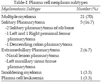
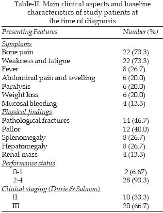
A total of 30 patients aged between 34-75 years with a diagnosis of plasma cell neoplasm (PCN) according to standard clinical and laboratory criteria over a period of 12 years were reviewed. These consisted of 20 males (66.7%) and 10 females (33.3%) with a male-to-female ratio of 2:1. The 30 patients of MM variants were identified out of a total of 395 haematological malignancies from the Oncology clinic attendance including autopsy findings within the study period. This gave an incidence of 7.6% of all the haematological cancers. MM constituted the major subtype number of patients (70%), followed by solitary plasmacytoma (16.7%) and extramedullary plasmacytoma (6.7%). Plasma cell leukaemia and Smoldering myeloma comprised 3.3% each (Table-I). All but the two of the 30 residents with myeloma diagnosed between 1992 and 2004 were recognized antemortem. None was known to have a previous monoclonal gammopathy of undetermined significance (MGUS) because tests were not done on them before they transformed to MM. The overall median age at presentation was 54 years with 13.3% of all MM patients less than 40 years. Table-II shows the clinical aspects at diagnosis. The main presenting symptoms were bone pain (73.3%) and anaemic symptoms (weakness and fatigue) (73.3%). Presence of fever was recorded in 26.7% of the patients while paralysis, weight loss and other symptoms were recorded in 20% each. The least presentation was mucosal bleeding in only four patients (13.3%). Pathological fractures (46.7%) and pallor (40%) formed the commonest physical findings. Baseline characteristics of study patients revealed that PS as assessed according to the ECOG scale that 93.3% of the patients were within the worst scale (2-4). The DS clinical staging system showed that 20 patients (66.7%) presented in the advanced stage III with 13.3% presenting with renal pathology while 10 patients (33.3%) presented in stage II.
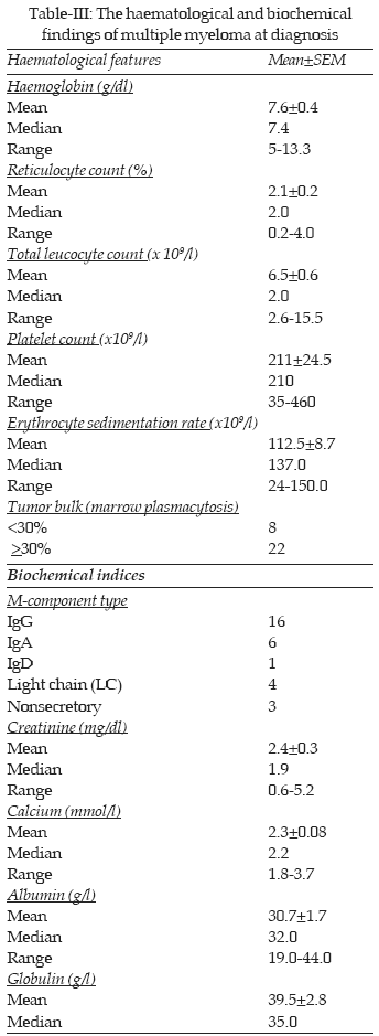
The blood findings at the time of diagnosis are shown in Table-III. Majority of the patients (66.7%) presented with severe anaemia with haemoglobin level less than the mean of 7.6g/dl and 93.3% of the patients less than 10g/dl. Presence of an adequate number of leucocytes and platelet counts within the normal limits was recorded, as granulocytopenia and thrombocytopenia are rare. The ESR was markedly elevated (>100mm/hr) in 73.3% of cases. Increased plasma cells (>30%) with abnormal morphology of flaming/mott myeloma cells in the bone marrow were seen in 73.3% of cases.
The serum protein immunoelectrophoretic pattern showed M-band in 53.3% with IgG, 20% IgA, 3.3% IgD, 13.3% Light chain (two kappa and 2 lambda type) while 10% of the cases were nonsecretory. Of note is that Bence Jones protein (BJP) and raised immunoglobulin were found mainly in the cases of MM. Renal function impairment (serum creatinine >1.3mg/dl) was observed only in 26.7% of cases. Remarkable serum calcium levels (>3.0mmol/l) were found in 6.7% of cases. High peak of abnormal globulin (>40g/l) was found in 33.3% of cases while marked hypoalbuminaemia (<30.0g/l) was also found in 33.3% of cases. The haematological and biochemical indices estimated by the one-way analysis of variance (ANOVA) for association was found to be statistically significant; P<0.0001 and P= 0.0193 respectively.
Therapy and survival: The most commonly used chemotherapeutic regimen were MP (melphalan 10mg/m
2 po Days 1-4 and prednisolone 1mg/kg/day po Days 1-4) for the elderly patients while VAD (Vincristine 0.4mg/day iv Days 1-4, Doxorubicin 10mg/m2/d iv Days 1-4 and 4-day pulses of high dose Dexamethasone 40mg/d) was used for the younger patients providing control of symptoms and/or tumor mass reduction. The median period of survival was three months (P<0.0001). Eight patients were seen up to six months and only six patients beyond one year while the remaining patients either died before three months or were lost to follow-up or voluntary cessation of clinic attendance. At two month of diagnosis, 53.3% of the patients had died from the disease related complications and were unable to purchase their cytotoxic drugs due to financial constraints. Overall survival (OS) was estimated to be only 33.3% at 12 months and 13.3% at 24 months.DISCUSSION
Multiple myeloma (MM) B cell malignancies of plasma cells with controversy about the origin of the plasma cells still remains incurable. Advances in high-dose chemotherapy and stem cell transplantation have improved overall survival and event-free disease, but relapses are inevitable.
5 Therapeutic novel agents are therefore being considered with a focus on immunomodulatory drugs, proteasome inhibitors and arsenic compounds.5MM accounted for approximately 7.6% of all haematological malignancies which is quite close to the 10% recorded in United States and is regarded as the second most frequently occurring haematological malignancy.
3 The higher incidence in some other geographic regions probably reflects the better and earlier detection of the disease. MM, which is the proto type of monoclonal malignant proliferation of plasma cells was the most frequent (70%). This was similar to the study in Ile-Ife, another major center in the South-Western rain forest area of Nigeria.9 Other studies in the diaspora10 have reported similar findings with the average specific incidence rates increasing sharply with age, independent of gender or race with a slightly lower rate being reported in UK, Eastern Europe, South America, India and Japan.11The median age of 54 years at presentation (age range 34-75years) was similar to the findings in the Western world including the Myeloma Research Group in Melbourne; Australia.
12 It is uncommon in persons younger than 40 years as the incidence increases with age.13 This was the observation in this study as less than 15% of our cases were 40 years and below which was similar to the studies of Salawu et al9 and Nossent et al.14 The male predominance found in this study is at variance with other studies that reported a male-to-female ratio of 1:115,16 but similar to the study of Salawu et al.9 This difference may be due to the poor economic empowerment of women in our environment which made it difficult to attend the hospital and also not being exposed to environmental pollution.Clinical presentation of MM patients generally results from tumor mass effects and from the proteins or cytokines secreted by tumor cells or normal accessory cells under the influence of tumor cell products. The severity of MM depends on the tumor load. The preponderance of lytic bone pain and anaemic symptoms in over 70% of the myeloma patients confers a less favorable prognosis and probably reflects the advanced stage of the disease. This lytic bone lesions are due to excessive osteoclast activating factor (OAF) activity exerted by cytokines.
11,17 Fever was the second biggest problem encountered. In a review by Nossent et al14 infection was found to be the immediate cause of death in 54% of cases. This was ascribed to granulocytopenia, immunoparesis and suppression of CD4+ cells.18 In addition, 20% of our patients also presented with neurological complications that may have resulted from wedge compression fracture of the vertebrae, mucosal bleeding (13.3%) that could result from platelet coating by the M-protein,19 though thrombocytopenia is rare. Detection of pathological fracture was the most common physical finding (46.7%).Majority of the patient population had advanced-stage disease. Twenty patients (66.7%) were in stage III while 10 patients (33.3%) were in stage II according to the DS clinical staging system. This system is actually a functional system which serves to evaluate the prognosis using various types of clinical and laboratory tests. Hence, it differs from the anatomic staging systems for solid tumors. However, the system is not optimal as the two recently studied systems of Kyle and Greipp, and the British Columbia Cancer Agency turned out to be the shortest and easiest of the systems.
20 The disease was usually disseminated at presentation and therefore majority of the patients were treated with systemic chemotherapy. This could be the reason why none of the patients was diagnosed as having MGUS. Also, lack of polymerase chain reaction (PCR) along with allele-specific-oligonucleotide (ASO) designed to detect the CDR3 sequence of the tumor in early stage/monitoring minimal residual disease may also be responsible.21,22 This was also reflected in the poor PS where 93.3% of the patients presented with the worst scale (2-4). Over 80% presented with some degree of anaemia but not all were symptomatic at the time of presentation. This has also being documented in other series.9,14 The key factors in the pathogenesis of anaemia is usually due to destruction of the bone marrow with invasion by MM cells, inhibitor of erythropoiesis by tumor factors, renal failure and plasma volume expansion secondary to M-protein.11The mean survival of 6.0 months (median; 3.0) obtained in this series is unacceptably low when compared with the 24 months of the population study on MM survival in a National Health Service in UK
23 and the 60 months of Blade et al study.24 The short survival of patients in this study is however not surprising in view of the unfavorable laboratory and clinical indices at presentation and lack of therapeutic novel agents present in developed countries. At the time of analysis only four patients (13.3%) survived beyond two years while the longest and only known survivor (a hospital matron) is still alive after six years. An important observation from this study was the high default rate which was due mainly to financial constraints and this is generally a problem in cancer management in Nigeria.25 This could be traced to the general poverty level in the society with very high cost of medical facilities despite being an oil producing country, religious beliefs and other strong limiting factors.In conclusion, of 30 patients of PCN studied in Edo state, Niger Delta region of Nigeria MM was the commonest subtype. Bone pains and anaemia with pathological fractures were the commonest characteristic features with a short three months median survival rate. If survival is to be improved, we advocate early referrals of patients with abnormal clinical features and laboratory tests (especially ESR) to the Haematologists for further follow-up. National Health Insurance Schemes and Health care planning with a view to better therapeutic drug availability is also emphasized.
REFERENCES
1. Sirohi B, Powles R. Multiple myeloma. Lancet 2004;363(9412):875-87.
2. Riedel DA, Pottern LM. The epidemiology of multiple myeloma. Hematol Oncol Clin North Am 1992;6(2):225-47.
3. Barlogie B, Shaughnessy J, Munshi N, Epstein J. Plasma cell myeloma. In: Beutler E, Lichtman MA, Coller BS, Kipps TI (eds). Williams Haematology 6th ed. New York. McGraw-Hills. 1996;1279-301.
4. Greenlee RT, Murray T, Bolden S, Wingo A. Cancer statistics 2000. CA- a cancer journal for clincians 2000;50:7.
5. Tariman JD. Understanding novel therapeutic agents for multiple myeloma. Clin J Oncol Nurs 2003;7(5):521-8.
6. San Miguel JF, Blade CJ, Garcia-Sanz R. Treatment of multiple myeloma. Haematologica 1999;84:36.
7. Osterborg A, Bjorkholm M, Bjoreman M, Brenning G, Carlson K, Celsing F, et al. Natural interferon- alpha in combination with melphalan/prednisolone versus melphalan/prednisolone in the treatment of multiple myeloma stages II and III: a randomized study from the Myeloma Group of Central Sweden. Blood 1993;81:1428-34.
8. Dacie JV, Lewis SM. Investigation of abnormal haemoglobins and thalasaemias: Practical Haematology 8th ed. Churchill Livingstone, Edinburgh 1994;249-86.
9. Salawu L, Durosinmi MA. Myelomatosis: Clinical and laboratory features in Nigerians. WAJM 2005;24(1):54-7.
10. Ucci G, Riccardi A, Luoni R, Ascari E. Presenting features of monoclonal gammopathies: an analysis of 684 newly diagnosed cases. Cooperative Group for the study and treatment of multiple myeloma. J Intern Medicine 1993;243:165-73.
11. Brian GM, Giles D, Giles F. Myelomatosis (multiple myeloma). In: Hoffbrand AV, Lewis SM, Tuddenham EGD (eds). Postgraduate Haematology. Butterworths-Heinemann. Oxford 1999:462-78.
12. Spencer A, Seldon M, Deveridge S, Cobcroft R, Cull G, Marlton P, et al. Induction with oral chemotherapy (CID) followed by early autologous stem cell transplantation for denovo multiple myeloma patients. Haemato J 2004;5:216-21.
13. Adam Z, Krahulova M, Spelda SS. Therapy of anaemia in patients with multiple myeloma. Acta Medica Austriaca 1995;22:59-64.
14. Nossent JC, Winkel CN, van Leeuwen JC. Multiple myeloma in the Afro-Caribbean population of Caracao. Netherlands J Med 1993;43:210-4.
15. Choo-kang E, Campell M. Biochemical abnormalities in multiple myeloma. West Indian Med J 1991;40:170-2.
16. Spasov E, Goranova V. Prognostic assessment of the Durie and Salmon staging system in patients with multiple myeloma. Folia Medica (Plovdiv) 1998;40:121-3.
17. Garett JR, Durie BGM, Nedwin GE. Production of lymphotoxin, a bone resorbing cytokine by cultured human myeloma cells. N Engl J Med 1989; 317: 526.
18. Schey SA, Linch DC. Myeloma and related disorders. In: Ludlam CA (ed). Clinical Haematology. Educational Low-Priced Books with Churchill Livingstone. Edinburgh. 1990;183-201.
19. Thiran C, Laloux P, Boucquey D, Brucher JM, Dooms G, Doyen C. Intracranial plasmacytoma manifesting as multiple myeloma: apropos of a case. Acta Neurologica Belgica 1992;92:278-88.
20. Ong F, Hermans J, Noordijk EM, Kluin-Nelemans JC. Is the Durie and Salmon diagnostic classification system for plasma cell dyscrasias still the best choice? Application of three classification systems to a large population-based registry of paraproteinaemia and multiple myeloma. Ann Hematol 1995;70(1):19-24.
21. Jonsson OG, Kitchens RL, Scott FC, Smith RG. Detection of minimal residual disease in acute lymphoblastic leukaemia using immunoglobulin hypervariable region specific oligonucleotide probes. Blood 1990;76:2072.
22. Billadeau D, Blackstadt M, Greipp P, Kyle RA, Okon MM, Kay N, et al. Analysis of B-lymphoid malignancies using allele-specific polymerase chain reaction. A technique for sequential quantitation of residual disease. Blood 1991;78:3021.
23. Phekoo KJ, Schey SA, Richards MA, Bevan DH, Bell S, Gillett D, et al. A population study to define the incidence and survival of multiple myeloma in a National Health Service Region in UK. Br J Haematol 2004;127(3):299-304.
24. Blade J, san Miguel JF, Fontanillas M. Survival of multiple myeloma patients who are potential candidates for early high-dose therapy intensification/auto-transplantation and who were conventionally treated. J Clin Oncol 1996;14:2167-73.
25. Durosinmi MA, Adediran IA. Cancer management under Structural Adjustment Programme (SAP): Experience in Ile-Ife; Nigeria. Nigerian Med J 1993;25:92-6.
HOME | SEARCH | CURRENT ISSUE | PAST ISSUES
Professional
Medical Publications
Room No. 522, 5th Floor, Panorama Centre
Building No. 2, P.O. Box 8766, Saddar, Karachi - Pakistan.
Phones : 5688791, 5689285 Fax : 5689860
pjms@pjms.com.pk