|
|
||||
|
Published by : PROFESSIONAL MEDICAL PUBLICATIONS |
||||
|
ISSN 1681-715X |
||||
|
||||
|
- |
||||
|
ORIGINAL ARTICLE |
||||
|
- |
||||
|
Volume 24 |
January - March 2008 |
Number 1 |
||
|
|
||||
|
|
||||
|
|
||||
|
Published by : PROFESSIONAL MEDICAL PUBLICATIONS |
||||
|
ISSN 1681-715X |
||||
|
||||
|
- |
||||
|
ORIGINAL ARTICLE |
||||
|
- |
||||
|
Volume 24 |
January - March 2008 |
Number 1 |
||
|
|
||||
|
|
||||
Frequency of carotid atherosclerosis
in cerebral infarctionMoazzam Ali Atif1, Hassan Ali2, Tariq Mahmood3
ABSTRACT
Objective: To know the frequency of carotid atherosclerosis in ischaemic stroke patients in our population using color Doppler ultrasound.
Methodology: This is an observational study conducted in Medical Unit-II of Jinnah Postgraduate Medical Centre (JPMC), Karachi from Oct 2003 to Feb 2004 in collaboration with Radiology Dept. All the patients admitted during this duration with CT scan proof of stroke were included in the study. Risk factors in all these patients were stratified. Doppler ultrasound was performed on all patients with ischaemic stroke according to the study protocol.
Results: A total of 100 patients were included in this study over a period of five months. 66% of these were having cerebral infarction. Hypertension (72%), diabetes (35%), smoking (29%) and obesity (20%) were the common risk factors. The frequency of significant carotid atherosclerosis in acute ischaemic stroke patients was 21%.
Conclusion: Carotid atherosclerosis is one of the most important indicators, predictors as well as an independent risk factor in the development of ischaemic stroke.
KEY WORDS: Carotid, Atherosclerosis, Doppler ultrasound.
Pak J Med Sci January - March 2008 Vol. 24 No. 1 69-73
1. Dr. Moazzam Ali Atif,
2. Dr. Hassan Ali,
3. Dr. Tariq Mahmood,
1-3: Department of Medicine,
Jinnah Postgraduate Medical Centre,
Karachi � Pakistan.
Correspondence
Dr. Moazzam Ali Atif,
Al-Hayat Hospital,
Hospital Road,
Rahim Yar Khan, Pakistan.
Email: moazzamatif@hotmail.com
* Received for publication: August 27, 2007
* Accepted: November 3, 2007
INTRODUCTION
Stroke is the third most common cause of death worldwide and chronic disability in elderly patients.
1-3 Physicians are trying to identify the stroke prone population in whom timely intervention might avert stroke and its accompanying disability.4 Carotid atherosclerosis is one of the well-known risk factors for ischaemic stroke.5Atherosclerosis is derived from a Greek word athero (meaning gruel or paste) and sclerosis (hardness). It involves deposits of fatty substances, cholesterol, cellular waste products, calcium and fibrin in the inner lining of an artery and causes stenosis/occlusion of a lumen. Atherosclerosis can affect the arteries of brain, heart, kidney, other vital organs and the arms and legs. When atherosclerosis develops in the arteries that supply the brain, stroke may occur.
4Various diagnostic modalities are available for evaluation of carotid atherosclerosis. Angiography was the first diagnostic imaging modality developed for the evaluation of vessels. The introduction of ultrasound in 1960�s, computed tomography in 1970�s, MRI in 1980�s and with their subsequent development, Color Doppler imaging, CT and MR angiography became available for the non-invasive evaluation of vascular system.
5Ultrasound is establishing its role in screening and diagnosis of carotid pathology because of patient comfort, lack of risks, low cost and accuracy in detecting carotid atherosclerosis.
4 The most recent in the series of major technological achievements in diagnostic ultrasonography is related to the re-evaluation in computer technology of 1980�s, which resulted in the development of color Doppler imaging.6For carotid ultrasound, a high frequency 5-10 MHz linear probe having facility for image steering is required.
7 B-mode gray scale imaging is used to identify and characterize the plaque. The addition of color helps in easy identification of vessels and accurate measurement of its lumen. The velocity measurements are used to grade the stenosis.In our study, we evaluated the extra cranial carotid system with the help of color Doppler ultrasound. The patients whose CT scan showed ischaemic strokes were selected for the study. The purpose of our study was to know the frequency of carotid atherosclerosis in ischaemic stroke patients. This will show the relationship of carotid atherosclerosis with ischaemic stroke.
PATIENTS AND METHODS
This study was conducted in Medical Unit II of JPMC Karachi from Oct 2003 to Feb 2004 in collaboration with Radiology Dept. JPMC is a tertiary care hospital dealing with all kinds of cases referred from main city of Karachi as well as from rest of Pakistan especially Sindh. Medical Unit II is a 50-bedded unit, which deals with a variety of medical cases.
All patients presenting with stroke were included in the study. A total of hundred patients (62 Male, 38 Female �1.6:1) were selected for this study without any age, sex, ethnic or socioeconomic discrimination. A detailed history and thorough physical examination of all patients were carried out on a questionnaire. Risk factors were also stratified. Every patient underwent a list of investigations including Lipid Profile, Electrocardiography (ECG), X-ray Chest (PA), Computed Tomography (CT) scan brain and Echocardiography to rule out any cardiac source of embolization.
Those patients whose CT scan showed evidence of cerebral infarction but without any cardiac cause of embolization were further subjected to carotid Doppler ultrasonography to look for the status of carotid arteries. Doppler study was performed by a radiologist using Toshiba Ecocce with a linear transducer of 7.5MHz. Examination started with the longitudinal survey of cervical carotid arteries with the transducer in a lateral position. Common carotid artery was identified at the clavicle and the transducer was moved cephaly along the artery until the carotid bifurcation was seen. After completion of longitudinal survey, the area of plaque formation was studied in detail. The extent of plaque, its morphological characteristics and especially degree of luminal narrowing were noted. After the lateral and posterolateral survey, the carotid arteries were re-examined from an anterior transducer position. The diameter of the residual lumen and the external diameter of the artery at the same level were measured and the degree of stenosis was calculated using the following relationship:
Percent stenosis
= D-d.100/DWhere D is vessel wall-to-wall diameter
D is vessel open diameter
Our "gold standard" has been angiography and the data that angiography gives us is diameter stenosis therefore, in ultrasound, we also use diameter stenosis. Results of the study were analyzed using SPSS ver 11.0. As this study deals with only frequency distribution of various factors, so no tests of significance were applied.
RESULTS
A total of 100 patients were included in this study following the above-mentioned protocol over a period of five months. 66% of these were having cerebral infarction. Sixty-two (62%) patients were male and thirty-eight (38%) were female. Overall mean age was 55 � 8 years. Two (2%) patients were less than 30 years, five (5%) patients fall between 30-40 years, twenty one (21%) patients were between 40-50 years, twenty eight (28%) patients were between 50-60 years, twenty eight (28%) were between 60-70 years and sixteen (16%) patients were above 70 years of age.
Twenty-eight (28%) patients were conscious at the time of presentation, twenty-two (22%) were drowsy, twenty-six (26%) were arousable while twenty-four (24%) patients were deeply comatose.
Thirty five (35%) patients presented with left sided weakness, fifty eight (58%) patients presented with right sided weakness while seven (7%) patients presented without any weakness which were either in deep coma or the area of brain involved was other than the motor area.
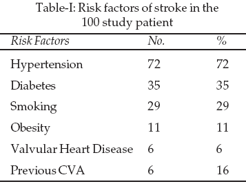
Seventy two (72%) patients were hypertensive, thirty five (35%) were diabetic, twenty nine (29%) gave the history of smoking, twenty (20%) patients were obese, sixteen (16%) had a previous attack of stroke, six (6%) patients had some valvular heart disease (mostly with mitral valve replacement) and one patient had Takayasu�s arteritis. Ten (10%) patients had carotid bruit; sixty (60%) patients had evidence of left ventricular hypertrophy as shown by ECG and cardiomegaly on chest x-ray.
CT scan of these patients showed that sixty six (66%) patients had cerebral infarction while thirty four (34%) patients had evidence of intracerebral or ventricular bleed. Mostly involved area of brain in all of these patients was left temporo-parietal region.
When carotid Doppler ultrasonography was done on patients with cerebral infarction, it showed that 32 out of 66 (48.5%) patients had some evidence of carotid atherosclerosis. Ten (31.3%) patients had right carotid artery involvement, thirteen (40.6%) had left sided involvement and nine (28.1%) had both carotid arteries involved.
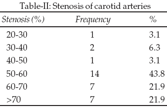
When luminal narrowing of these involved carotid arteries was calculated, it showed that 14 (21%) had significant stenosis (more than 60% stenosis of lumen as it is considered as cut off point for carotid endarterectomy).
DISCUSSION
The stroke patients admitted in our unit were not representative of any specific area or socioeconomic class belonging mainly to middle to lower socioeconomic class. The study included a total of hundred consecutive patients, all presented with recent stroke (involving both ischaemic as well as haemorrhagic stroke) proven by CT scan.
The male preponderance (62 out of 100) is in accord with most of the local as well as international studies. A M Siddiqi et al have shown a 1.5: 1 male to female ratio in their study conducted at Lahore.
8 Numan et al have shown 1.6:1 male: female ratio.9 Piravej K et al has documented male: female ratio of 1.2:1 in his study conducted at Thailand.10 Most of our study subjects were in the age group of 50-70 years (n=56). This is again in accordance with the data already available. Ansari et al, Vohra E et al, Intiso D et al and Piravej K have documented in their respective studies higher incidence as well as poor outcome in elderly people.10-13Main risk factors involved in this study were hypertension (72%), diabetes mellitus (35%), smoking (29%), obesity (20%) whereas 16% had a history of previous CVA and 6% had some valvular heart disease (mostly mitral valve replacement). Incidence of hypertension is more in our study (72%) as well as in other studies carried out in Pakistan as compared to world data. Numan A et al, Zaidi K et al, Ansari AK et al and many others have also shown the same higher incidence of hypertension in our population.
9,10,14 The reason behind this may be the sedentary life style along with improper diet. Baena Diez et al and Intiso D et al, in their studies carried out in the West have shown the same frequency of these risk factors.13,15CT scan findings in this study showed that 66% were cerebral infarcts and 34% were intracerebral or ventricular hemorrhages. This higher frequency of hemorrhage is consistent with other studies carried out in Pakistan.
8,9,16 The reason of this much high frequency of cerebral hemorrhage could be better explained by the higher incidence of uncontrolled hypertension as discussed above. Hemorrhagic strokes are more common in Asia as compared to west.10 Infarction constitutes 80% of strokes over there.2Carotid Doppler ultrasonography performed on patients with ischaemic strokes showed that thirty-two (48.5%) out of 66 patients have involvement of carotid arteries (right, left or both). Out of these, fourteen (21%) patients had significant stenosis of carotid arteries. Razzaq A et al have shown 25% incidence of severe stenosis.
17 Bogousslavsky et al have shown 20%, Pessin et al has documented 39%, Balow et al has shown 33%, Colin P Derdeyn has shown 30% incidence of severe stenosis in their respective studies which were all done on symptomatic population.18-21 These all studies have been conducted on patients having symptoms of involvement of carotid arteries like stroke. Similarly, a lot of studies have been done on asymptomatic patients, which have documented a very high incidence of involvement of carotid arteries. Hennerici et al, Alexandrove et al, Ahn et al, Luisiani et al and Punjia et al have documented 32.8%, 17%, 14%, 11% and 3.8 % incidence of more than 50% involvement of carotid arteries.22-25This high incidence of carotid atherosclerosis in symptomatic as well as in asymptomatic individuals calls for a need of screening of all high risk individuals by carotid Doppler ultrasonography as it is a non-invasive technique. Severe carotid stenosis is associated with high risk for cerebrovascular events. The evidence does not support the routine use of serial sonography to determine the risk of stroke in unselected and asymptomatic carotid disease.
CONCLUSION
It is concluded that carotid atherosclerosis is one of the most important indicators, predictors as well as an independent risk factor in the development of ischaemic stroke. It can be prevented or controlled by keeping good control of other major risk factors like hypertension, diabetes, dyslipidemia, smoking and obesity. Doppler ultrasound is accurate, non-invasive, safe and cost-effective modality for evaluation of carotid vessels.
REFERENCES
1. Ali L, Hamida J, Alam S. Risk factors in stroke. J Coll Phys Surg Pak 1996;7(1):7-10.
2. Allen CMC, Leuck CJ. Cerebrovascular disease. Haslett C, Chilvers ER, Hunter JAA, Boon NA. Davidson�s Principles and Practice of Medicine.18th ed. London: Churchill Livingstone; 1999;974-83.
3. Charles RAC. Cerebrovascular disease and stroke. In: Kumar P, Clarke M, Jane A, Lawrence RI, Nigel B, Carol M, et al. Clinical Medicine. 5th ed. London: WB Saunders 2002.1163-73.
4. Taylor KJW, Burn PN, Wells PNT. Clinical application of Doppler ultrasound. Raven Ltd. New York 1995;1-128.
5. Pohl MI. Carotid artery disease. Optom-Clin 1994;3(4):157-74.
6. Christopher RB, Meritt MD. Doppler color imaging. Churchill Livingstone Inc 1992;1-95.
7. Roger CS, Clinical sonography: a practical guide. 3rd ed, 1998.
8. Siddiqi AM, Ali A, Masrur S, Monga MA, Tauqeer A, Rehman KU. Clinical audit of patients with CVA in medical unit-I, Jinnah Lahore. Ann King Ed Med Coll 2001;7(2):79-82.
9. Numan A, Nasrullah M. An audit of stroke patients and total admissions in year 2000 Neurology Department, Mayo Hospital, Lahore. Pak J Neurol 2001;7(1):1-5.
10. Piravej KW, Watkul W. Risk factors for stroke in Thai patients. J Med Assoc Thai 2003;86:291-8.
11. Ansari AK, Akhund IA, Sheikh A. Stroke in elderly; Identification of risk factors. J Ayub Med Coll. Abottabad 2001;13(3):11-3.
12. Vohra E, Ahmad W, Ali M. Aetiology and prognostic factors of patients admitted for stroke. J Pak Med Assoc 2000;50(7):234-6.
13. Intiso D, Stampatore P, Zarrelli MM, Guerra GL, Arapia G, Simone P et al. Incidence of first ever ischaemic and hemorrhagic stroke in a well-defined community of Southern Italy, 1993-1995. Eur J Neurol 2003;10(5):559-65.
14. Zaidi K, Ara J. Stroke in young patients. Pak J Neurol 2001;7(2):43.
15. Baena DJM, Toma PJ, Mersino AM, Arboix A, Ellacuria TA, Garcia LM, et al. Modifiable risk factors for non-cardioembolic transient ischaemic attacks. Case control studies in general population. Rev Neurol 2003;37(3):206-10.
16. Akhtar M, Ahmad M, Shahid M, Sial H, Naheed T. Risk factors in stroke. Pak J Neurol 1998;4(1):55-8.
17. Razzaq A, Jadoon C, Baig S. Carotid Doppler ultrasonography in young stroke patients. J Pak Med Assoc 1999;49(4):97-9.
18. Bogousslavsky J, Hachinski VC, Boughner DR, Fox AJ, Vinnela F, Barnett JM. Cardiac and arterial lesions in carotid ischemic attacks. Arch Neurol 1986;43:223-8.
19. Pessin MS, Duncan GW, Mohr JP, Poskanzev DC. Clinical and angiographic features of carotid transient ischemic attacks. N Engl J Med 1977;296:358-62.
20. Balow J, Alter M, Resch JA. Cerebral thromboembolic stroke; clinical appraisal of 100 cases. Neurology 1996;16:559-64.
21. Collin P, William JP, Moran CJ, Cross DT, Allen BT. Role of Doppler ultrasound in screening for carotid atherosclerotic disease. Radiology 1995;197:635-43.
22. Hennerice M, Aulich A, Sandmann W, Freund HJ. Incidence of asymptomatic extra cranial arterial disease. Stroke 1981;12:750-58.
23. Alexandrova NA, Gibson WC, Maggisano R. Carotid artery disease and peripheral vascular disease. Stroke 1995;26:175.
24. Ahn SS, Baker JD, Walden K, Moore WS. Which asymptoamtic patients should underg routine screening carotid duplex scan. Am J Surg 1991;162:180-3.
25. Luisiani L, Visona A, Pagnan A. A non-invasive study of arterial hypertension and carotid atherosclerosis. Stroke 1990;21:410-14.
26. Pujia A, Rubba P, Spencer MP. Prevalance of extracranial carotid artery disease detectable by echo-Doppler in elderly population. Stroke 1992;23:818-22.
HOME | SEARCH | CURRENT ISSUE | PAST ISSUES
Professional
Medical Publications
Room No. 522, 5th Floor, Panorama Centre
Building No. 2, P.O. Box 8766, Saddar, Karachi - Pakistan.
Phones : 5688791, 5689285 Fax : 5689860
pjms@pjms.com.pk