|
|
||||
|
Published by : PROFESSIONAL MEDICAL PUBLICATIONS |
||||
|
ISSN 1681-715X |
||||
|
||||
|
- |
||||
|
ORIGINAL ARTICLE |
||||
|
- |
||||
|
Volume 24 |
July - September 2008 |
Number 4 |
||
|
|
||||
|
|
||||
|
|
||||
|
Published by : PROFESSIONAL MEDICAL PUBLICATIONS |
||||
|
ISSN 1681-715X |
||||
|
||||
|
- |
||||
|
ORIGINAL ARTICLE |
||||
|
- |
||||
|
Volume 24 |
July - September 2008 |
Number 4 |
||
|
|
||||
|
|
||||
Imunochromatographic strip test detection of anti-rK39
antibody for the diagnosis of kala-azar in an
endemic zone of Bangladesh
Md. Abdus Salam1
ABSTRACT
Objectives: A rapid immunochromatographic dipstick test (ICT) has become available for the qualitative detection of anti-Leishmania antibody using recombinant rK39 antigen. This study was carried out at the department of Microbiology of Rajshahi Medical College, Bangladesh, in order to evaluate the diagnostic potential of immunochromatographic dipstick test.
Methodology: A total of one hundred cases including 60 admitted patients with strong clinical suspicion of kala-azar and 40 healthy controls were investigated for the performance of the ICT. Splenic smears were examined for microscopic detection of Leishman Donovan (LD) bodies obtained from the admitted patients only and smear-positive cases were considered as gold standard as well as confirmed cases of kala-azar.
Results: Out of 60 suspected patients, fifty three (88.33%) were found smear-positive and fifty nine (98.33%) were positive for immunochromatographic strip test. All smear-positive cases were also positive for strip test. Voluntary healthy controls (40), which included twenty persons from the endemic zone and twenty from non-endemic zone of kala-azar, were found all negative for the strip test. The sensitivity and specificity of immunochromatographic strip test were found to be 100.00% and 86.95% respectively.
Conclusion: Present study findings again reinforce that the immunochromatographic strip test is a simple, reliable and easy-to-perform non-invasive diagnostic tool for visceral leishmaniasis in the endemic area of Bangladesh.
KEY WORDS: Kala-azar, rK39-Immunochromatographic strip test (ICT), Splenic smear, LD body, Sensitivity, Specificity.
Pak J Med Sci July - September 2008 Vol. 24 No. 4 497-501
How to cite this article:
Salam MA. Imunochromatographic strip test detection of anti-rK39 antibody for the diagnosis of kala-azar in an endemic zone of Bangladesh. Pak J Med Sci 2008;24(4):497-501.
1. Dr. Md. Abdus Salam,
Assistant Professor of Microbiology,
Rajshahi Medical College,
Rajshahi-6000
Bangladesh.
Correspondence
Dr. Md. Abdus Salam,
E-mail: salamrmc@yahoo.com
* Received for Publication: October 2, 2007
* Revision Received: February 3, 2008
* 2nd Revision Received: July 11, 2008
* Final Revision Accepted: July 12, 2008
INTRODUCTION
Visceral leishmaniasis (VL), also known as Indian kala-azar is a protozoan parasitic disease caused by Leishmania donovani complex. Sandfly belonging to the species Phlebotomus argentepes is the proven vector for Indian kala-azar. The disease is geographically and ecologically widespread, occurring in tropical and subtropical regions on all continents except Australia and threaten 350 million people. Around five hundred thousand new cases of visceral leishmaniasis occur each year with an estimated 75,000 deaths. Surprisingly, India, Bangladesh and Nepal together account for 60% of all reported VL cases in the world.
1A review of literature reveals that since its recognition, kala-azar had been endemic in Bangladesh with epidemic outbreaks of ten years duration occurring every 15 to 20 years.
2 Available reports show that many parts of the country are at present at least hyperendemic for the disease and the number of cases detected in certain northern districts indicates that kala-azar is probably in the epidemic proportion in these areas.3The diagnosis of VL is complex because its clinical features are shared by a host of other commonly occurring diseases. At present the routine diagnosis of VL is done by direct microscopy of aspirated tissue material taken from the patient or by culture. The microscopic detection of Leishmania amastigotes in smears prepared from spleen, bone marrow or lymph node aspirates is relatively simple and cheap, but performance of spleen aspiration may be dangerous under field conditions while bone marrow and lymph node aspirates are of limited sensitivity
4,5 and retrieval of such samples is inconvenient for the patient. Isolation of parasites by culture is time-consuming, expensive and difficult.6 Because of these drawbacks, the development of diagnostic tests for improved case management of VL has been rated as one of the most needed among the infectious diseases prevalent in the developing world.7With the consequences several tests based on immunological principle are currently in practice for the diagnosis of kala-azar. A promising ready-to-use immunochromatographic dip stick using rK39 antigen has now become available commercially specially for use in field conditions.
8,9 A kinesin-related protein-encoding gene has been discovered in Leishmania chagasi that contains a repetitive 117-bp sequence encoding 39 amino acid residues (K39) conserved at the C-terminal end in all of the VL-causing isolates examined so far.10 The recombinant product of K39 (rK39) has proven to be very sensitive and specific antigen used in the immunochromatographic dipstick for the serodiagnosis of VL in different endemic foci. The test is simple, rapid (10 minutes), relatively inexpensive, requires no other reagents or instruments and can be performed in the field by the paramedics. The estimated sensitivity and specificity of rK39 dipstick test in several Indian studies done in the recent years have been shown to be 100% and 93 to 98% respectively.11,12In the present study, we investigated a total of one hundred cases comprising of both suspected patients of kala-azar and healthy controls to evaluate the diagnostic role of the rK39 immunochromatographic dipstick test in an endemic area of kala-azar in Bangladesh.
METHODOLOGY
The protocol of this study was approved by the ‘Institutional Review Committee’ of Rajshahi Medical College, Bangladesh for ethical issues related to this research. Informed written consent was obtained from each patient and control or from the legal guardian before invasive procedures like splenic aspiration and venipuncture for collection of blood.
Clinically suspected 60 patients of visceral leishmaniasis of different age and sex admitted at different Medical and Paediatric wards of Rajshahi Medical College Hospital (RMCH), Bangladesh who underwent splenic aspiration for microscopic detection of LD bodies were included as study cases.
Twenty (20) healthy non-endemic and twenty (20) endemic controls of comparable age and sex without having past history of prolong fever, splenomegaly or other clinical features suggestive of visceral leishmaniasis were selected from Dhaka city and Godagari Thana of Rajshahi, Bangladesh respectively.
Case definition: Patient with splenic smear-positive for LD bodies was considered as confirmed case of kala-azar.
Splenic aspiration and microscopic examination for LD bodies: Before splenic aspiration from admitted patients, bleeding time (BT), clotting time (CT), platelet count and prothrombin time (PT) were estimated for each patient. Splenic aspiration was done only when results of these tests were found within normal limits. Following proper aseptic precautions and standard techniques, splenic aspiration was done by the Assistant Registrar of the concerned ward/unit and at least three smears were prepared on clean glass slides at bed side. The slides were air-dried and stained by Leishman stain at the department of Microbiology on the same day. Two good quality smears from each patient were examined under oil-immersion lens of Olympus CH-20 microscope for the presence of intra or extra cellular amastigotes.
Immunochromatographic dipstick test (ICT) for kala-azar: Immunochromatographic dipstick tests were done for all sixty patients and forty controls. Two (2.0) ml of venous blood was drawn from each case after taking all aseptic precautions and poured into a sterile test tube without anticoagulant for separation of serum. 20µl of separated serum from each sample was used for performing rK39 Immunochromatog-raphic dipstick test (ICT) for kala-azar on the same day of collection.
Principle of the test: The Kalazar Detect
TM test (InBios International, Inc., Seattle, WA.) is a qualitative, membrane based immunoassay for the detection of antibodies to visceral leishmaniasis in human serum. The membrane is pre-coated with a novel recombinant VL antigen on the test line region and chicken anti-protein A on the control line region. During testing, the serum sample reacts with the dye conjugate (protein A-colloidal gold conjugate) which has been pre-coated in the test device. The mixture then migrates upward on the membrane chromatographically by capillary action to react with recombinant VL antigen on the membrane and generates a red line. Presence of this red line indicates a positive result, while its absence indicates a negative result. Regardless of the presence of antibody to VL antigen, as the mixture continues to migrate across the membrane to the immobilized chicken anti-protein A region, a red line at the control line region will always appear. The presence of this red line serves as verification for sufficient sample volume and proper flow and as a control for the reagents.Test procedure
* Test strip, the Kalazar Detect
TM was removed from the foil pouch* 20µl of serum was added to the test strip in the area beneath the arrow
* The test strip was then placed into a test tube so that the end of the strip was facing downward as indicated by the arrows on the strip
* 2-3 drops (150µl) of the Chase Buffer solution were added
* Results were read in 10 minutes
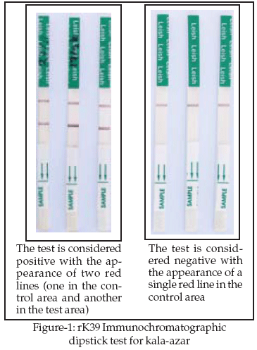
Interpretation of Results: Results of the ICT were noted as positive (a control line and test line appeared in the test area) or negative (only the control line appeared) (Fig-1).
RESULTS
Study population was divided into five different age groups viz., up to 10 years, 11-20 years, 21-30 years, 31-40 years and above 40 years. Among the patients, 11-20 years age group constituted the highest number (30.00%) followed by 21-30 years (23.33%). There were only 04 (06.67%) patients over 40 years of age in this group. Similarly highest number of cases was also noted within 11-20 years and 21-30 years age groups in both endemic and non-endemic control groups. Only 02 (10.00%) persons in the endemic and 01 (05.00%) person in the non-endemic controls were above 40 years of age.
Out of 60 suspected patients of kala-azar, LD bodies were detected in 53 (88.33%) cases and remaining 07 (11.37%) were negative for LD bodies.
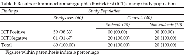
Results of the Immunochromatographic dipstick test (ICT) are shown in Table-1. Among suspected patients, fifty nine (98.33%) were found ICT-positive and one (1.67%) was negative, while all controls from both endemic and non-endemic areas were found negative for dipstick test. All smear-positive cases were also found positive for ICT. The estimated sensitivity and specificity of immunochromatographic dipstick test when compared to the confirmed cases of kala-azar and healthy controls were found to be 100.00% and 86.95% respectively.
DISCUSSION
Visceral leishmaniasis continues to be an important public health problem with significant morbidity and mortality in the endemic areas. The clinical and epidemiological findings of leishmaniasis are non pathognomonic and these can mimick several other conditions. Detection of parasite for the diagnosis of any infectious disease remains beyond above controversy but failure to detect parasite does not always rule out the existence of an infection. There are many diagnostic options currently available for kala-azar with proven merits and demerits in each case. Practice of diagnostic modalities varies greatly among different centres for obvious reasons of available facilities. There is always a need for improved serological tests for kala-azar which will be diagnostically accurate, simple and cheap enough to be used in developing countries as well as suitable for field use too.
We have performed rK39 Immunochro-matographic dipstick test (ICT) for all of our study population. There were fifty nine (98.33%) ICT-positive cases out of sixty clinically suspected VL cases. On the contrary, ICT was found negative among all endemic and non-endemic controls (Table-I). All smear-positive cases (confirmed cases of kala-azar) were also found positive for ICT indicates its sensitivity to be 100%. There have been many studies carried out on ICT in the recent years and the results of the present study are very much consistent with majority of those studies.
10,13-15 However, it is important to point out that strip test for anti-rK39 antibody may not be equally sensitive among patients of different geographical regions.8,16 Studies have shown that, Indian VL is associated with high titres of circulating anti-rK39 antibody17 and particularly well suited for serodiagnosis by strip testing. It is also speculated that, a few cases might have been diagnosed as false positive by ICT as a part of its potential inherent limitations. Since it detects IgG anti-leishmanial antibodies in the serum that might persist for an extended period after successful treatment for VL may bring controversy in detection of active cases.It is evident from our study that this simple, cheaper and non-invasive test has very high diagnostic accuracy. More over, alike other tools, its use are free from all those limiting factors like costly equipments, trained manpower, electricity etc. The ICT can be performed even by the paramedics at fields and the results are easily interpretated. Considering all these points in favour, ICT is already being used as a versatile diagnostic tool for the population at risk in the endemic areas of many countries.
Finally, the ICT results of present study and similar studies carried out in neighboring countries have encouraged us to recommend this highly sensitive, non-invasive, rapid, easy to perform, low-cost diagnostic test to be used anonymously in the field for the serodiagnosis of kala-azar. In fact, WHO in its South East Asia regional meeting for the prevention and control of Dengue, Japanese Encephalitis and Kala-azar, has recommended Immunochroma-tographic dipstick test to be used for screening people at risk in the endemic areas of this region.
18REFERENCES
1. Murray CJL, Lopez AD. Global Health Statistics: A Compendium of Incidence, Prevalence and Mortality Estimates for Over 200 Conditions. Murray CJL, Lopez AD, eds. Global Burden of Disease and Injury Series. 1996, Volume II. Boston: Harvard University Press.
2. Manson-Bahr PEC, Bell DR. Manson’s Tropical Diseases, 19th ed. London: Bailliere Tindall (ELBS), 1987;87-113.
3. Laboratory diagnosis of Kala-azar (Visceral Leishmaniasis) with the Direct Agglutination Test (DAT) - A training module for laboratory Technician, Published by M&PDC and IEDCR, DGHS, Dhaka, Bangladesh. 1996.
4. Siddig AM, Ghalib HW, Shillington DC, Peterson EA. Visceral leishmaniasis in Sudan: comparative parasitological methods of diagnosis. Trans R Soc Trop Med Hyg 1989;82:66-8.
5. Zijlstra EE, Siddig AM, El-Hassan AM, El-Toum IA, Satti M, Ghalib HW et al. Kala-azar: A comparative study of parasitological methods and the direct agglutination test in diagnosis. Trans R Soc Trop Med Hyg 1992;86:505-7.
6. Weigle KA, de Davalos M, Heredia P, Molineros R, Saravia NG. Diagnosis of cutaneous and mucocutaneous leishmaniasis in Colombia: A comparison of seven methods. Am J Trop Med Hyg 1987;36:489-6.
7. Mabey D, Peelin RW, Ustianowsk A, Perkin MD. Tropical infectious diseases: diagnostics for the developing world. Nat Rev Microbiol 2004;2:231-40.
8. Jelinek T, Eichenlaub S, Oscher T. Sensitivity and specificity of a rapid immunochromatographic test for diagnosis of visceral leishmaniasis. Eur J Clin Microbiol Infect Dis 1999;18:669-70.
9. Kumar R, Pai K, Sundar S. Enzyme-linked immunosorbent assay for recombinant K39 antigen in diagnosis and prognosis of Indian visceral leishmaniasis. Clin Diag Lab Immunol 2001;8:1220-4.
10. Burns JM Jr, Shreffler WG, Benson DR, Ghalib HW, Badaro R, Reed SG. Molecular characterization of a kinesin-related antigen of Leishmania chagasi that detects specific antibody in both African and American visceral leishmaniasis. Proc Natl Acad Sci USA 1993;90:775-90.
11. Sundar S, Reed SG, Singh VP, Kumar K, Murray HW. Rapid accurate field diagnosis of visceral leishmaniasis. Lancet 1998;351:563-5.
12. Sundar S, Pai K, Sahu M, Kumar V, Murray HW. Immunochromatographic strip test detection of anti k39 antibody in Indian visceral leishmaniasis. Ann Trop Med Parasitol 2002;96:19-23.
13. Bern C, Jha SN, Joshi AB, Thakur GD, Bista MB. Use of the recombinant K39 dipstick test and the direct agglutination test in a setting endemic for visceral leishmaniasis in Nepal. Am J Trop Med Hyg 2000;63:153-7.
14. Sarker CB, Chowdhury KS, Siddiqui NI, Jamal MF, Rahman S, Momen A, et al. Clinical profile of kala-azar in adults: As seen in Mymensingh Medical College Hospital, Mymensingh, Bangladesh. Mymensingh Med J 2003;12(1):41-4.
15. Ritmeijer K, Melaku Y, Mueller M. Evaluation of a new recombinanat K39 rapid diagnostic test for Sudanese visceral leishmaniasis. Am J Trop Med Hyg 2006;74(91):76-80.
16. Zijlstra EE, Nur Y, Desjeux P, Khalil EA, El-Hassan AM, Groen J. Diagnosing visceral leishmaniasis with the recombinant K39 strip test: experience from the Sudan. Trop Med Int Health 2001;6:108-13.
17. Singh S, Gilman-Sachs A, Chang KP, Reed SG. Diagnostic and prognostic value of K39 recombinant antigen in Indian leishmaniasis. J Parasitol 1995;81:1000-3.
18. WHO Fifty fifth session of SEA regional meeting for the Prevention and Control of Dengue, Japanese Encephalitis and Kala-azar, held in Jakarta, Indonesia from 11-13 September 2002.
HOME | SEARCH | CURRENT ISSUE | PAST ISSUES
Professional
Medical Publications
Room No. 522, 5th Floor, Panorama Centre
Building No. 2, P.O. Box 8766, Saddar, Karachi - Pakistan.
Phones : 5688791, 5689285 Fax : 5689860
pjms@pjms.com.pk