|
|
||||
|
Published by : PROFESSIONAL MEDICAL PUBLICATIONS |
||||
|
ISSN 1681-715X |
||||
|
||||
|
|
||||
|
- |
||||
|
ORIGINAL ARTICLE |
||||
|
- |
||||
|
Volume 25 |
July - September 2009 |
Number 4 |
||
|
|
||||
|
||||
|
|
||||
|
Published by : PROFESSIONAL MEDICAL PUBLICATIONS |
||||
|
ISSN 1681-715X |
||||
|
||||
|
|
||||
|
- |
||||
|
ORIGINAL ARTICLE |
||||
|
- |
||||
|
Volume 25 |
July - September 2009 |
Number 4 |
||
|
|
||||
|
||||
Clinico-Pathological study of
abdominal hysterectomies
Riffat Jaleel1, Ayesha Khan2, Nargis Soomro3
ABSTRACT
Objectives: To correlate indications of abdominal hysterectomy with the histo-pathological findings, in order to determine the percentage of pre-operative diagnosis that was confirmed on histopathology and to determine the frequency of unexpected pathologies.
Methodology: This cross sectional study was conducted in the Department of Obstetrics and Gynecology, Unit II, Civil Hospital Karachi, during January 1995 to December 1996 and Department of Obstetrics and Gynecology, Unit V, Dow Medical College and Lyari General Hospital, Karachi, during August 2005 and October 2008. One hundred sixty six patients undergoing abdominal hysterectomy for gynecological disease, were studied. Data was recorded on proformas, including demographic characteristics and clinical features. Indication for the procedure was documented. Surgical specimens were sent for histopathology and reports were analyzed and compared with the indications of surgery.
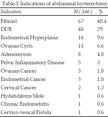
Results: Commonest indication for hysterectomy was fibroid in 40% followed by dysfunctional uterine bleeding (DUB) in 29% cases. Histopathological confirmation of pre-operative diagnosis was 100% for malignancy, endometrial hyperplasia, endometritis and hydatidiform mole, 94% for fibroids, 83% for adenomyosis, 60% for pelvic inflammatory disease and 14.1% for DUB. Majority of cases (65%) pre-operatively diagnosed as DUB were found to have adenomyosis. One case of undifferentiated uterine sarcoma was discovered on histopathology.
Conclusions: Histo-pathological analysis correlates well with the pre-operative diagnosis / indication for hysterectomy. Histo-pathology is mandatory for ensuring diagnosis and thus management, in particular of malignant disease.
KEY WORDS:
Hysterectomy, Indications, Histo-pathology.Pak J Med Sci July - September 2009 Vol. 25 No. 4 630-634
How to cite this article:
Jaleel R, Khan A, Soomro N. Clinico-Pathological study of abdominal hysterectomies. Pak J Med Sci 2009;25(4):630-634.
1. Dr. Riffat Jaleel, FCPS, Assistant
Professor,
2. Professor Ayesha Khan, FRCOG,
Department of Obstetrics & Gynecology, Unit V,
1-2: Dow Medical College & Lyari General Hospital
3. Professor Nargis Soomro, DA, FRCOG,
Department of Obstetrics & Gynecology, Unit II,
Dow Medical College & Civil Hospital
1-3: Dow University of Health Sciences,
Karachi – Pakistan.
Correspondence:
Dr. Riffat Jaleel,
C – 53, Block-I, North Nazimabad,
Karachi – 74700, Pakistan.
Email: drriffatjaleel@yahoo.com
* Received for Publication: January 10, 2009
* Accepted: June 22, 2009
INTRODUCTION
Hysterectomy is the most commonly performed major gynecological surgery throughout the world. It is performed in 560 / 100,000 women per year in the US
1 and 414 / 100,000 women per year in Finland.2Hysterectomy is a successful operation in terms of symptom relief and patient satisfaction. It provides definitive cure to many diseases involving uterus as well as adnexae, eg., fibroids, DUB, adenomyosis, endometriosis, pelvic inflammatory disease, pelvic organ prolapse and malignancy.
Histo-pathological examination of surgical specimens carries ethical, legal, diagnostic and therapeutic significance. A variety of conditions in gynecological practice require removal of a uterus that may show no gross or microscopic pathology when examined by the pathologist. Removal of a normal uterus may be indicated and permitted in the treatment of ovarian, fallopian tube and vaginal cancer, pelvic inflammatory disease, endometriosis, DUB, pelvic organ prolapse, pelvic pain and pelvic tuberculosis.
3The diagnostic value of histo-pathological examination is well explained in patients with genital cancer where adjuvant treatment is dependent upon grade and extent of invasion of disease. Similarly diagnosis of adenomyosis is only established by histo-pathological examination, while DUB is a diagnosis of exclusion. Conversely, many patients may be suspected of having a malignancy on pre-operative assessment eg., those with postmenopausal bleeding and histo-pathological examination may aid to rule out this suspicion.
The purpose of this study was therefore, to correlate various indications of abdominal hysterectomy with the histo-pathological findings of the specimens, thus determining the percentage of the pre-operative clinical diagnoses that were confirmed on histo-pathological examination. We also wanted to determine the frequency of unexpected disease, thus high lighting the need for subjecting each specimen for histo-pathological examination. Failing this may result in sub-optimal care or treatment and over treatment of certain diseases, in particular the malignant conditions.
METHODOLOGY
This cross sectional study was conducted in the Department of Obstetrics and Gynecology, Unit II, Civil Hospital Karachi, during January 1995 to December 1996 and Department of Obstetrics and Gynecology, Unit V, Dow Medical College and Lyari General Hospital, Karachi, during August 2005 and October 2008.
The study included all women undergoing planned abdominal hysterectomy. Data was recorded on proformas, including demographic characteristics and clinical features. Only one dominant diagnosis was considered and documented as the indication for the procedure.
Hysterectomy specimens were saved in 10% formalin and sent to the Department of Pathology, Dow Medical College, Karachi. Histopathology reports were analyzed and compared with the indications of surgery. Any hysterectomy specimen was considered to be normal if it showed no abnormality except functional ovarian cysts or metaplasia / inflammation of the cervix.
RESULTS
One hundred sixty six abdominal hysterectomies were studied. Of this 50.8% women belonged to the age group between 45 – 55 years, 28.8% between 41 – 44 years, 17% were < 40 years and 3.4% > 55 years of age 62.7% had parity >5, while 5.1% were nulliparous and 2.4% patients were unmarried.
Presenting complaint was menorrhagia and pain in 18% of cases and post-menopausal bleeding in 7.8% patients. One patient presented with hydatidiform mole. Her hysterectomy was decided due to multiparity and age >40 years. Another patient had cervico-vesical fistula and underwent hysterectomy along with repair of urinary bladder.
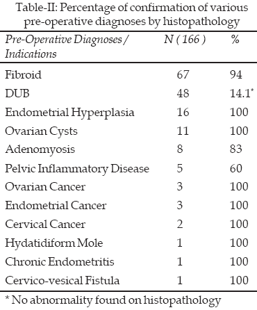
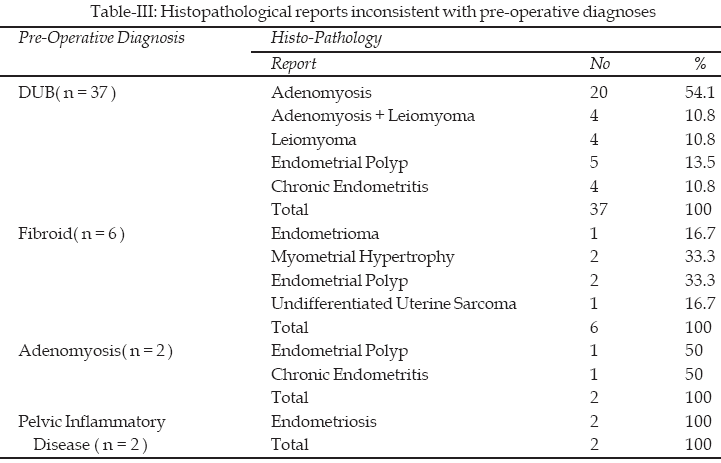
Various indications for hysterectomy are listed in Table-I. Commonest indication was fibroid, followed by DUB. Table-II shows percentage of confirmation of pre-operative diagnoses by histo-pathology. Table-III shows histo-pathology reports which did not match the pre-operative clinical diagnosis. Majority of them were cases of DUB.
DISCUSSION
Indications for abdominal hysterectomy vary from benign to malignant diseases. For purpose of clarity, we chose only one dominant pre-operative diagnosis as indication for hysterectomy for each case. Gambone and associates have pointed out that the process of using only a single designated indication and reviewing only two documents in the record ie., the surgeon’s pre-operative notes and the pathology report, greatly simplified the quality assurance process in order to monitor the justification for hysterectomy.
4The surgical specimens were examined by pathologists of a single department. Therefore, their criteria for diagnosis of any pathology were believed to have only negligible variation and thus no effect on our results.
The mean age at hysterectomy in this study was 45.7 years. In a study in Nepal, the mean age of women undergoing hysterectomy was 46.3 years.
5 Only four hysterectomies were carried out in unmarried women. Three of them had large fibroids with severe menorrhagia, while one of them had endometrial cancer. All of them were older than 40 years of age. Majority of women were parous, with mean parity 5.9. Lee NC found a man parity of 3.1.6 This difference can be explained by the lower use of contraceptive methods in our country as compared to Western countries.The commonest presenting symptom in the study population was menorrhagia with or without pelvic pain. It is well known that perimenopausal age group and high parity are associated with these symptoms. This was also seen by Shergill SK, who found that abnormal menstrual flow was the commonest compliant seen in 66% cases.
7The indications for abdominal hysterectomy in our study were consistent with other studies. Commonest indication was fibroid (34%) and DUB (26%) in the study by Shergill SK.
7 Jha R found that leiomyoma was the indication in 24.9%, ovarian tumour in 14.9% and DUB in 7.7% cases.5 Similar results have been reported by Pokras and Hufnagel.8 Clarke A has reported the commonest indication to be DUB (58%), followed by fibroids (23.2%).9Upon review of histopathology reports, leiomyoma was the most common diagnosis in our study, followed by adenomyosis. Sobande AA also found that fibroid was the most common pathology seen in 25.8% of hysterectomy specimens followed by adenomyosis (22.7%).
10Only few studies have compared pre-operative diagnoses with the histopathology of hysterectomy specimens. We have found that majority of pre-operative diagnoses of our cases were confirmed on histo-pathology. Lee NC found that of 1283 women studied, 80% of the pre-operative diagnoses were confirmed in the potentially confirmable group.
6 Miller studied 246 hysterectomy specimens and found that clinical diagnoses were confirmed in 50% cases.11Because we had based pre-operative diagnoses of endometrial hyperplasia, endometritis, endometrial cancer and cervical cancer on diagnostic curettage, 100% confirmation rate of these diseases was expected. In Lee NC study, endometrial hyperplasia was confirmed in 95% and cervical intraepithelial neoplasia in 89% cases.
6 Similarly, clinical features of leiomyomas as well as hydatidiform mole are highly suggestive of their diagnosis, so a high confirmation rate in our study was not surprising and is compatible with Lee NC study.6Conversely, women with adenomyosis and pelvic inflammatory disease often have nonspecific physical findings. Therefore, the confirmation rates were lower. Lee NC also reported that pelvic inflammatory disease was confirmed in 75% and adenomyosis in 48% cases only.
6DUB is a diagnosis of exclusion. It was confirmed in 14.1% of total cases and 22.5% of cases with pre-operative diagnosis of DUB. Shergill SK
7 showed that DUB was confirmed in 30.8%, while Lee NC 6 found that DUB was confirmed in 38% cases. Miller 11 reported that 31% specimens were normal on histo-pathology. Foster found that 16.9% uteri were histopathologically normal.12 Rest of our cases pre-operatively diagnosed as DUB, were actually found to show some pathology, the commonest being adenomyosis. Same result is reported by Shergill SK, where adenomyosis, leiomyoma, polyps and endometritis were found on histo-pathology of DUB cases.7 Transvaginal ultrasound is the preferred choice for diagnosis of adenomyosis which is also dependent on sonographers’ skills.13 As we only used transabdominal ultrasound for clinical diagnosis, adenomyosis was under-diagnosed in our series.These lastly discussed cases also constituted the major chunk of reports which were inconsistent with the pre-operative diagnoses. In this study 9.6% other cases were found to have histo-pathology reports differing from pre-operative diagnosis. Notable amongst them was a case of undifferentiated uterine sarcoma, incidentally diagnosed on histopathology. This sufficiently highlights the importance of histopathological examination of all surgical specimens.
CONCLUSIONS
While confirming the pre-operative diagnoses by histopathological examination, high confirmation rates were found for endometrial hyperplasia, malignancy, leiomyomas, ovarian tumours and hydatidiform mole. Majority of patients pre-operatively diagnosed as DUB were found to have adenomyosis. Histopathology is mandatory for confirming diagnosis and thus ensuring optimal management, in particular of malignant disease.
REFERENCES
1. Farquhar CM, Steiner CA. Hysterectomy rates in the United States 1990 – 1997. Obstet Gynecol 2002;99:229-234.
2. Vuorma S, Teperi J, Hurskainen R, Keshimaki I, Kujansuu E. Hysterectomy trends in Finland in 1987 – 1995 – a register based analysis. Acta Obstst Gynecol Scand 1998;77:770-776.
3. Thompson JD. Hysterectomy. In: Thompson JD, Rock JA, eds. Te Linde’s Operative Gynecology. 7th edition. JB Lippincott Company, Philadelphia, 1992. 663-738.
4. Gambone JC, Lench JB, Slesinski MJ. Validation of hysterectomy indications and the quality assurance process. Obstet Gynecol 1989;75:1045.
5. Jha R, Pant AD, Jha A, Adhikari RC, Syami G. Histopathological analysis of hysterectomy specimens. J Nepal Med Assoc 2006 Jul-Sep;45(163):283-290.
6. Lee NC, Dicker RC, Rubin G, Oray HW. Confirmation of the pre-operative diagnosis for hysterectomy. Am J Obstet Gynecol 1984;150(3):283-287.
7. Shergill SK, Shergill HK, Gupta M, Kaur S. Clinicopathological study of hysterectomies. J Indian Med Assoc 2002;100(4):238-239,246.
8. Pokras R, Hufnagel VG. Hysterectomy in the United States, 1965 – 1984. AJPH 1988;78:852.
9. Clarke A, Black N, Rowe P, Mott S, Howle K. Indications for and outcome of total abdominal hysterectomy for benign disease: a prospective cohort study. Br J Obstet Gynaecol 1995;102:611-620.
10. Sobande AA, Eskandar M, Archibong EI, Damole IO. Elective hysterectomy: a clinicopathological review from Abha catchment area of Saudi Arabia. West Afr J Med 2005;24(1):31-35.
11. Miller NF. Hysterectomy: therapeutic necessity or surgical racket? Am J Obstet Gynecol 1946;51:804.
12. Foster HW. Removal of the normal uterus. South Med J 1976;69:13.
13. Duoholm M, Lundorf E. Transvaginal ultrasound or MRI for diagnosis of adenomyosis. Curr Opin Obstet Gynecol 2007 Dec;19(6):505-512.
HOME | SEARCH | CURRENT ISSUE | PAST ISSUES
Professional
Medical Publications
Room No. 522, 5th Floor, Panorama Centre
Building No. 2, P.O. Box 8766, Saddar, Karachi - Pakistan.
Phones : 5688791, 5689285 Fax : 5689860
pjms@