|
|
||||
|
Published by : PROFESSIONAL MEDICAL PUBLICATIONS |
||||
|
ISSN 1681-715X |
||||
|
||||
|
- |
||||
|
CASE REPORT |
||||
|
- |
||||
|
Volume 22 |
October - December 2006 |
Number 4 |
||
|
|
||||
|
||||
|
|
||||
|
Published by : PROFESSIONAL MEDICAL PUBLICATIONS |
||||
|
ISSN 1681-715X |
||||
|
||||
|
- |
||||
|
CASE REPORT |
||||
|
- |
||||
|
Volume 22 |
October - December 2006 |
Number 4 |
||
|
|
||||
|
||||
Hydatidosis of tibia
M. Fakoor1, Marashi-Nejad SA2, S. Maraghi3
ABSTRACT
A 71 year old female from a village was admitted to Ahwaz Imam Khomeini hospital in southwestern part 7 years ago with pain and swelling at the upper and middle third of right tibia. Operation was performed and permanent sinus secretion of the midportion occurred. After 7 years the patient returned to hospital recently with warmness, swelling and edema and sinus secretion in middle and upper of right leg. Radiological examination revealed osteolytic metaphyseal and diaplyseal lesion with honey combs appearance transitional zone was narrow without reactive bone and incomplete fractures were also noted. Abdominal sonography was normal. Wound discharge culture indicated Klebsiella, Proteus and E.coli. After antibiotic therapy the patient was put under general anesthesia and complete excision of middle part of tibia from proximal metaphysic to distal metaphysis was performed. During the operation laminated layers and daughter cysts of hydatid cysts were seen. The lesion was washed with 0.5% of NO3Ag and 3cm under the tibia plateau to 7cm of tibia planoid was debrided and cement spacer was used for filling the spaces. Histopathologic examination of the resected specimen confirmed the hydatid cyst. The patient was prescribed albendazole and discharged from the hospital. She is being followed up now.
KEY WORDS: Hydatid cyst , Tibia , Bone.
Pak J Med Sci October - December 2006 Vol. 22 No. 4 468-470
1. Dr. M. Fakoor
Department of Orthopedy2. Dr. Marashi-Nejad
Department of Orthopedy3. Dr. S. Maraghi
Department of Mycoparasitology1-3: Imam Khomeini Hospital, School of Medicine,
Jundi-Shapour University of Medical Sciences,
Ahwaz, Iran.Correspondence:
Dr. M. Fakoor,
E-Mail: m_fakoor@ajums.ac.ir* Received for Publication: January 21, 2006
* Accepted : May 26, 2006
INTRODUCTION
Hydatid cyst is a parasitic infection caused by larval stage of Echinococcus granulosus in man and domestic animals.
1,2 The adult worm resides in the intestine of canine which functions as definitive hosts. Ingestion of ova is passed with the faeces of definitive host by man and domestic animals as intermediate hosts hatch in the small intestine and enter the blood circulation and locate in different tissues and produce hydatid cyst.3The incidence of hydatid cyst in liver is from 59-75%, in lung is 27%, the kidneys and brain each account for 3 and 1-2% of cases respectively.
4 Hydatid cyst in bone is extremely low as most of the larvae are trapped by the liver and lungs upon release of embryos into the portal blood stream.1-3 Skeletal hydatid cyst is found in only 0.5-2% of cases. The growth of cyst in bone is slow and might lie dormant for as many as 20 years.5-7 The diagnosis of osseous hydatidosis is still primarily based on roentgenography findings.3,6 Plain radiography, Computed Tomography (CT), and Magnetic Resonance Imaging (MRI) are helpful in the diagnosis of Skeletal hydatidosis.6,7CASE REPORT
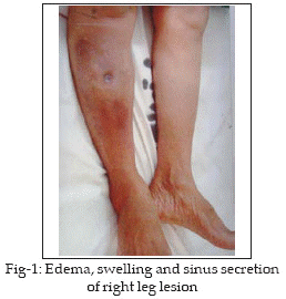
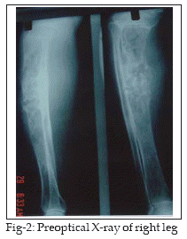
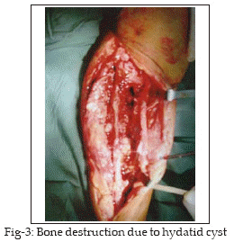
A 71 year old female from a village near by Ahwaz city southwestern part of Iran was admitted to Imam Khomeini hospital in 2003 with swelling, edema and sinus secretion in middle and front of right leg. The patient had an operation 7 years ago because of pain and swelling at the upper and middle third of right tibia. After the operation she had permanent sinus secretion in the middle of her leg, but she has not any information from previous operation. She was under antibiotic therapy. Recent admission was because of warmness, edema, swelling, and sinus secretion in middle and upper of right leg (Fig-1). Radiologic examination revealed osteolytic honeycomb appearance lesions with no reactive bone and incomplete fractures were also noted (Fig-2). Abdominal sonography was normal. Laboratory findings in the first day of admission are shown in Table-1.
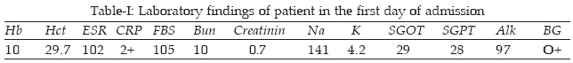
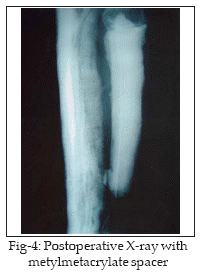
Culture of wound discharge indicated Klebsiella, Proteus and E.coli. Antibiotic therapy was prescribed and the patient received Keflin Amp (1gramQID), Gentamycin Amp (80mg TID) and Albendazol tab (4g BID) before operation and anemia was corrected by blood transfusion. The patient was operated under general anesthesia. Complete excision from proximal to distal metaphysis of tibia was performed (Fig-3). During the operation laminated layers of hydatid cysts and daughter cysts were observed. The cavities were washed with 0.5% NO3Ag and 3cm under the tibia plateau to 7cm of proximal part of tibial plafond was debrided and metyl metacrylate was used for filling the dead spaces. After operation clindamycin (300mg TID) was added to the previous medicines (Fig-4).
Histopathological examination of the resected specimen confirmed the hydatid cyst. The patient left the hospital and continued the treatment using 3 course of 400 mg Albendazol for 4 weeks with two weeks interval and checking the CBC and liver function tests. She has been followed up till now.
DISCUSSION
Osseous hydatidosis is a rare occurrence of hydatid cyst.
8 The hydatid cyst may lie dormant in the bone for many years and most of skeletal hydatid cases have been adults. Skeletal cystic echinococcosis lesions may be single or multiple.5 The lesions though usually secondary to hepatic or pulmonary hydatidosis may on occasion occur as a primary disease. Cases of bone hydatid cyst have been reported in vertebrate, femur, tibia and pelvis.2,7,9-12 Intraosseous lesion usually start at the epiphysis and may be either polycystic or less often in the form of a solitary hydatid cyst.13 As the hydatid cyst of bone remain asymptomatic over a long period, it is usually detected after a pathologic fracture or secondary infection, or following the onset of compressive myelopathy in cases of vertebral lesions. However a definite pre-operative diagnosis without histologic examination is often difficult as there are no pathogonomic signs, radiologic findings may be confused with those of other tumoral lesions and serological tests are of limited value.14-16 The unusual feature in the present case was the ignoring of hydatid cyst in first admission. Although no evidence of liver or lung hydatid cysts revealed even though hydatid cysts were present in tibia and diagnosis occurred 7 years after no cureness and during histological examination of resected specimen. This case emphasizes the importance of considering osseous hydatidosis in the differential diagnosis of destructive tumoral bone lesions and rare diaphyseal involvement.ACKNOWLEDGEMENT
We would like to thank the staff of orthopedic ward, operation room of Imam Khomeini hospital & Mr. A. Jelowdar for his critical help.
REFERENCES
1. Rong SH, Nie ZO. Hydatid disease of bone. Clin Radiology 1985; 36:301-5.
2. Morris BS, Madiwale CV, Grag A, Chavhan GB. Case report: Hydatid disease of bone. A mimic of other skeletal pathologies. Aust Radiol 2002; 46(4): 431-4.
3. Zlitni M, Ezzaouia K, Lebib H, Karray M, Kooli M, Mestiri M. Hydatid cyst of bone: diagnosis & treatment. World J Surg 2001; 26(1): 75-82.
4. Fazel F, Ghanbary H. Hydatid cyst of the orbit. Journal Isfahan Med School 2002; 20(65).
5. Merkle E, Schulte M, Vogel T. Musculoskeletal involvement in cystic echinococcasis. Report of eight cases and review of the literature. Am J Rhinology 1997; 168: 1531.
6. Edeiken J, Dalinka M, Karasick D. Edeiken,s Roentgen diagnosis of disease of bone. 1990; Vol 1, 4th edn. William and Wikins. Baltimore.
7. Madiwale C, Shenoy A, Joshi A, Vora I, Hemmadi SS, Bhosale PB. Hydatid cyst of tibia. J Postgrad Med 1992; 38(4), 194-5.
8. Patond KR, Srivastava SK, Kumar N, Musculoskeletal hydatidosis. Indian Pract 1991, 54: 299-302.
9. Voustinas S, Sayakos J, Srnymis P. Echinococcus infestation complicating total hip replacement. J Bone Joint Surg 1987; 69(3): 1456-8.
10. Karray S, Fowles JV, Zourai O, Slimme N, Kassab MT, Rosset, P. Vertebral hydatidosis and paraplegia. J Bone Joint Surg 1990; 72(13): 84-8.
11. Knudsen C, Marks R, Learmonth GM. Orthopedic hydatid disease. J Bone Joint Surg 1988; 70(13): 504.
12. Fanian H, Karimian MM. A case report of hydatid disease in long bone. J Res Med Sci 2005; 10(2): 101-4.
13. Pintilie DC, Panoza GH, Hatman D, Faher M. Echinococcus of the humerus, treatment by resector and bone drafting: A case report. J Bone Joint Surg 1966; 45-A: 957-61.
14. Resnick D. Diagnosis of bone and joint disorders.Vol 4, 3rd edn. WB Saunders, Philadelphia 1995.
15. Akyar G, Berksun A, Ouz T. Aggressive hydatid disease of the foot and ankle. Aust Radiol 1997; 41: 41-3.
16. Morris Dl. Musculoskeletal hydatid disease. In: Goombs R, Fitz Gerald RH Jr, Editors, Infections in the orthopedic patients. London, Butterworth and Co Ltd 1989; pp.314-22.
HOME | SEARCH | CURRENT ISSUE | PAST ISSUES
Professional
Medical Publications
Room No. 522, 5th Floor, Panorama Centre
Building No. 2, P.O. Box 8766, Saddar, Karachi - Pakistan.
Phones : 5688791, 5689285 Fax : 5689860
pjms@pjms.com.pk