|
|
||||
|
Published by : PROFESSIONAL MEDICAL PUBLICATIONS |
||||
|
ISSN 1681-715X |
||||
|
||||
|
- |
||||
|
ORIGINAL ARTICLE |
||||
|
- |
||||
|
Volume 23 |
October - December 2007 (Part-I) |
Number 5 |
||
|
|
||||
|
|
||||
|
|
||||
|
Published by : PROFESSIONAL MEDICAL PUBLICATIONS |
||||
|
ISSN 1681-715X |
||||
|
||||
|
- |
||||
|
ORIGINAL ARTICLE |
||||
|
- |
||||
|
Volume 23 |
October - December 2007 (Part-I) |
Number 5 |
||
|
|
||||
|
|
||||
Bacteriologic study of
diabetic foot ulcer
Seyed Mohammad Alavi1, Azar D. Khosravi2,
Abdulah Sarami3,
Ahmad Dashtebozorg4, Effat Abasi Montazeri5
ABSTRACT
Objective: To study the relative frequency of bacterial isolates cultured from diabetic foot infections and assess their in vitro susceptibility to the commonly used antibacterial agents.
Methodology: In total 32 hospitalized diabetic patients with foot infections were investigated. Deep tissue biopsies were inoculated into freshly prepared Thioglycollate broth medium. Bacterial agents were identified by conventional bacteriologic methods. Sensitivity tests were performed according to standard disc diffusion method of Kirby & Bauer.
Results: Clinical grading and bacteriological study of 32 patients with diabetic foot lesions revealed polymicrobial etiology in 16 (50%) and single etiology in 10 (31.2%) and six negative cultures. Aerobic Gram-positive bacteria accounted for 42.9%. Staphylococcus aureus was the most frequent microorganism yielded (26.2%), and Staphylococcus epidermidis was regularly associated with the lesions (14.3%). Gram-negative rods accounted for 54.8%. Escherichia coli was the most predominant gram negative organism (23.8%). No anaerobes were isolated from the ulcers. All the microorganisms isolated showed high resistance to used antibiotics, amongst them, Staphylococcus aureus and Pseudomonas aeruginosa were the most resistant bacteria in present study.
Conclusion: Staphylococcus aureus, Escherichia coli, Staphylococcus epidermidis and Proteus vulgaris were the most common causes of diabetic foot infections in present study. And the rate of antibiotic resistance was 65% among the isolates. Due to polymicrobial infection and antibiotic resistance, surgical intervention must be concerned.
KEY WORDS:
Diabetic foot, Bacteriology, Antibiotic resistance, Infection.Pak J Med Sci October - December 2007 (Part-I) Vol. 23 No. 5 681-684
1. Seyed
Mohammad Alavi MD,
2. Azar D. Khosravi, Ph.D,
3. Abdulah Sarami MD,
4. Effat Abasi Montazeri MSc.
5. Ahmad Dashtebozorg MD,
1,3, 5: Razi Hospital, Iran.
2,4: Department of Microbiology, School of Medicine,
Ahwaz Jondishapour University of Medical Sciences,
Ahwaz – Iran.
1,2: Infectious and Tropical Diseases Research Center,
Ahwaz Jondishapour University of Medical Sciences,
Ahwaz – Iran.
Correspondence
Dr. Azar D. Khosravi,
E-mail: khosraviaz@yahoo.com
* Received for Publication: April 9, 2007
* Revision Received: July 14, 2007
* Revision Accepted: July 16, 2007
INTRODUCTION
Foot ulcers are a significant complication of diabetes which are the most common cause of nontraumatic lower extremity amputations in the industrialized world. The risk of lower extremity amputation is 15 to 46 times higher in diabetics than in persons who do not have diabetes mellitus.
1,2 Furthermore, foot complications are the most frequent reason for hospitalization in patients with diabetes.Careful inspection of the diabetic foot on a regular basis is one of the easiest, least expensive and most effective measures for preventing foot complications. Appropriate care of the diabetic foot requires recognition of the most common risk factors for limb loss. Many of these risk factors can be identified based on specific aspects of the history and a brief but systematic examination of the foot.
3,4Foot infections are the most common complications of diabetic foot and plays a main role in the development of moist gangrene.
5 Pseudomonas spp., Enterococcus spp. & Proteus spp. carry a special role and are responsible for continuing and extensive tissue destruction with the poor blood circulation of the foot.6 A high frequency of anaerobic infection has also been reported.7 Patients with diabetes also can have a combined infection involving bone and soft tissue called fetid foot. This extensive soft tissue and bone infection causes a foul exudate, is chronic, and usually requires extensive surgical debridement and/or amputation. In general, people with diabetes have infections that are more severe and take longer to cure than equivalent infections in other people.The infection leads to the early development of complication even after a trivial trauma, the disease progresses and becomes refractory to antibacterial therapy.
8,9 It is essential to assess the magnitude of bacterial infection of the lesions to avoid further complications and save the diabetic foot. Early diagnosis of micorbial infections is aimed to institute the appropriate antibacterial therapy and to avoid further complications.7,10 However, these infections are difficult to treat because these patients have impaired microvascular circulation, which limits the access of phagocytic cells to the infected area and results in a poor concentration of antibiotics in the infected tissues. For this reason, cellulitis is the most easily treatable and reversible form of foot infections in patients with diabetes. Deep skin and soft tissue infections also usually are curable, but they can be life threatening and result in substantial long-term morbidity.11In terms of the infecting microorganisms and the likelihood of successful treatment with antimicrobial therapy, acute osteomyelitis in people with diabetes is essentially the same as in those without diabetes. Chronic osteomyelitis in patients with diabetes mellitus is the most difficult infection to cure. Adequate surgical debridement, in addition to antimicrobial therapy, is necessary to cure chronic osteomyelitis.
12 To study the relative frequency of bacterial isolates cultured from diabetic foot infections and assess their in vitro susceptibility to the commonly used antibacterial agents, a prospective microbiological study was carried out and results are presented here.PATIENTS AND METHODS
A total of 32 hospitalized patients with diabetic foot lesions from Infectious Disease unit of Razi Medical School hospital, Ahwaz, Iran, were screened between March 2004 and March 2006. These patients were clinically assessed and the foot lesions were graded depending on the severity of lesions with 3 to 5 as grade 3 - deep ulcer, abscess formation and bone involvement; grade 4 - localized gangrene and grade 5 - gangrene of whole foot,
13 and based on twice fasting blood sugar test of more than 126mg/dl.Pus aspirates from the abscesses and debrided necrotic materials were collected for aerobic and anaerobic culture using punch biopsy. A Gram stained direct smear of the specimen was examined. The specimens were cultured on blood agar, MacConkey agar, Thioglycollate broth and Robertson’s cooked meat media for aerobic and anaerobic culture. The bacterial isolates were identified by conventional biochemical tests.
14 Antimicrobial susceptibility testing was performed by Kirby Bauer’s disc diffusion method according to National Committee for Clinical Laboratory Standards (NCCLS) guidelines.15 The patients were treated with antibacterial agents according to culture and antibacterial susceptibility pattern.RESULTS
Among 32 patients with diabetic foot, 17 were male and 15 were female patients and the age ranged from 35 to 65 years with mean age being 47 years. Clinical grading and acteriological study of patients revealed polymicrobial aetiology in 16 (50%) and single etiology in 10 (31.2%). Bacteriology culture yielded negative results in six patients. These were patients with burn graded 3 and 4. The variety of bacteria in rest of patients was similar. The number of types of bacterial isolates are given in Table-I.
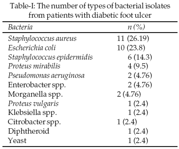
Aerobic Gram-positive bacteria accounted for 42.9%. Staphylococcus aureus was the most frequent microorganism yielded (26.2%), and Staphylococcus epidermidis was regularly associated with the lesions (14.3%). Gram-negative rods accounted for 54.8%. Escherichia coli was the most predominant gram negative organism (23.8%). The other most frequent isolated gram negative aerobic bacilli were Proteus mirabilis (9.5%), Pseudomonas aeruginosa, Enterobacter spp. and Morganella spp. (4.76 each). No anaerobic bacteria were isolated using standard anaerobic culture system.
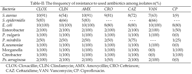
Antibacterial susceptibility testing revealed that, Staphylococcus aureus isolates were resistant to all tested antibiotics except for Ciprofloxacin and Amikacin, which the sensitivity rates were 91% and 80% respectively. All the gram negative isolates were resistant to Cloxacillin, Amoxycillin, Clindamycin and Vancomycin except that 50% of isolates of Proteus mirabilis which were susceptible to these antibiotics. The gram negative isolates showed high resistance to Cephazolin and Ceftriaxone as well. Psudomonas aeruginosa was the second most resistant isolate with resistance to all antibiotics used expect sensitivity of 100% to Ciprofloxacin and 50% to Ceftriaxone. The frequencies of resistance of isolates are given in Table-II.
DISCUSSION
Foot ulcers are a significant complication of diabetes and often precede lower extremity amputation. The most frequent underlying etiologies are neutropathy, trauma, deformity, high plantar pressures, and peripheral arterial disease.
16 Although infection is rarely implicated in the etiology of diabetic foot ulcers, the ulcers are susceptible to infection once the wound is present.All the patients in this study were hospitalized due to the severity of their foot ulcers which categorized into grade 3 to 5. We could not find any significant differences between the variety of isolated organisms and the grade of ulcers but the load of the same organisms were higher in patients with grade 5. S. aureus was the commonest isolate, which was in agreement to studies of Tahaway
17 and Unachukwu et al.18 Sixteen patients revealed two or three types of mixed bacterial infection; (a) in seven patients S. aureus was recovered along with either E. coli, Klebsiella spp. or S. epidermidis; (b) in nine patients E. coli was recovered along with S. aureus, S. epidermidis or other gram negative bacilli. Single type of bacteria was isolated in 10 patients. These findings was in favour to other studies showing the polymicrobial infections in patients with diabetic foot.9,19Based on the results from susceptibility testing, the isolated bacteria showed 65% resistance to used antibiotics. This was a higher resistance compared to similar work of Hartemann et al. which they yielded 18% multidrug resistance.
20 S. aureus showed high resistance to Cloxacillin (91%), Amoxycillin (91%), Ceftazidime (72%), Vancomycin (63%) and Clindamycin (54%), which the resistance was higher compared to study of Pathare et al, as they reported 40% resistance in this organism to similar antibiotics.9 S. aureus showed good sensitivity to Ciprofloxacin as the similar results were reported previously by Tahawy.17 All the gram negative isolates showed 100% resistance to used antibiotics except for Proteus mirabilis which the resistance rate was 50%. Besides isolates of Klebsiella, Proteus vulgaris and Pseudomonas aeruginosa were fully sensitive to Ciprofloxacin.It seems that the status of multidrug resistance among the majority of isolates in present study, was not associated with patient characteristics (age, sex, type and complications of diabetes), wound duration or wound type (neuropathic or ischaemic), while a history of previous hospitalization for the same wound was very important in emergence of resistant organisms.
In conclusion Staphylococcus aureus, Escherichia coli, Staphylococcus epidermidis and Proteus vulgaris were the most common causes of diabetic foot infections in present study. And the rate of antibiotic resistance was 65% among the isolates. Due to polymicrobial infection and antibiotic resistance, surgical intervention must be concerned.
REFERENCES
1. Sarkar PK, Ballantyne S. Management of leg ulcers. Postgrad Med J 2000;76:674-82.
2. Fahey T, Sadaty A, Jones W. Diabetic impaires the late inflammatory response to wound healing. J Surg Res 1991;50:308-13.
3. Mandell G, Bennet J, Dolin R. Cellulitis and soft tissue infection. Principles and practice of infectious diseases. Sixth ed. Pennsylvania: Churchill Livingstone 2005;2:1046-7.
4. Frykberg RG. Diabetic foot ulcers: pathogenesis and management. Am Fam Physician 2002;66:1655-62.
5. Smith JMB, Payne JE, Berue TV. Diabetic foot lesions of skin and soft tissue infections of surgical importance. Chapter 14. In: The surgeons Guide to Antimicrobial Chemotherapy 2002;218-21.
6. Armstrong DG, Lavery LA. Diabetic Foot Ulcers: Prevention, Diagnosis and Classification. Am Farm Physician 1998;57:1325-32.
7. Bailey TS, Yu HM, Rayfield EJ. Patterns of foot examination in a diabetic clinic. Am J Med 1985;78:371-4.
8. Pittet D, Wyssa B, Herter-Clevel C, Kursteiner K, Vaucher J, Lew PD. Outcome of diabetic foot infections treated conservatively a retrospective cohort study with long term follow up. Arch Inter Med 1999;159:851-6.
9. Pathare NA, Sathe SR. Antibiotic combinations in polymicrobic diabetic foot infections. Indian J Med Sci 2001;55:655-62.
10. Frykberg RG. Diabetic foot ulcers: current concepts. J Foot Ankle Surg 1998;37:440-6.
11. Caputo GM, Cavanagh Pr, Ulbrecht JS, Gibbons GW, Karchmer AW. Assessment and management of foot disease in patients with diabetes. N Engl J Med 1994;331:854-60.
12. Lipsky BA. Osteomyelitis of the foot in diabetic patients. Clin Infect Dis 1997;25:1318-26.
13. Wagner FW. The diabetic foot. Orthopedics 1987;10:163-72.
14. Forbes BA, Sahm DF, Weissfeld AS. Bailey & Scott’s Diagnostic Microbiology. 11th Ed., Mosb Inc.y: St. Louis, USA, 389-97.
15. Performance standards for antimicrobial susceptibility testing. 12th informational supplement. NCCLS document M100-S12,22, No.1,Pennsylvania, USA, 2002.
16. Frykberg RG, Armstrong DG, Giurini J, Edwards A, Kravette M, Kravitz S, et al. Diabetic foot disorders: A clinical practice guidline. American College of Foot and Ankle Surgeons. J Foot Surg 2000;39(5 Suppl):51-60.
17. Tahawy AT. Bacteriology of diabetic foot. Saudi Med J 2000;21:344-7.
18. Unachukwu CN, Obunge OK, Odia OJ. The bacteriology of diabetic foot ulcers in Port Harcourt, Nigeria. Nigr J Med 2005;14:173-6.
19. Anandi C, Alaguraja D, Natarajan V, Ramanathan M, Subramaniam CS, Thulasiram M, et al. Bacteriology of diabetic foot lesions. Indian J Med Res 2004;22:175-8.
20. Hartemann-Heurtier A, Robert J, Jacqueminet S, Havan G, Golmard JL, Jarlier V, et al. Diabetic foot ulcer and multidrug resistant organisms; Risk factor and impact. Diabet Med 2004;21:710-5.
HOME | SEARCH | CURRENT ISSUE | PAST ISSUES
Professional
Medical Publications
Room No. 522, 5th Floor, Panorama Centre
Building No. 2, P.O. Box 8766, Saddar, Karachi - Pakistan.
Phones : 5688791, 5689285 Fax : 5689860
pjms@pjms.com.pk