Results of fracture union in closed
reamed
interlocking nail in fractures of femur
Muhammad Kamran Shafi1, Naeem Ahmed2,
Arif Hassan Khan3, Amer Aziz4
ABSTRACT
Objective: To evaluate the results of fracture
union in closed reamed interlocking nail in fractures of femur.
Methodology: A descriptive study conducted at
Department of Orthopaedic surgery Ittefaq Hospital Lahore from March 2003 to
July 2004. Fifty patients were recruited from Accident & Emergency and out
patient department having close fracture of femoral shaft. All patients were
operated under general or spinal anesthesia and closed reamed interlocking
nailing was done. All patients were followed for nine month.
Results: Out of fifty patients, Forty seven
patients underwent union in 90 to 150 days with a mean of 110.68 days. Ten
patients had dynamization within six weeks because of obvious fracture gap
in radiograph. There was one patient who had non-union, and two patients had
delayed union which was treated with dynamization.
Conclusion: Close reamed interlocking
intramedullary nail in femoral shaft fractures is the treatment of choice.
Patient rehabilitation is early, hospitalization is short, and fracture
healing response is good.
KEY WORDS: Close reamed interlocking nail, Femoral
shaft fractures, Union, Dynamization.
Pak J Med Sci October - December 2008
(Part-I) Vol. 24 No. 5 698-701
How to cite this article:
Shafi MK, Ahmed N, Khan AH, Aziz A. Results of fracture union in closed
reamed interlocking nail in fractures of femur. Pak J Med Sci
2008;24(5):698-701.
1. Dr. Muhammad Kamran Shafi (FCPS-Orth.)
2. Dr. Naeem Ahmed (FCPS-Orth),
Assistant Prof. of Orthopaedics
3. Dr. Arif Hassan Khan (FRCS Orth),
Consultant Orthopaedic Surgeon,
Ittefaq Hospital Trust
4. Dr. Amer Aziz (FRCS, FCPS Orth, M.Sc. Orth),
Prof. of Orthopaedics,
Lahore Medical and Dental College,
Gurki Trust Teaching Hospital,
Jallo More, Lahore - Pakistan.
Correspondence
Dr. Muhammad Kamran Shafi,
Department of Orthopaedics & Spinal Surgery,
Lahore Medical & Dental College /
Ghurki Trust Teaching Hospital,
Jallo More, Lahore - Pakistan.
E-mail: shafi_75@yahoo.com
* Received for Publication: June 23, 2008
* Revision Received: August 1, 2008
* Revision Accepted: August 2, 2008
INTRODUCTION
Fractures of the shaft of femur are a major cause of
morbidity and mortality in patients with lower extremity injuries. Most
fractures are sustained in young adults during high velocity injuries.
1
Fractures of the shaft of femur
can be life threatening due to an open wound, fat embolism, ARDS (Adult
Respiratory Distress Syndrome) or resultant multiple organ failure. Even
with survival after initial trauma, disability usually results from femoral
shortening, fracture malalignment or prolonged immobilization of the
extremity by traction or casting in an attempt to maintain fracture length
and alignment during early phases of healing.2
Shortening and malalignment of the leg can lead to a limp and post-traumatic
arthritis.
There is considerable debate regarding the best method of
treating femoral fractures.
3
The unattainably perfect method of fracture treatment would safely fix the
fracture so firmly that soft tissue and joints could be mobilized early and
continuously during healing and offer sufficient strength to maintain
fracture alignment4.
A method closely approaching this perfection is intramedullary interlocking
nails. Interlocking intramedullary nailing of femur greatly improves
rotational stability and can be used for axially unstable diaphyseal
fractures.5
This treatment modality has been the subject of controversy since its
introduction because of concerns of damage to the medullary circulation
possibilities of fat embolism6,7
and complications of misapplication of the technique because of a lack of
understanding of the biomechanical principles of intramed ullary nail
fixation, radiation exposure8
and the equipment required.
Radiation exposure, cost of procedure and nonavailability
of equipment (image intensifier, full range of nails, power reamers etc) are
few of reasons why this treatment has not become popular in Pakistan. Our
institute is one of few centers in Pakistan where we routinely perform
closed reamed intramedullary interlocking femoral nail. To evaluate its use
we decided to study the role of this mode of treatment in achieving the
ultimate goal of fracture fixation i.e. union. We hope that this study will
help in eliminating the controversy which exists regarding this technique
especially in Pakistan.
METHODOLOGY
This descriptive study was carried out at Orthopaedic
Department of ITTEFAQ Hospital Lahore. Forty patients were male and ten
patients were females. All the patients were explained about treatment plan,
cost of operation, hospital stay after surgery, complications of anaesthesia
and their follow up after operation till the time of union. Examination of
patients was done thoroughly at the time of admission to exclude other
injuries.
Patient was laid supine on the fracture table with
traction pin in condyles of fractured femur. The fracture was reduced by
traction and manipulation under image intensification. After preparing the
femur in standard manner, an oblique skin incision was given on the proximal
tip of greater trochanter upto 6-8cm proximally and posteriorly. The fascia
of gluteus maximus incised in line with its fibers. Subfascial plane of the
gluteus maximus identified and piriformis fossa palpated. Bone awl was used
to locate exact entry point. Guide rod was introduced and advanced into the
center of the distal fragment until the tip reached the epiphyseal scar.
Reaming with power reamers was done, 2mm over the selected diameter of nail
and then. Nail was introduced. Proximal locking was done with jig and distal
locking with free hand technique. Wound was closed with suction drain in
standard manner and antiseptic dressing was done. Drain was removed on 2
nd
post-operative day. Touch down weight bearing was started on 2nd
post-operative day. Patient was discharged on 5th
post-operative day and called for stitchs removal on 14th
post-operative day. These patients were assessed clinically and
radiologically for union timing at nine months following surgery.
RESULTS
There were fifty patients in this study, ten were female
and forty were male. The patients were divided in three groups according to
their age for simplicity. Young age group included those patients whose age
was less than forty years. In this group there were five females and twenty
five males. Middle age group included patients, who were between the ages of
40-60 years. This group included five females and ten males. Old age group
included patients older than sixty years. This group consisted of five
males. Six male patients were diabetics and four of them were taking
insulin. Five female patients were diabetics and were taking oral
hypoglycemic.
The clinical results of our study were rated on the basis
of the criteria of union, nonunion, delayed union or malunion. The patients
were followed according to their clinical status. Forty seven patients had
union in 90 to 150 days with a mean of 110.68. Ten of our patients had
diabetes. Union was achieved in eight patients in 95-109 days with a mean of
103.38.
We allowed our patients to start touch down walking with
crutches on the 2
nd
day of operation as they feel comfortable. All patients except two started
partial weight bearing on 6th
week and full weight bearing on 12th
week. These two patients had non weight bearing ambulation till the callus
became visible on radiographs. They had comminution at fracture site (Winquest
and Hensen type III).
All of our patients had full range of motion of there
knees and hips. Three patients out of forty seven complained post operative
knee pain, which was spontaneously resolved in two weeks. Our ten patients
needed dynamization within six weeks because of obvious gap at the fracture
site in subsequent radiographs. This was due to over distraction of fracture
during operation. They were dynamized before starting partial weight
bearing. The screw of less critical stability was determined (the screw
which was away from the fracture) and it was removed in local anaesthesia.
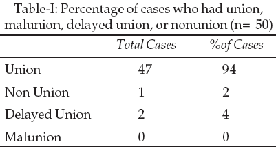
There were two i-e 4% delayed unions which were treated by dynamization. In
our study only one of our patients (2%) was labeled as nonunion and was
treated by exchange nailing.
DISCUSSION
Interlocking intramedullary nailing has been advocated as
the treatment of choice for femoral shaft fractures by many international
centers. Complications after dynamic or simple nailing of an unstable
fracture pattern include shortening (average of 2cm) and malrotation that
frequently requires reoperation. To confirm the clinical observations that
statically locked fractures heal and to prevent the complications from not
locking unstable fractures, Brumback etal.,
9
prospectively treated one hundred femoral fractures with statically locked
Russell �Taylor nails, regardless of comminution. All fractures united, and
only two required dynamization for union. Lepore L, Lepore S, Maffulli N
reported Five out of 43 patients who underwent traditional nailing required
dynamization to achieve union.
In our center we routinely perform the locking in static
mode. In this study all the fractures were treated with statically locked
intramedullary nail. Forty seven out of 50 patients were united with out any
complication. Ten of our patient needed dynamization i-e removal of proximal
or distal screw before starting partial weight bearing.
Two more patients showed no radiological signs of union
at 4
th
month. They were dynamized and were encouraged to walk with bearing full
weight. One of our patients had nonunion. He had comminution at fracture
site i-e Winquest and Hensen type-II. He was treated with exchange nailing.
In our study union rate is 94% which is very close to the reported series.
Numerous studies have documented 97-100% union rate after reamed locked
nailing of femoral shaft fractures. Kropfl etal10
reported a union rate of 100% in a study of 81 femoral shaft fractures with
locked intramedullary nailing. Imran, et al,11
reported a study of 37 cases in which union rate was 100% without any
complication. Javaid, et al reported in their study (Closed intramedullary
nailing versus dynamic compression plating for femoral shaft fractures in 85
adults) that reamed interlocking nailing is the treatment of choice in
femoral shaft fractures and they had 100% union rate without any
complication.12
In our study union occurred in 90 to 150 days with a mean of 110.68 days
which is very close to other studies.
Brumback et al,
13
tested the safety and efficacy of immediate weight-bearing after locked
intramedullary nailing of femoral fractures and found that this was safe if
the construct had relatively high fatigue strength using two locking screws
distally instead of one.
In our study we did nailing after reaming the canal. This
is a routine protocol in our center. There is reported literature which
shows increased blood loss, increased operating time, increased risk of
pulmonary embolism and adult respiratory distress syndrome after reamed
interlocking nail. We have a reasonable policy to wait and stabilize the
patient who has certain risk factors such as associated chest trauma and
anaemia etc. When the patient is stable and fit for surgery we perform close
reamed statically locked intramedullary nail.
The functional out come of patients with femoral shaft
fracture is probably the most important consideration when deciding on the
best mode of treatment for a particular fracture pattern. Successful early
fracture union in femoral shaft fracture is the most demanding out come for
good function in addition to other factors.
CONCLUSION
We have concluded that close reamed interlocking
intramedullary nail in femoral shaft fractures is the treatment of choice,
because patient rehabilitation is early, hospitalization is short, and
fracture healing response is good. If proper equipments are available,
pre-operative assessment and per-operative care is taken, it is safe method
for treating femoral shaft fractures.
REFERENCES
1. Salminen ST, Philajamaki HK, Avikainen VJ, Bostman OM.
Population based epidemiologic and morphologic study of femoral shaft
fractures. Clin Orthop 2000;372:241.
2. Catagni MA, Mendlick RM. Femoral fractures. Tech
Orthop 1996;11:160.
3. Anderson RL. Conservative treatment of fractures of
the femur. J Bone Joint Surg (Am) 1967;49-A:1371.
4. Zickel RE. Fractures of the adult femur excluding the
femoral head and neck: a review and evaluation of current therapy. Clin
Orthop 1980;147: 93.
5. Sultan S. Interlocked nailing of comminuted fractures
shaft of femur. J Ayub Med Coll Abottabad 2001;13(3):14.
6. Bone LB, Babikian G, Stegemann PM. Femoral canal
reaming in the poly- trauma patient with chest injury: a clinical
perspective. Clin Orthop 1995;318: 91.
7. Norris BL. Pulmonary dysfunction in patients with
femoral shaft fracture treated with intramedullary nailing. J Bone Joint
Surg (Am) 2001;83-A:1162.
8. Kohlhass AR, Howard R. Radiation protection during
interlocking Kuntscher nailing. Orthop Rev 1982;11:83.
9. Brumback RJ, Toal TR Jr, Murphy-Zane MS. Immediate
weight bearing after treatment of a comminuted fracture of femoral shaft
with a statically locked intramedullary nail. J Bone Joint Surg (Am) 1999;
81:1538.
10. Kropfl A, Naglik H, Primavesi C, Hertz H. Unreamed
intramedullary nailing of femoral fractures. J Trauma 1995;38:717.
11. Imran F, Tussadaq N, Ahmed Z, Khan AR, Malik G.
Interlocking nail for long bone fractures. Fauji Found Health J
2001;2(1):14.
12. Zubair M Javaid, Mateen MA, Hussain A. Closed
intramedullary nailing versus dynamic compression plating for femoral shaft
fractures in adults. J Pakistan Inst Med Sci 1997;7(2)8(1,2):499-504.
13. Brumback RJ, Ellison TS, Poka A. Intramedull ary nailing of femoral
shaft fractures, long term effects of static interlocking fixation. J Bone
Joint Surg(Am) 1990;74-A:106.