|
|
||||
|
Published by : PROFESSIONAL MEDICAL PUBLICATIONS |
||||
|
ISSN 1681-715X |
||||
|
||||
|
- |
||||
|
ORIGINAL ARTICLE |
||||
|
- |
||||
|
Volume 24 |
October - December 2008 (Part-I) |
Number 5 |
||
|
|
||||
|
|
||||
|
|
||||
|
Published by : PROFESSIONAL MEDICAL PUBLICATIONS |
||||
|
ISSN 1681-715X |
||||
|
||||
|
- |
||||
|
ORIGINAL ARTICLE |
||||
|
- |
||||
|
Volume 24 |
October - December 2008 (Part-I) |
Number 5 |
||
|
|
||||
|
|
||||
Determination of visual status of Iranian
veterans 17-25 years after injuryMohammad Ghassemi-Broumand1, Zohreh Amiri2
ABSTRACT
Objective: To investigate the current visual status of Iranian veterans.
Methodology: Eight hundred veterans, with a history of penetrating ocular injuries were examined 17-25 years after injury during Iraq - Iran war. Their age, gender, the injured eye, the ocular components involved and their final visual acuity were recorded.
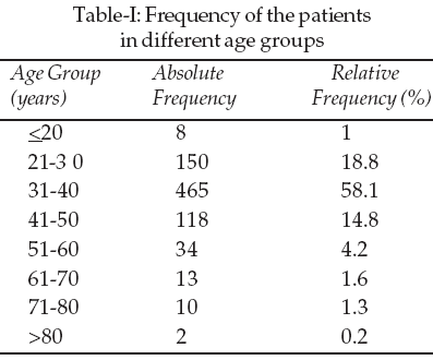
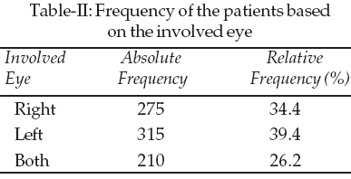
Results: Ninety nine percent of there veterans were male and 58.1% were in the age of 31-40 years. The left eye was involved in 39.4%, the right in 34.4% and 26.2% had bilaterally eye involvement. The cornea was the most commonly involved component (23.4%) and optic nerve the least (1%). The results showed 33.5% were enucleated.
Conclusion: Ocular penetrating injuries can lead to devastating visual outcomes of blindness. Prompt management of these injuries by adequately trained surgeons can lead to better outcomes. As a preventive measure, the use of eye protectives is also recommended.
KEY WORDS: Penetrating eye trauma, Visual acuity, Warfare patients.
Pak J Med Sci October - December 2008 (Part-I) Vol. 24 No. 5 673-677
How to cite this article:
Ghassemi-Broumand M, Amiri Z. Determination of visual status of Iranian veterans 17-25 years after injury. Pak J Med Sci 2008;24(5):673-77.
1. Mohammad Ghassemi-Broumand, MD,
Associate Professor,
2. Zohreh Amiri, PhD,
Assistant Professor,
1-2: Shahid Beheshti University (M.C),
Damavand Ave,
Tehran – Iran.
Correspondence
Dr. Mohammad Ghassemi-Broumand,
Associate Professor,
Faculty of Rehabilitation,
Shahid Beheshti University (M.C),
Damavand Ave, Tehran – Iran.
E- mail: m_ghbr_oph@yahoo.com
* Received for Publication: November 10, 2007
* Revision Received: July 1, 2008
* Revision Accepted: July 25, 2008
INTRODUCTION
During the war between Iraq and Iran, a lot of Iranian soldier’s eyes were injured by bullets and bomb particles. Awareness of these injuries and the visual status of the veterans can help us prioritize our strategies dealing with therapeutic needs and health care service of these patients including the problems they have regarding loss of visual acuity and blindness, etc.
Trauma is one of the most common causes of visual loss.
1 Furthermore, 75% of patients experiencing ocular trauma will have at least one blind eye.1 Despite all efforts to prevent ocular traumas, this is still an important ocular problem. Every year, 2.4 million ocular traumas occur in the United States.2,3 The left eye is more commonly involved than the right one, because majority of the people are right-handed and right-hand punches usually hit the left eye.3 In developed countries, a variety of organizations and databases have been registered in this regard such as "National Eye Trauma System Registry (NETS)" to collect the data regarding penetrating eye traumas. In 1985, a uniform algorithm was introduced to deal with penetrating ocular traumas. All the data is sent to NETS.3Worldwide, 19 million people suffer from unilateral visual disorders due to trauma, 2.3 million people have bilateral visual disorders and 1.2 million people are blind.
4 The majority of traumas are unilateral.4 Most traumas occur at home in the United States and Great Britain.4,5 The other places include office, outdoor activities, fights, and self mutilation.6The frequency of trauma is equal in males and females in age range of above 70 years.
7,8 Penetrating trauma can result in rupture of anterior and posterior segment.9 Traumas can involve one or both eyes.10 Moreover, sympathetic ophthalmia has to be considered in penetrating traumas.11 This is caused by the trauma itself in 50% of the cases and is also the complication of the surgery in the remaining of the cases.11 Penetrating ocular traumas might warrant a variety of therapeutic procedures and might even end up in enucleation, even though therapeutic measures of anterior segment, vitreous and retina have been improved a lot in the past 50 years.12Considering the above mentioned issues, we decided to evaluate the visual status of 800 veterans to assess the procedures carried out for them and the results thereof.
METHODOLOGY
We enrolled 800 traumatic ophthalmic patients from Iraq-Iran war injured soldiers (1010 eyes). The data of the cases including age, gender, site of involvement, presence of unilateral or bilateral involvement, number of ocular lesions, number of surgeries and visual acuity were recorded. Complete ophthalmic examination was done. It is noteworthy that none of the patients had a history of underlying ophthalmic disorders. The results were presented using frequency tables and descriptive statistics.
The visual acuity of the patients was tested by Snellen chart from six meters distance. Informed consent was taken from all the patients and the study was approved by ethical committee of Shahid Beheshti University (M.C).
RESULTS
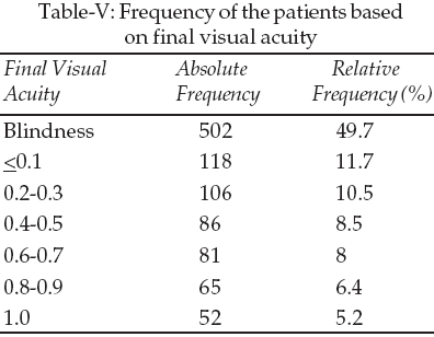
The patients included 790 males (99%) and 10 females (1%). The frequencies of the patients based on age groups, involved eye (right, left, or both), involved component, treatment outcome and final visual acuity are presented in Tables I-V respectively. One-thousand five- hundred and eighty six ocular injuries were found (table III). Totally, 2149 surgeries were carried out on these patients (mean of 2.7 operations per patient ranging from 0 to 12 operations).
DISCUSSION
The study showed that penetrating ocular trauma due to warfare results in severely impaired visual status. Between the years 1985 and 1991, 2939 cases of penetrating ocular trauma were reported to (NETS) National Eye Trauma System Registry from 48 centers in 28 different states. The mean age of the cases was 29.2 years (1-92 years) and 83% of the cases were male. Sixty two percent of the cases had hand movement sight. Primary sight was more than 20/200 in 23%.
3
Werner9 showed that the frequency of penetrating ocular trauma was 48%. In our study all patients suffered from penetrating trauma. The study performed by Wong et al.10 showed that explosive particles caused bilateral injury in 15-25% of the cases, resulting in open or closed lesions both in anterior or posterior segments of the eye. Endophthalmitis was the most serious involvement in these patients. The rate of bilateral involvement in our study was as high as 26 .2%.
The results of Gasch11 indicated that sympathetic ophthalmia occurred in 0.28-1.9% with penetrating trauma and 0.01-0.05% of surgeries. Ninety percent of cases of sympathetic ophthalmia occurred in the first year (5 days to 66 years after trauma). Generally, 50% of cases of sympathetic ophthalmia are due to trauma itself.11 The rate of this complication was as low as 0.4% in our study.
In the study performed by Danneberg et al. 13 in 1992, penetrating ocular trauma due to fights ranged from 1-53% and it was remarkably common in men of 20-40 years of age.
Esmaeli14 showed that treatment outcome was likely to be favorable in patients with primary visual acuity of more than 20/200, presence of the lesion in anterior segment and length of the lesion below 10 millimeter in trauma with a sharp object.
Ninety five percent of patients with primary visual acuity of above 1/10 gained final visual acuity of above 3/10 and there were no cases of enucleation in these patients.14 The study of Spraul and Grossniklaus15 evaluated 24,444 cases of surgery due to ocular trauma in a 55 year period. The results showed that 40.9% of the cases had to undergo enucleation due to following causes: penetration (42.2%), contusion (26.7%), rupture (17%) and perforation (3%). In a study by Groessl et al16 in 1993, Alcohol and substance abuse was reported to be involved in 50-70% in penetrating injuries.
The patients in the study by Shoja and Ardakanian17 were between the age of 2-83 years. Left eye was involved in 57%, and right eye in 43%, and 74.7% of the patients were male. The traumas were 79.4% penetrating.
However, all our cases had experienced penetrating trauma. Moreover, the results of Shoja indicated that 40.6% of traumas occurred in first decade of life. Ninety six point nine percent of our cases were in their 2nd or 3rd decades of life.
The results of Shoja and Ardakanian 17 showed that all patients with primary visual acuity of equal to or more than 0.6 experienced a final visual acuity of more than 0.6. Moreover, primary visual acuity of hand movement (HM) to light perception (LP) ended up in final visual acuity of LP to 5/200 in 38.1% of the cases. Sixty three point three percent of cases of perforation occurred in corneal limbus and 12.7% occurred in sclera. Likewise the results of Pieramici18 showed that 52% of the patients had left eye involvement; 80% were male; and the mean age of the patients was 31.2 years (1-89 years). A study by Nik-Eghbali19 showed that 92% of the traumas were penetrating. Only 24% of their cases were below 10 years of age while 88% were male. Another study performed by Shoja20 in 1996 reported a male/female ratio of 4:1. The age group with the highest frequency was 5-9 years old (24.9%). The traumas in this study were 70.5% penetrating.20 The results of our study showed that 99% were male and the patients’ ages ranged from 17 to 90 years. Left eye involvement was observed in 39.4%, right eye in 34.4% and both eyes in 26.2%. All our cases had experienced penetrating trauma. Generally, all studies showed that penetrating ocular involvement was more common in males, and in left eye. Moreover, the results of Shoja & Ardakanian indicated that 40.6% of traumas happened in first decade of life but in our study 76.9% of our cases were in their 2nd or 3rd decades of life.
Pieramici18 reported the frequency of visual acuity of higher than 0.5 to be 48% in penetrating cases and in 33.5% ended in enucleation. The results of the present study showed visual acuity of above 0.6 in 19.6% and enucleation in 33.5%. The incongruence of our results might indicate that warfare traumas have a more severe nature than other traumas. The results of Pieramici18 were very similar by reporting a 88% rate of presence of final visual acuity of 0.5 and more in patients with primary visual acuity of 0.5 and more. They also showed that 43% of patients with primary visual acuity of HM and LP ended up with final visual acuity of LP to 5/200.18 Their results also showed that none of the cases with primary visual acuity of 5/200 and more resulted in enucleation.18 A study carried out by Gilbert et al.21 indicated that 60% of the cases of perforations posterior to insertion point of rectus muscles ended up in enucleation. Different studies have established a significant relationship between the length of rupture and its outcome22,23 Thompson24 showed that delays of more than 24 hours in repair of perforations increased the risk of endophthalmitis by four times. The study carried out by Roper-Hall25 in 1959 reported the rate of enucleation to be 45% which was dramatically higher than the above mentioned studies which is most probably due to development of new surgical and therapeutic methods and equipments.
CONCLUSION
Generally, final visual acuity and outcome in the patients of our study were not favorable. This can be because of different reasons including severity of warfare trauma, lack of a uniform protocol to approach these patients, very long intervals between the trauma and initiation of appropriate therapy which was due to the distance between the site of injury and hospitals.
Considering the importance of ophthalmic trauma and their unfavorable outcome, application of proper preventive measures (including helmets, eye shield, etc.) in this field is of great importance. Establishment of an NGO to collect data related to different kinds of ocular penetrating trauma is strongly recommended.
ACKNOWLEDGMENT
The investigators wish to thank the veterans for participation in this study and Mr. Mohammad-Ebrahim Mahdavi and Ms Paria Gassemi-Broumand for assisting in preparation of the manuscript.
REFERENCES
1. Operational research department. National Society to Prevent Blindness. Vision problem in US: A statistical analysis. National Society to Prevent Blindness. New York; 1980;1-46.
2. Parver LM. Eye trauma. The neglected disorder. Arch Ophthalmol 1986;104:1452-3.
3. Parver LM. Characteristics and causes of penetrating eye injuries reported to the National Eye Trauma Registry. 1985-1991. Pub Health Rep 1993;108:625-32.
4. MacEwen CJ. Ocular injuries. J R Coll Surg Edinb 1999;44:317-23.
5. May DR. The epidemiology of serious eye injuries from the United States Eye Injury Registry. Graefe’s Arch Clin Exp Ophthalmol 2000;238:153-7.
6. Dunn ES. The epidemiology of ruptured globes. Ann Ophthalmol 1992;24:405-10.
7. Tielsch JM. Frequency and consequences of ocular trauma. A population perspective. Ophthalmol Clin NA 1995;8:559-67.
8. Maecwen CJ. Eye injuries a prospective survey of 5671 cases. Br J Ophthalmol 1989;73:888-94.
9. Werner M. Predictors of occult scleral rupture. Ophthalmology 1994;101:1941-4.
10. Wong TY, Seet B, Ang CL. Eye injuries in twentieth century warfare: A historical perspective. Sur Ophthalmol 1997;41:433-59.
11. Gasch AT. Postoperative sympathetic ophthalmia. Int Ophthalmol Clin 2000;40:69-84.
12. Hersh PS, Zagelbaum BM, Shingleton BJ, Kenyon KR. Anterior segment trauma. In: Jakobiec, Albert, editors. Principles and practice of ophthalmology. Philadelphia: WB Saunders; 2000;(5)3340-83.
13. Dannenberg AL, Parver LM, Fowler CJ. Penetrating eye injuries related to assault. The National Eye Trauma System Registry, Arch Ophthalmol 1992;110:849-52.
14. Esmaeli B. Visual outcome and ocular survival after penetrating trauma. A clinicopathologic study. Ophthalmology 1995;102:393-400.
15. Spraul CW, Grossniklaus HE. Analysis of 24,444 surgical specimens accessioned over 55 years in an ophthalmic pathology laboratory. Int Ophthalmol 1998;21:283-304.
16. Groessl S, Nanda SK, Mieler WF. Assault-related penetrating ocular injury. Am J Ophthalmol 1993;116:26-33.
17. Shoja M, Ardakanian. Evaluation of open globe injury and final visual acuity in Yazd Railway Hospital from 1997-1998. (Iranian) Bina J Ophthalmol 2000;5:256-66.
18. Pieramici DJ. Open-globe injury. Ophthalmology 1996;103:1798-1803.
19. Nikeghbali M. Scleral Rupture due to trauma in Rasoul Acram(S) Hospital. Iranian J Ophthalmology 1995;7:64-5.
20. Shoja M. Evaluation of penetrating eye injury in Yazd Raiway Hospital from 1992-1995, Shaheed Sadoughi Uni J1996;56-60.
21. Gilbert CM, Soong HK, Hirst LW. A two year prospective study of penetrating ocular trauma at the Wilmer Ophthalmological Institute. Ann Ophthalmol 1981;19:104-6.
22. DeJuan E Jr. Penetrating ocular injuries; type of injuries and visual results. Ophthalmology 1983;90:1318-22.
23. Stanberg P Jr, DeJuan E Jr, Auere MRG. Multivariate analysis of prognostic factors in penetrating ocular injuries. Am J Ophthalmol 1984;98:467-72.
24. Thompson JT. Infectious endophthalmitis after penetrating injures with retained (IOFB). Ophthalmology 1993;100:1468-74.
25. Roper-Hall MJ. Treatment of ocular injures. Trans Ophthalmol Soc UK 1955;79:57-92.
HOME | SEARCH | CURRENT ISSUE | PAST ISSUES
Professional
Medical Publications
Room No. 522, 5th Floor, Panorama Centre
Building No. 2, P.O. Box 8766, Saddar, Karachi - Pakistan.
Phones : 5688791, 5689285 Fax : 5689860
pjms@pjms.com.pk