|
|
||||
|
Published by : PROFESSIONAL MEDICAL PUBLICATIONS |
||||
|
ISSN 1681-715X |
||||
|
||||
|
- |
||||
|
CASE REPORT |
||||
|
- |
||||
|
Volume 24 |
October - December 2008 (Part-I) |
Number 5 |
||
|
|
||||
|
|
||||
|
|
||||
|
Published by : PROFESSIONAL MEDICAL PUBLICATIONS |
||||
|
ISSN 1681-715X |
||||
|
||||
|
- |
||||
|
CASE REPORT |
||||
|
- |
||||
|
Volume 24 |
October - December 2008 (Part-I) |
Number 5 |
||
|
|
||||
|
|
||||
Intramuscular Hydatid cyst of the
superior rectus muscleMohammed Etaiwi1, Loai Mabreh2
Summary
In endemic areas hydatid cyst should be included in the differential diagnosis of focal intramuscular orbital cystic lesion. In addition, the presence of undulating membrane within the cyst is a feature that can exist in intramuscular OHC. We report a unique case of intramuscular hydatid cyst located in the superior rectus muscle and it’s MRI appearance. T2W MRI images showed undulating hypointense membrane diagnostic of hydatid cyst.
KEY WORDS: Hydatid, Superior rectus muscle, MRI.
Pak J Med Sci October - December 2008 (Part-I) Vol. 24 No. 5 762-764
How to cite this article:
Etaiwi M, Mabreh L. Intramuscular Hydatid cyst of the superior rectus muscle. Pak J Med Sci 2008;24(5):762-64.
1. Mohammed Etaiwi, MD,
2. Loai Mabreh, MD,
1-2: Department of Diagnostic Radiology,
King Hussein Medical Center,
Amman, Jordan.
Correspondence
Loai Mabreh,
E-mail: Loai_Mabreh@yahoo.com
* Received for Publication: April 12, 2008
* Accepted: July 28, 2008
INTRODUCTION
Hydatid disease is prevelant in South America, Australia, the Middle East, and miditeranian countries.
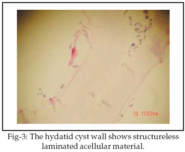
1,2 However,with increased travel,isolated cases can be seen anywhere in the world.1 Hydatid disease in humans is caused by tape-worms, Echinococcus granulosus, and the larval stage is known as the hydatid cyst.3 E.granulosus are most commonly seen in liver and lungs, it may also involve almost every organ or tissue in the body via the portal and systemic circulation.1,3 However, orbital infestation constitutes less than 1% of all hydatid disease cases.1,4-6CASE REPORT
A 27 year old previously healthy female presented with left eye proptosis and inferiorly displaced eyeball, diplopia maximum on upgaze was also present. Physical examination revealed limitaion of upward occular movements and signs of left optic nerve compression.
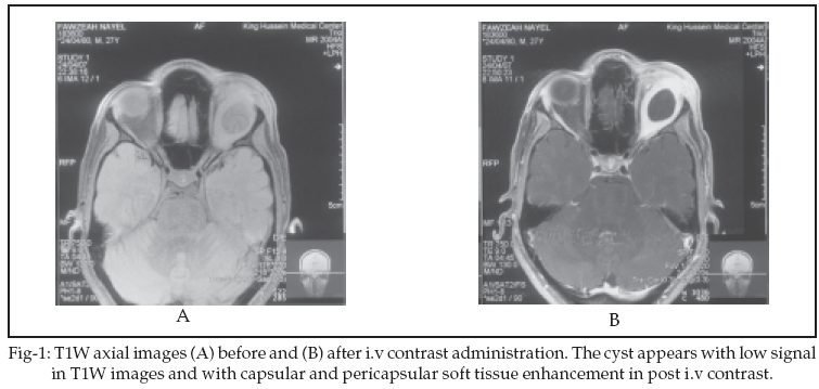
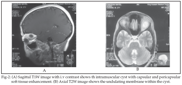
Orbital MRI was done which showed a 2 X 2cm well circumscribed, cystic lesion located solely in the superior rectus muscle. It appeared hypointense in T1W (Figure-1A) and hyperintense in T2W images with internal undulating hypointense membrane (Figure-2B). The internal undulating hypointense membrane is the characteristic appearance of hydatid cyst anywhere in the body. Following I.V contrast injection, the cyst showed thick enhancement of the capsule (Figure-1B, Figure-2A). The patient underwent surgical removal of the cyst by transcranial surgical approach. Post operative examination revealed no proptosis, residual limitation of upgaze occular movement. Postoperative MRI shows no evident of cyst residue.
DISCUSSION
Orbital Hydatid Cyst (OHC) are almost invariably situated in the superolateral and superomedial angles of the orbit, either within or outside the muscle cone.
7 Because of their superior location, they may erode the orbital roof and become intracranial.8,9
Inferiorly located cysts are very rarely seen.
1 In addition, more uncommon locations such as retro-bulbar have been reported.1,4,10 In our computer-based search for intramuscular hydatid cysts within the orbit, only one case of OHC within the medial rectus muscle was found.11 Our case is the first described intramuscular hydatid cyst located in the superior rectus muscle.The main clinical feature of the disease is progressive unilateral proptosis. Other clinical findings include mechanical restriction of occular movements, visual impairment, occular tension or pain, lid edema, papilloedema, and optic atrophy.
7,8,12 Upon MRI examination, the cystic lesion appeared low signal on T1W images, high signal on T2W images, capsular and pericapsular soft tissue enhencement, and the capsule was seen as a hypointense rim surrounding the mass on T2W images. Those features were also reported in previous cases.4,5 However, we have also detected an undulating membrane within the hydatid cyst which represent a typical characteristic of the echinococcal cysts in other organs.In adults, the most common cause of extraocular muscle enlargement is Grave’s disease, followed by myositic nonspecific inflammation, arteriovenous malformations, acromegaly, tumors at the orbital apex, rhabdomyosarcoma, lymphoma, and metastatic tumors,with the aid of imaging studies the differential diagnosis of focal intramuscular cystic lesions can be narrowed to include Trichinella, Cysticercosis, hemorrhagic cysts, and epithelial inclusion cysts.
13Preoperative early diagnosis is important to avoid cyst rupture so that severe allergic inflammatory reactions,seeding and recurrence of the disease can be avoided.
5The only definite treatment of hydatid cyst is surgical removal. However, due to the restricted area, total extirpation of the cyst without rupture is almost impossible and this will lead to the spillage of the contents of the cysts which leads to secondary dissemination with local recurrence, so, post operative treatment is of great importance. Mebendazole and albendazole have been shown to be effective in such cases.
8,14,15ACKNOWLEDGMENTS
The authors would like to thank University of Jordan, (Amman, Jordan) and Jordan University of Scince and Technology, (Irbid, Jordan) for support in literature search. They would like also to thank Mrs Rufaida Al-Zoubi for technical support.
REFERENCES
1. Aksoy F, Tanrikulu S, Kosar U. Inferiorly located retrobulbar hydatid cyst: CT and MRI features. Computerized Medical Imaging and Graphics 2001;25:535.
2. Mahesh L, Biswas J, Subramanian N. Role of ultrasound and CT-scan in diagnosis of hydatid cyst of the orbit. Orbit 2000;19(3):179.
3. Diren H, Ozcanli H, Boluk M, Kilic C. Unilocular orbital, cereberal and intraventricular hydatid cysts: Neuroradiology 1993;35:149.
4. Ciurea A, Giuseppe G, Machinis T, Coman T, Fountas K. Orbital hydatid cyst in childhood: A report of two cases. Southern Med J 2006;99(6):620.
5. Gokcek C, Gokcek A, Bayar M, Tanrikulu S, Buharali Z. Orbital hydatid cyst: CT and MRI. Neuroradiology 1997;39:512.
6. Rastogi A, Arora R, Chaturvedi K. Orbital hydatid cyst: an unusual presentation. Orbit 1998;17(2):107.
7. Turgut A, Turgut M, Kosar U. Hydatosis of the orbit in Turkey: results from review of the literature 1963-2001. International Ophthalmology 2004;25:193.
8. Jimenez-Mejias M, Alarcon-Cruz J, Marquez-Rivas F, Palomino-Nicas J, Montero J, Pachon J. Orbital hydatid cyst: treatment and prevention of reccurence with Albendazole plus Praziquantel. The British Infection Society 2000;105.
9. Fink A, Newman D, Stringer D. Pediatric Case of the Day. Radiographics 1995;15:731.
10. Jeblaoui Y, Bougila J, Yacoub K, Bouguila H, Ben Neji N, Besbes G. Orbital hydatid cyst. STOMAX 2008;143. (Article in Press).
11. Betharia S, Sharma V, Pushker N. Ultrasound findings in orbital hydatid cyst. Amer J Ophthalology 2003;135(4):568.
12. Alparslan L, Kanberoglu K, Peksayar G, Cokyuksel O. Orbital hydatid cyst: assessment of two cases. Neuroradiology 1990;32:163.
13. Kýratli H, Bilgic S, Ozturkmen C, Aydin O. Intramuscular hydatid cyst of the medial rectus muscle. Amer J Ophthalmol 2003;6:98.
14. Ergun R, Okten A, Yuksel M, Gul B, Evliyaoglu C, Ergungor F, et al. Orbital hydatid cysts: report of four cases. Neurosurg Rev 1997;20:33.
15. Selcuklu A, Ozturk M, Kulahli I, Dugan H. Successful syrgical management of an intraorbital hydatid cyst through a transmaxillary approach: case report. Skull Base 2003;13(2):101.
HOME | SEARCH | CURRENT ISSUE | PAST ISSUES
Professional
Medical Publications
Room No. 522, 5th Floor, Panorama Centre
Building No. 2, P.O. Box 8766, Saddar, Karachi - Pakistan.
Phones : 5688791, 5689285 Fax : 5689860
pjms@pjms.com.pk