|
|
||||
|
Published by : PROFESSIONAL MEDICAL PUBLICATIONS |
||||
|
ISSN 1681-715X |
||||
|
||||
|
- |
||||
|
ORIGINAL ARTICLE |
||||
|
- |
||||
|
Volume 24 |
October - December 2008 (Part-II) |
Number 6 |
||
|
|
||||
|
|
||||
|
|
||||
|
Published by : PROFESSIONAL MEDICAL PUBLICATIONS |
||||
|
ISSN 1681-715X |
||||
|
||||
|
- |
||||
|
ORIGINAL ARTICLE |
||||
|
- |
||||
|
Volume 24 |
October - December 2008 (Part-II) |
Number 6 |
||
|
|
||||
|
|
||||
Ocular volume determination in Nigerians
Osesogie Usuale Ogbeide1, Afekhide Ernest Omoti2
ABSTRACT
Objective: To determine the ocular volume in healthy Nigerian eyes.
Methodology: This was a cross-sectional descriptive study of volunteers, staff and students of University of Benin Teaching Hospital (UBTH) and University of Benin, Nigeria. Measurements of the eyeball were taken on the B-mode image using a Medison’s Sonoace 1500 ultrasound machine using a 6.5 MHz curvi-linear transducer placed over the closed eyelid. The vertical, horizontal and axial diameters of the eyes of healthy subjects were measured and used to compute the ocular volumes. The age and sex of the subjects were also recorded.
Results: Two hundred subjects comprising 125 females (62%) and 75 males (38%) were included in the study. The age range was 3-92 years and overall mean age was 41.48 ± 23.26 years for both sexes. The mean eyeball volume was slightly larger for males than for females. (Males: Right eye=10.64ccm3, Left eye=10.37ccm3; Females: Right eye=10.59 ccm3, Left eye=10.19 ccm3). The difference was not statistically significant (P= 0.229). The right eyeball volume was higher on the right for both males and females but the difference was not statistically significant (P= 0.198). There was a gradual increase in eyeball volume with age.
Conclusions: This study was able to generate normal eyeball volumes in a sample of Nigerians which hopefully will serve as reference for normal values in Black Africans.
KEY WORDS: Ocular volumes, Ultrasound, Nigerians.
Pak J Med Sci October - December 2008 (Part-II) Vol. 24 No. 6 808-812
How to cite this article:
Ogbeide OU, Omoti AE. Ocular volume determination in Nigerians. Pak J Med Sci 2008;24(6):808-12.
1. Osesogie Usuale Ogbeide MBBS,
FMCR
Department of Radiology,
2. Afekhide Ernest Omoti MBBS, FMC (OPH), FWACS
Department of Ophthalmology,
University of Benin Teaching Hospital,
Pmb 1111, Benin City, Nigeria.
Correspondence
Osesogie Usuale Ogbeide MBBS, FMCR
Department of Radiology,
University of Benin Teaching Hospital,
PMB 1111, Benin City, Nigeria.
E mail:
drosesogieogbeide@yahoo.com
* Received for Publication: May 8, 2008
* Accepted: October 2, 2008
INTRODUCTION
Ultrasonography of the eyeball has become increasingly important as a diagnostic tool in clinical practice because it is a rapid, safe and atraumatic method of examination. The use of B-scan technique in ophthalmology was first described by Baum et al.
1 and introduced their two-dimensional B-mode ultrasound application in ophthalmology, where they stated that their apparatus simulates a slit lamp, except that a pulsed ultrasound beam is substituted for the light beam.Ossoinig
2,3 reviewed the reliability and accuracy of ultrasonic diagnosis of one hundred ophthalmologic cases. Their conclusion was that ultrasonic evaluation provided the surgeon with maximum information available prior to surgical exploration as to the size and position of an orbital tumour. Advances in orbital sonography have led to prominence being attached to its benefits in the determination of eyeball biometry. Also due to the global acceptance of eyeball ultrasonography, there is a need for local values for eyeball biometry in our environment to serve as a reference.Ocular volume may be increased pathologically in congenital glaucoma (buphthalmos), ectasias, staphylomas and high myopia, or may be decreased in microphthalmia, phthysis bulbi; or it may vary physiologically or as a racial difference.
This study aimed at determining the sonographic range of normal eyeball volumes in Nigerians which could serve as reference values for Africans.
METHODOLOGY
This was a cross-sectional descriptive study. The study population was made up of volunteers, staff and students of University of Benin Teaching Hospital (UBTH) and University of Benin. Demographic data, including the age and sex were obtained. Informed consent was obtained from the subjects before the commencement of the investigation. Ethical approval was obtained from the Ethical Committee of the University of Benin Teaching Hospital, Benin City, Nigeria.
Exclusion criteria:
Subjects with positive ophthalmic pathology like tumours or fractures affecting the facial bones, proptosis and refractive errors such as hypermetropia and myopia were excluded. Also excluded were subjects with any previous history of ophthalmic surgery, subjects with orbital cellulites or past history of chronic glaucoma and subjects whose age could not correctly be ascertained. Subjects with trauma to the eye where contact with the transducer was impossible were also excluded. Similarly subjects with other systemic disorders such as toxic goitre, hypertension and diabetes mellitus were also not included in the study.Procedure for eye examination:
With the subject lying in the supine position, they were asked to close the eyelid and coupling gel was applied over the lid. The subjects were instructed to fix their gaze at the ceiling. The 6.5 MHz curvi-linear transducer of the Medison’s Sonoace 1500 ultrasound machine was placed over the closed eyelid and scanning was done in the transverse and vertical or cranio-caudal planes of the eye. This is known as the direct contact scanning technique.During the examination, those found to have previously undetected ocular lesions were excluded from the study. Frozen images or static scan protocol was taken which enabled accurate measurements to be carried out. Both eyes were assessed with two readings taken for each measurement. The mean values of both eyes were used to compute the results. Eyeball volume was determined by measuring the following: The maximum distance between the corneal interface to the vitreo-retinal interface to the macula. The anterior corneal interface echo was clearly visible as was the posterior lens interface echo and the optic nerve. This gave the axial length or the antero-posterior diameter.
Note that the vertical or cranio-caudal diameter and axial length are approximately equal; both measurements being done from the corneal interface to the macula, the vertical diameter was not recorded in this study. Extreme care was taken by the sonologists to ensure that there was no indentation of the anterior surface of the cornea. Basic statistics were performed using the Statistical Analysis System (SAS) and the analysed data were expressed in descriptive statistics such as frequency tables, percentages, mode, median and mean. Correlative analysis, Students’ t-test, was used to test for significant differences. A statistical significance level of p-value of <0.05 was adopted.
RESULTS
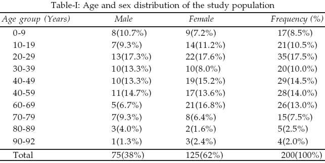
The sex distribution of the study population showed that of the two hundred subjects who completed the study, 125 (62%) were females and 75 (38%) were males, (Table-I). The age range was 3-92 years and overall mean age was 41.48 ± 23.26 years for both sexes. The mean age was 42.22 ± 23.04 years for males and 40.24 ± 23.73 years for females. There was no statistically significant difference in the mean age between both sexes (P=0.521). Subjects within the age group 20-29 years constituted the majority (n=35 or 17.5%) of the study population, followed by those within the age group 40-49 years (n=29 or 14.5%).
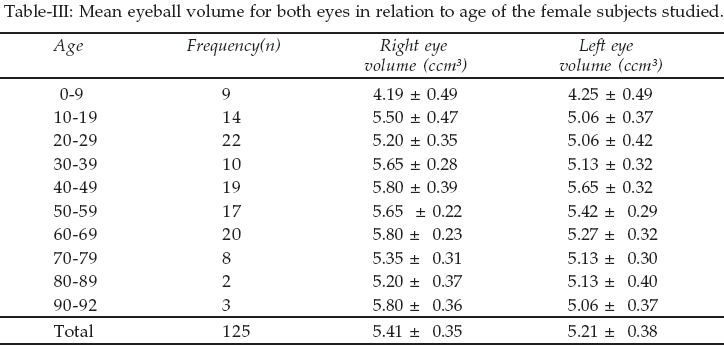
The mean eyeball volume for both sexes in relation to age is presented in Tables-II & III. The mean eyeball volume was shown to be slightly larger for males than for females. (Males: Right eye=10.64ccm
3, Left eye=10.37ccm3; Females: Right eye=10.59 ccm3, Left eye= 10.19 ccm3). The difference was not statistically significant (P= 0.229).
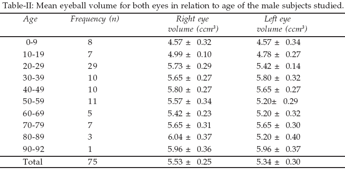
The right eyeball volume was also found to be higher on the right for both males and females. However, the difference was not statistically significant (P= 0.198). The values for eyeball volume showed similar pattern for both males and females.
DISCUSSION
Ultrasonography was used in this study to ascertain the biometry of the eye due to the following reasons. The eye is located at the body surface and during ultrasonography it normally reflects only a few echoes that can easily be identified.
4-8 Ultrasonography is cheap, readily available, easy to perform, and can be carried out easily in the clinic setting. Ultrasonography is able to demonstrate the existence and composition of the eyeball, presence of intra-ocular and extra-ocular masses, as well as monitor changes in dimensions or appearance of such masses.The evaluation of eyeball volume was done in this study electronically using the measurements from the two eyeball diameters measured. The mean eyeball volume reported in this study was less than what has been reported in several studies in Caucasian populations.
8-12 Variation in eyeball volume may be due to racial differences in eyeball sizes, method of eyeball diameter measurement (contact or non-contact A- or B- mode ultrasonography), age group studied or a combination of all these factors. Babalola et al10 showed that the eyeball dimensions for the West African emmetropic eye was consistently lower than Caucasian values in their study.This study showed that there was a gradual increase in eyeball volume with age, with the older age group having the highest values in both males and females. This can be explained by continued growth of the eyeball with age. The finding was more prominent in the males. A possible explanation for this finding is that the eyeball is a pressurized chamber with the corneo-scleral coat of the eyeball confining the intraocular pressure. This preserves the eye’s dimensions with the uveal circulation as the source of the intraocular fluid. This results in a steady ocular pressure throughout life, even in the elderly.
13,14 The overall effect was a consistent preservation of adult eyeball diameter once the maximum value has been attained throughout life or even a gradual increase of these values in the elderly subjects.The finding of an increase in eyeball dimensions with age was also reported in other studies,
15-17 which stated that the growth of the eyeball was similar to the growth curve of the brain and central nervous system rather than to the general body growth and not related to height, diet, skeletal, genital or lymphoidal growth. The finding further stated that during the period of active growth, the eye shares with the brain the peculiarity of having a precocious growth. In contrast to this finding, Lim et al15 reported a decrease in eyeball diameters and volume occurring at an older age group. It also stated that this change was more prominent in females than males, attributing these changes to potential influence of systemic endocrine or metabolic factors.However the measurement of eyeball volume in all the studies has not been done using a universal method. The difference being that the eyeball has been assumed to be either slightly ellipsoidal or spherical for eyeball volume estimation. Furthermore, the eyeball volume has generally been estimated with measurement of only one or two or three eyeball diameters. This explains the different mathematical formulae used to calculate eyeball volume in different studies and the resultant difficulty to corroborate the findings in these studies. Studies done have showed that the calculation of all three diameters have a better result in estimation of eyeball volumetry.
18-20In conclusion, this study has provided eyeball volumes in a sample of Nigerians that can be used as reference values for Nigerians. These values are lower than what has been reported in Caucasian eyes and there is a gradual increase in ocular volume with age.
Limitations of the study:
Clinical application of the study and its usefulness has not been discussed in this study which could have further enhanced its usefulness.REFERENCES
1. Baum C, Greenwood J. The application of ultrasonic locating techniques of ophthalmology II Ultrasonic visualization of soft tissues. Ach Ophthalmol 1958;60:263-79.
2. Ossoinig K. Clinical Echo-ophthalmology. Current Concepts of Ophthalmology 1972;3:101-30.
3. Ossoinig K. Ultrasound diagnosis in Ophthalmology. Weiner Medizinisha Ocheaschrifc 1968;118:362-7.
4. Baxter GM. Ultrasound investigation. Br J Ophthalmol 1979;63:531–2.
5. Coleman OJ, Konig WF, Katz L. A hand–operated ultrasound system for ophthalmic evaluation. Am J Ophthalmol 1969;68:255–63.
6. Coleman DJ, Weininger R. Ultrasound M-Mode technique in Ophthalmology. Arch Ophthalmol 1974;82:475-6.
7. Scammon RE, Wilmer HA. Growth of components of the human eyeball. Arch Ophthalmol 1950;43:580-1.
8. Goyal R, North RV, Morgan JE. Comparison of Laser interferometry and ultrasound A–scan in the measurement of axial length. Acta Ophthalmol Scand 2003;81:331-5.
9. Stenstrom S. Investigation of the variation and the correction of the optical elements of the Human eye. Transl. by Woolf D in Annals Am Academy Ophthalmol 1948;25:218-496.
10. Babalola J, Szajnzicht E. Ocular characteristics in West African and Europeans: A comparison of two Groups. Brit J Phys Ophthalmol 1960;17:27-32.
11. Ramani KK, Mani B, Ronnie G, Joseph R, Lingam V. Gender variation in ocular biometry and ultrasound biomicroscopy of primary angle closure suspects and normal eyes. J Glaucoma 2007;16;122-8.
12. Fanny A, Ouattara A, Aka J, Coulibaly F, Gbe K, Boni S, et al. Ocular biometric values of the black African patient and theoretical consideration of the role of these values in various pathologies : Analysis of 325 eyes. J Fr Ophtalmol 2007;30:68-72.
13. Hirsh MJ, Weymouth FW. Notes on ametropia-A further analysis of Stenstrom’s data. Annals Am Academy Ophthalmol 1947;24:601-2.
14. Fledelius HC, Christensen AC. Reappraisal of the human ocular growth curve in fetal life, infancy and early childhood. Br J Ophthalmol 1996;80:918-21.
15. Lim KJ, Hyung SM, Youn DH. Ocular dimensions with aging in normal eyes. Korean J Ophthalmol 1992;6:19-31.
16. Strakhov VV, Mineeva LA, Buzykin MA. Involutional changes in the human eye accommodation apparatus as evidenced by ultrasound biometry and biomicroscopy. Vestn Oftalmol 2007;4:32-5.
17. Santodomingo–Rubido J, Mallen EA, Gilmartin B, Wolffsohn JS. A new non-contact optical device for ocular biometry. Br J Ophthalmol 2002;86:458-62.
18. Forbes G, Gehring DG, Gorman CA, Brennan MD, Jackson IT. Volume measurements of normal orbital structures by computed tomographic analysis. AJR 1985; 145:149-54.
19. Bremond-Gignac D, Cussenot O, Deplus S. Computation of eyeball growth by magnetic resonance imaging. Surg Radiol Anat 1994;16:113-5.
20. Hann FJ, Chu WK. Ocular volume measured by CT scans. Neuroradiology 1984;26:419-20.
HOME | SEARCH | CURRENT ISSUE | PAST ISSUES
Professional
Medical Publications
Room No. 522, 5th Floor, Panorama Centre
Building No. 2, P.O. Box 8766, Saddar, Karachi - Pakistan.
Phones : 5688791, 5689285 Fax : 5689860
pjms@pjms.com.pk