|
|
||||
|
Published by : PROFESSIONAL MEDICAL PUBLICATIONS |
||||
|
ISSN 1681-715X |
||||
|
||||
|
- |
||||
|
CASE REPORT |
||||
|
- |
||||
|
Volume 24 |
October - December 2008 (Part-II) |
Number 6 |
||
|
|
||||
|
||||
|
|
||||
|
Published by : PROFESSIONAL MEDICAL PUBLICATIONS |
||||
|
ISSN 1681-715X |
||||
|
||||
|
- |
||||
|
CASE REPORT |
||||
|
- |
||||
|
Volume 24 |
October - December 2008 (Part-II) |
Number 6 |
||
|
|
||||
|
||||
Laparoscopic fundoplication with mesh
repair of a large type III hiatal hernia
Chinnusamy Palanivelu1, Muthukumaran Rangarajan2,
Gokul Kruba Shanker3, Madhupalayam Velusamy Madankumar4
SUMMARY
Mixed (type III) hiatal hernias – with sliding and paraesophageal components – are uncommon, accounting for 10-15% of all hiatal hernias. Symptoms are that of gastroesophageal reflux disease. The paraesophageal hernia may get incarcerated and present as an emergency. We present a 50-year old patient with symptoms of severe reflux disease who was diagnosed with type III hiatus hernia. Laparoscopic mesh repair with fundoplication was performed, as the defect was large. Postoperatively, patient’s symptoms were almost completely relieved. Follow up manometry was done, which showed normal pressure range. Thoracotomies or laparotomies were the traditional methods of surgery for type III hiatal hernias, though now thoracoscopy and laparoscopy are becoming popular. We favor the laparoscopic approach as the hernial sac can be dealt with and crurorraphy with fundoplication can also be done. The hernial sac may or may not be completely excised. Some feel that if it is not excised, the residual sac may be filled with fluid and it can present as paracardiac cyst. Hence it is preferable to excise the hernial sac. Though controversial, we feel mesh repair of hiatus is advisable in case of large hiatal defects. Laparoscopic repair of type III hiatal hernias is more or less well established and carries all the benefits of minimally invasive surgery.
KEYWORDS: Wide hiatus, Paraesophageal hernia, Hernioplasty.
Pak J Med Sci October - December 2008 (Part-II) Vol. 24 No. 6 872-875
How to cite this article:
Palanivelu C, Rangarajan M, Shanker GK, Madankumar MV. Laparoscopic fundoplication with mesh repair of a large type III hiatal hernia. Pak J Med Sci 2008;24(6):872-75.
1. Dr. Chinnusamy Palanivelu MNAMS, MS, MCh, FRCS,
FACS,
Director
2. Dr. Muthukumaran Rangarajan MS, DipMIS(Fr), FACS,
Professor of Surgery
3. Dr. Gokul Kruba Shanker MS,
Registrar in Surgical Gastroenterology
4. Dr. Madhupalayam Velusamy Madankumar MS,
Registrar in Surgical Gastroenterology.
1-4: GEM Hospital, 45-A,
Pankaja Mill Road, Ramnathapuram,
Coimbatore – 641045,
India.
Correspondence
Dr. Muthukumaran Rangarajan,
GEM Hospital, 45-A,
Pankaja Mill Road, Ramnathapuram,
Coimbatore – 641045, India.
E-mail: rangy68@gmail.com
* Received for Publication: April 15, 2008
* Accepted: September 15, 2008
INTRODUCTION
Paraesophageal hernia (PEH) combined with sliding hiatal hernia is a specific form of hiatal hernia (type III) where there is a diaphragmatic defect at the esophageal hiatus, typically anterior and lateral to the esophagus. The incidence is 10-15% of hiatal hernias.
1 Akerlund was the first to use the term paraesophageal hernia in 1926 for acquired hiatal hernia. This type of hernia likely results from progression of a simple type I hernia with rolling of the fundus up alongside the gastroesophageal junction. Most patients are symptomatic, with dysphagia and heartburn being extremely common problems. The gastric fundus is the most common intra-abdominal organ to herniate through the hiatus into the chest, rolling cephalad alongside the distal esophagus. They generally occur in elderly individuals with more medical comorbidities than the typical patient undergoing fundoplication for gastroesophageal reflux disease (GERD). Displacement of the gastroesophageal junction into the chest may produce symptomatic GERD. Up to one third of patients may present with complications such as bleeding, obstruction, gastric volvulus, strangulation, or perforation.2 Here we report a case of symptomatic type III hiatal hernia who underwent mesh reinforcement hiatoplasty and fundoplication.METHODOLOGY
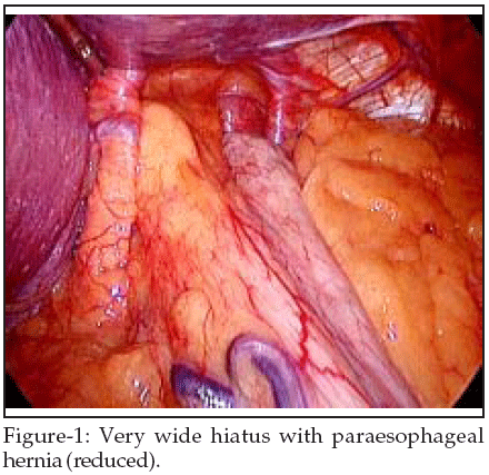
The patient was a 50-year old gentleman presenting with retrosternal burning, regurgitation of food particles and belching for two years. He underwent a complete course of anti-reflux drugs but only got temporary relief. Evaluation included a barium esophagogram, chest radiogram, upper gastrointestinal endoscopy and manometry (LES pressure – 8-12mmHg). A type III hiatal hernia was identified and laparoscopy was planned. The patient was positioned supine on the operating table, and the surgeon works from the right side with the assistant on the left. Four 5mm and one 10mm laparoscopic ports were placed in the upper abdomen. The left lateral segment of the liver was retracted anteriorly with a 5mm flexible retractor. The intrabdominal pressure was kept below 14mmHg to allow easy reduction of hernial contents. After exposing the hiatus, the herniated stomach was reduced into the abdomen using atraumatic graspers (Fig-1). Dissection was started by dividing the gastrohepatic ligament and exposing the right crus of the diaphragm using the Harmonic scalpel (Ethicon, USA). The phreno-esophageal ligament was divided.
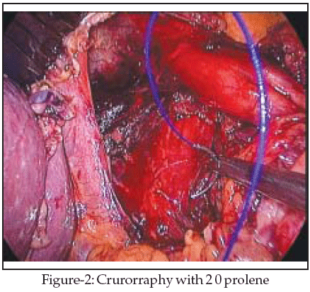
Next, the gastrosplenic ligament was divided along with the posterior attachments to the fundus. Dissection was continued to expose the junction of the right and left crura at the retroesophageal space. The hernial sac and the gastroesophageal fat pad were carefully dissected out, sweeping the anterior vagus nerve to the right of the esophagus with the fat pad. A combination of sharp dissection with the Harmonic scalpel and blunt dissection with graspers was used to disconnect the hernial sac from the mediastinum. The wide hiatus was closed with interrupted 20 prolene intracorporeal sutures. Crurorraphy was done by approximating the right and left crura with interrupted 2 0 prolene sutures (Fig-2).
A Gore-Tex
TM (W. L. Gore & Associates, Inc., Flagstaff, AZ, U.S.A.) mesh of size 7x 5cm was fixed over the sutured defect (Fig-3).

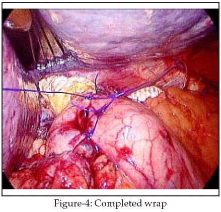
The esophagus was retracted caudally using an umbilical tape tied around the esophagogastric junction. Posterior esophageal window was created and the fundus was wrapped around the esophagogastric junction. Three sutures were taken with 2 0 prolene to fix the wrap (Fig-4). Sutures were also taken with the esophagus and right crus to further prevent wrap migration.
RESULTS
Barium swallow was done on the 1
st postoperative day (POD). Since there was no leak, the nasogastric tube was removed and the patient was started on liquids and soft diet on the next day. He was discharged on the 4th POD. Intravenous analgesics were needed only for the 1st POD. At discharge, patient had mild retrosternal discomfort and dysphagia, no regurgitation and no pain. Follow up surgery, the patient was reviewed at one, six, twelve and twenty four months; barium swallow and manometry were done at each follow up. Lower esophageal sphincter pressure was 15-20mmHg at the 1st follow up and continued to be so during all the follow up visits. He had reflux symptoms at the 12th month follow up, which was relieved with drugs. There was no recurrence of the paraesophageal component either.DISCUSSION
Many controversies exist about repair of large type III hiatal hernias. Some authors think that PEHs are at risk of becoming incarcerated or strangulated, and the mere presence of one is considered to be an indication for surgical repair in the patient medically fit for operation.
3 We feel that all Type III hiatal hernias should be repaired to relieve symptoms or to prevent future complications.Traditionally, repair of large PEHs has been performed through an open laparotomy or left thoracotomy. They have recently given way to laparoscopic repair as the procedure of choice for many surgeons.
4-6 Because of its recent application; long-term outcome data for the laparoscopic approach are not available. Initial short-term studies have shown similar recurrence rates in open and laparoscopy groups.5 One of the controversies is dealing with the hernial sac. Some authors prefer sac excision as it results in fewer fluid collections that accumulate in the ‘dead space’. Most surgeons feel the sac should not be excised, disconnection alone would suffice. The extensive mobilization of the hiatus required to perform the repair may result in gastroesophageal reflux even if it is not present before surgery. It is therefore prudent to perform fundoplication. There are some technical points of controversy regarding the use of prosthetics to bolster the repair and esophageal lengthening.4,7 Mesh is needed if the defect is large. Also, some surgeons prefer not to close the larger defects, instead fix a mesh for a ‘tension-free’ repair. We prefer to close the defect as long as there is no undue tension.The largest case series of laparoscopic PEH repair, with excellent intermediate term results has been reported by Luketich et al.,
6 Frantzides et al reported the only randomized trial comparing primary repair with polytetrafluoroethylene (PTFE) mesh.8 Primary repair of a large hiatal hernia is associated with a published recurrence rate of up to 10%. The use of PTFE reinforcement for primary repair of large hiatal hernias may result in a lower rate of recurrent herniation compared to primary repair alone.9 Use of prostheses is associated with the risk of a foreign body in close proximity to the esophagus eroding directly into the esophagus or causing dysphagia. If a prosthetic patch is required, ePTFE is currently the prosthetic material of choice. The assessment of esophageal length is important. There is a high incidence of recurrent herniation if the intrabdominal esophagus is <2.5cm after mobilization of the GE junction.10CONCLUSION
Laparoscopic repair of large type III hiatal hernias is an appropriate method with less morbidity. Although most PEH may be repaired laparoscopically, the procedure is typically more difficult to perform than routine antireflux operations.
REFERENCES
1. Ellis FH, Crozier RE, Shea JA. Paraesophageal hiatus hernia. Arch Surg 1986;121:416-20.
2. Dahlberg PS, Deschamps C, Miller DL, Allen MS, Nichols FC, Pairolero PC. Laparoscopic repair of large paraesophageal hiatal hernia. Ann Thorac Surg 2001;72:1125-9.
3. Kuster GG, Gilroy S. Laparoscopic technique for repair of paraesophageal hiatal hernias. J Laparoendosc Surg 1993;3:331-8.
4. Frantzides CT, Carlson MA. Prosthetic reinforcement of posterior cruroplasty during laparoscopic hiatal herniorrhaphy. Surg Endosc 1997;11:769-71.
5. Gantert WA, Patti MG, Arcerito M. Laparoscopic repair of paraesophageal hiatal hernias. J Am Coll Surg 1998;186:428-33.
6. Luketich JD, Raja S, Fernando HC. Laparoscopic repair of giant paraesophageal hernia: 100 consecutive cases. Ann Surg 2000;232:608-18.
7. Carlson MA, Richards CG, Frantzides CT. Laparoscopic prosthetic reinforcement of hiatal herniorrhaphy. Dig Surg 1999;16:407-10.
8. Frantzides CT, Richards CG, Carlson MA. Laparoscopic repair of large hiatal hernia with polytetrafluoroethylene. Surg Endosc 1999;13:906-8.
9. Gantert WA, Patti MG, Arcerito M, Feo C, Stewart L, Fujino Y, et al. Laparoscopic repair of parae ophageal hiatal hernias. J Am Coll Surg 1998;186:428-33.
10. Hashemi M, Peters JH, DeMeester TR. Laparoscopic repair of large type III hiatal hernia: objective follow-up reveals high recurrence rate. J Am Coll Surg 2000;190:553-61.
HOME | SEARCH | CURRENT ISSUE | PAST ISSUES
Professional
Medical Publications
Room No. 522, 5th Floor, Panorama Centre
Building No. 2, P.O. Box 8766, Saddar, Karachi - Pakistan.
Phones : 5688791, 5689285 Fax : 5689860
pjms@pjms.com.pk