|
 |
|
Published
by : PROFESSIONAL MEDICAL PUBLICATIONS |
|
ISSN 1681-715X |
|
|
|
- |
|
ORIGINAL
ARTICLE |
|
- |
|
Volume 25 |
October - December 2009
(Part-II) |
Number 6 |
|
|
|
Effects of adenosine on the organ injury and
dysfunction caused By Hemorrhagic Shock
Mona M. Soliman1
ABSTRACT
Objectives: Adenosine has been shown in animal and
human studies to decrease the post-ischemic myocardial injury by lowering the
levels of tumor necrosis factor-a. The objectives of the study was to examine
the protective effects of adenosine on the organ injury (liver, kidney,
pancreas) associated with hemorrhagic shock in rats.
Methodology: The study was conducted at
Cardiovascular Physiology laboratory, King Saud University, Riyadh in
2007-2008. Anesthetized male Sprague- Dawley rats were assigned to hemorrhage
and resuscitation treated with 20mM adenosine , untreated, or similar time
matched control groups (n=6 per group). Rats were hemorrhaged for one hour
using a reservoir model. Arterial blood pressure was monitored for one hour,
and maintained at a mean arterial blood pressure of 40 mmHg. Adenosine 20mM
was injected intra-arterially, before resuscitation in the adenosine treated
group. Resuscitation was performed by reinfusion of the sheded blood for 30
minutes. Arterial blood samples were analyzed for biochemical indicators of
multiple organ injury: 1) liver function: aspartate aminotransferase (AST),
alanine aminotransferase (ALT), 2) renal function: urea and creatinine, 3)
pancreatic function: amylase.
Results: In the control group there was no
significant rise in the serum levels of (i) urea and creatinine, (ii)
aspartate aminotransferase (AST) and alanine aminotransferase (ALT), (iii)
amylase. While in the adenosine treated group, resuscitation from one hour of
hemorrhagic shock resulted in significant rises in the serum levels of (i)
urea and creatinine , (ii) aspartate aminotransferase (AST) and alanine
aminotransferase (ALT), (iii) amylase. Treatment of rats with 20mM adenosine
before resuscitation following one hour of hemorrhagic shock decreased the
multiple organ injury and dysfunction caused by hemorrhagic shock.
Conclusion: Adenosine attenuated the renal, liver
and pancreatic injury caused by hemorrhagic shock and resuscitation in rats.
Thus, the inflammatory response to shock may contribute to the multiple organ
failure developed after hemorrhagic shock and resuscitation.
KEY WORDS:
Adenosine, hemorrhagic shock, multiple organ failure, TNF-a, inflammation.
Pak J Med Sci October - December 2009
(Part-II) Vol. 25 No. 6 890-894
How to cite this article:
Soliman MM. Effects of adenosine on the organ injury and dysfunction caused
By Hemorrhagic Shock. Pak J Med Sci 2009;25(6):890-894.
1. Dr. Mona M. Soliman, MBBS, MSc., PhD
Assistant Professor, Dept. of Physiology (29)
College of Medicine, King Khalid University Hospital,
Riyadh 11461, Saudi Arabia.
Correspondence:
Dr. Mona M. Soliman, MBBS, MSc., PhD
Email: monaslmn@yahoo.com
* Received for Publication: May 21, 2009
* Revision Received: October 12, 2009
* Accepted: October 21, 2009
INTRODUCTION
Multiple organ failure (MOF) is recognized as the leading
cause of death following traumatic injury.
1-3
Most trauma deaths result from either insufficient tissue perfusion due to
excessive blood loss, or the development of inflammation, infection and organ
injury following resuscitation.4
In the past decade, there have been no effective interventions for post-injury
MOF.5
Despite the improvement in intensive care medicine, the mortality of
hemorrhagic shock remains very high.6
Trauma victims who survive their initial injuries face a risk of death of
multiple organ failure7
Thus, there is still a great need for new approaches to improve therapy and
outcome for patients with hemorrhagic shock.
The exact mechanism of MOF following hemorrhagic shock is
unclear. Recent evidence suggest that the overproduction of pro-inflammatory
cytokines may mediate the progression of shock to MOF and death.
8-10
Many factors may be involved, as the marked increased production of oxygen
free radicals which in turn triggers cytokines and TNF-a production and
development of organ injury.10,11
However, little is known regarding the role of these radicals in hemorrhagic
shock. Research is directed toward the protection and maintenance of liver,
cardiac, intestinal and renal function following hemorrhagic shock.
There are multiple therapeutic interventions that lower the
inflammatory response to shock. Adenosine, an endogenous nucleoside, possesses
several properties that could be valuable in protection following hemorrhagic
shock insult. Adenosine may redistribute blood flow and increase vital organ
perfusion. Adenosine can decrease whole body and myocardial oxygen consumption
12
and improve recovery of energy metabolism after intestinal
ischemia-reperfusion.13
Adenosine has been shown in animal and human studies to lower the
post-ischemic myocardial injury.14-16
The exact mechanism is not known. However, it may be mediated via attenuating
the inflammatory response to shock. Adenosine may produce anti-inflammatory
effects by inhibiting neutrophil adhesion to endothelial cells and inhibiting
neutrophil superoxide anion generation and de-granulation.17
Moreover, adenosine reduces the release of pro-inflammatory cytokine.18
Despite the intensive research that has been done on the role of adenosine in
protecting the heart following ischemic insults, little is known regarding the
protective effects of adenosine following hemorrhagic shock and resuscitation.
The present study evaluated the effects of adenosine on the
organ injury (liver dysfunction, kidney dysfunction and pancreatic
dysfunction) caused by hemorrhagic shock and resuscitation in rats.
METHODOLOGY
The study was conducted at the Cardiovascular Physiology
laboratory at King Khalid University Hospital, College of Medicine, King Saud
University, Riyadh, Saudi Arabia in 2007-2008. Sixteen male Sprague Dawley
rats weighing 400-500 gm were used. Rats were anesthetized with urethane
(125mg/kg i.p.) and cannula was placed in the carotid artery for measurement
of arterial blood pressure, for withdrawal of blood and resuscitation. Heparin
sodium (2000 I.U.) was then injected intra-arterially. Rats were randomly
assigned to 4 groups: (1) hemorrhagic shock untreated, (2) hemorrhagic shock
treated with adenosine, (3) sham hemorrhage untreated and (4) sham hemorrhage
treated with adenosine. After 15 minutes stabilization, rats were hemorrhaged
using a reservoir (a 10 ml syringe). Blood was withdrawn from the carotid
artery until MAP reached 35-40 mmHg, over a period of 60 minutes. At 90
minutes, rats were resuscitated by reinfusion of sheded blood, together with
saline when required to achieve normotension (80-110mmHg). If the blood
pressure drop below 30 mmHg or failed to be restored to normal, experiments
were excluded. The same surgical procedures were performed for the sham
hemorrhage groups except that rats were not hemorrhaged.
In the treated groups, rats were injected with adenosine
(20µM) intra-arterially via the carotid artery cannula before resuscitation,
followed by 30 minutes resuscitation as described above.
After one hour of hemorrhagic shock and 30 minutes
resuscitation, 1.5 ml of blood was collected into a serum gel S/1.3 tube (Sarstedt,
Germany) from the catheter placed into the carotid artery. The blood sample
was centrifuged (1610 x g for 3 minutes at room temperature) to separate
plasma. All plasma samples were analyzed within 24 hours at the central
Biochemestry laboratory at King Khalid University Hospital. The following
marker enzymes were measured in the plasma as biochemical indicators of
multiple organ injury/dysfunction: (1) Renal dysfunction was assessed by
measuring the rises in plasma levels of creatinine (as an indicator of
impaired renal function) and urea (as indicator of impaired excretory function
of the kidney)
19;
(2) Liver function was assessed by measuring the rise in plasma levels of
aspartate aminotransferase (AST, a non-specific marker for hepatic injury),
alanine aminotransferase (ALT, as indicator of hepatic parenchymal injury)5;
(3) Pancreatic injury was assessed by the rises in serum levels of lipase, a
specific indicator for the development of pancreatic injury and (4)
Neuromuscular injury was assessed by measuring the levels of creatinine kinase.
Statistical analysis: Data are presented as means and
standard deviations. Data was analyzed with analysis of variance (ANOVA). Data
were considered statistically significant when yielding a P- value less
than 0.05.
RESULTS
As previously reported, hemorrhagic shock induced acute
renal injury as evidenced by the significant rise in the serum levels of urea
and creatinine (Fig-1 and Table 1), compared to the sham controls.
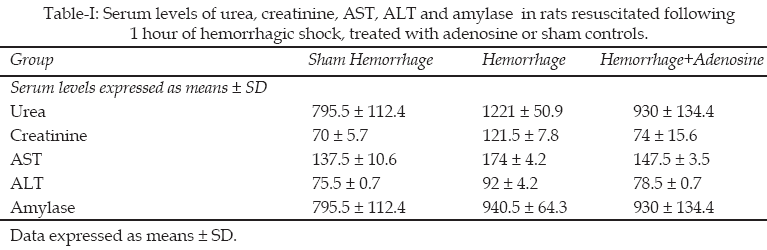
The protective effect of treatment with adenosine before
resuscitation was evidenced by markedly lowering the serum levels of urea and
creatinine in the hemorrhage treated group as compared to the hemorrhage
untreated one(Figure-1).
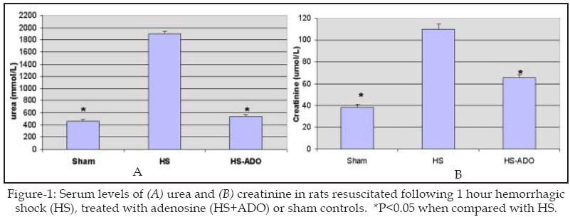
As compared to sham hemorrhage group, resuscitation from
one hour of hemorrhagic shock resulted in significant rise in the serum levels
of AST and ALT compared to sham group, demonstrating the development of he
patocellular injury triggered by hemorrhage and resuscitation. Treatment with
adenosine before resuscitation lowered the levels of AST and ALT in the
hemorrhage treated group (Figure-2).
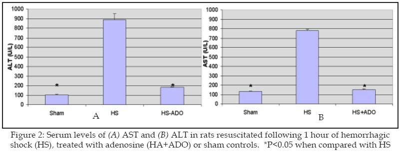
Resuscitation following one hour of hemorrhagic shock did
result in non-significant rises in the serum levels of amylase, demonstrating
the possibility of pancreatic injury (Figure 3). Pretreatment of rats with
adenosine non-significantly decreased the levels of amylase (Figure 3).
DISCUSSION
In this study, we have evaluated the effects of treatment
with adenosine on multiple organ failure associated with hemorrhagic shock in
rats. Hemorrhage for 60 minutes followed by resuscitation with shed blood for
30 minutes resulted in (i) liver dysfunction, (ii) renal dysfunction and (iii)
pancreatic injury. We have previously confirmed that
20
the model of hemorrhagic shock used here results in myocardial contractile
dysfunction and myocardial injury. We reported that treatment with 20µM
adenosine (intra-arterially) before resuscitation of hemorrhagic shock
protected the myocardium against post-resuscitation injury and dysfunction by
improving the myocardial contractile function and preserving the myocardial
structure.21
The present study showed that the treatment with 20µM adenosine
(intra-arterially) before resuscitation of hemorrhagic shock abolishes (i) the
liver injury, (ii) the renal dysfunction and (iii) the pancreatic injury
caused by hemorrhage and resuscitation.
Hemorrhagic shock and resuscitation leads to injury to
target organs including the heart, liver, brain and kidney.
22
The progression of hemorrhagic shock to multiple organ failure is associated
with an increase in mortality5
Hemorrhagic shock has been shown to result in an inflammatory response with
the activation of neutrophils and the release of a number of inflammatory
mediators.10
Adenosine has been shown to protect the organs following
ischemic insults by an anti-inflammatory effects via inhibition of neutrophil
infiltration to the myocardium and lowering the levels of the inflammatory
mediators.
23
Another possibility could be due to the vasodilator effect of adenosine on the
coronary bed as it will increase blood flow and enhance oxygen delivery to the
organs.24
Despite the research that has been done on the role of adenosine in protection
against post-ischemic insults, little is known about the role of adenosine in
protection following hemorrhagic shock and resuscitation. In our study we have
shown that adenosine protected the liver, kidney and pancreas against
post-resuscitation dysfunction. Our results are consistent with the previous
reports3
that hemorrhage and resuscitation leads to organ ischemia.2,22
In conclusion, this study demonstrated that adenosine is
protective of the renal, liver and pancreatic function if given before
resuscitation of hemorrhagic shock. Our result is consistent with the previous
hypothesis that hemorrhages and resuscitation leads to organ injury and
dysfunction.
REFERENCES
1. Baker CC, Oppenheimer L, Stephens BG, Lewis FR, Trunkey
DD. Epidemiology of trauma deaths. Am J Surg 1980;140(1):144-50.
2. McCord J. Oxygen-derived free radicals in post-ischemic
tissue injury. N Engl J Med 1985;312(3):159-63.
3. McDonald MC, Filipe HM, Thirmermann C. Effects of
inhibitors of the activity of poly (ADP-ribose) synthetase on the organ injury
and dysfunction caused by haemorrhagic shock. Br J Pharmacol
1999;128(6):1339-45.
4. Wu R, Wang P. Preclinical studies with adrenomedullin
and its binding protein as cardiovascular protective agents for hemorrhagic
shock. Cardiovasc Drug Rev 2006;24(3-4):204-13.
5. Baue AE. Multiple organ failure, multiple organ
dysfunction syndrome, and the systemic inflammatory response syndrome-where do
we stand? Shock 1994;2:385-97.
6. Morgan WM, O’Neill J. Hemorrhagic and obstructive shock
in pediatric patients. New Horiz 1998;6(2):150-4.
7. Vincent JL. Prevention and therapy of multiple organ
failure. World J Surg 1996;20(4):465-70.
8. Loppnow H, Libby P. Cytokine induction by
lipopolysaccharide (LPS) corresponds to lethal toxicity and is inhibited by
nontoxic Rhodobacter capsulatus LPS. Infect Immun 1990;58:3743-50.
9. Anderson BO, Harken AH. Multiple organ failure:
inflammatory priming and activation sequences promote autologous tissue
injury. J Trauma 1990;30:S44-9.
10. Akkose S, Ozqurer A, Bulut M, Koksal O, Ozdemir F,
Ozquc H. Relationships between markers of inflammation, severity of injury,
and clinical outcomes in hemorrhagic shock. Adv Ther 2007;24(5):955-62.
11. Lloyd SS, Chang AK, Taylor FB, Janzen EG, McCay PB.
Free radicals and septic shock in primates: the role of tumor necrosis factor.
Free Radic Biol Med 1993;14:233-8.
12. Karimi A, Ball KT, Power GG. Exogenous infusion of
adenosine depresses whole body O2 use in fetal/neonatal sheep. J Appl Physiol
1996;81(2):541-7.
13. Wang ZQ, Todani T, Watanable Y, Toki A. The effects of
adenosine on the energy metabolism of the reperfused intestine in rats. Surg
Today 1998;28(2):178-83.
14. Lasley RD, Mentzer RMJ. Protective effects of adenosine
in the reversibly injured heart. Ann Thorac Surg 1995;60(3):843-6.
15. Lasley RD, Mentzer RMJ. Adenosine improves recovery of
postischemic myocardial function via an adenosine A1 receptor mechanism. Am J
Physiol 1992;263:H1460-H5.
16. Lerman BB, Ellenbogen KA, Kadish A, Platia E, Stein KM,
Markowitz SM, et al. Electrophysiologic effects of a novel selective adenosine
A1 agonist (CVT-510) on atrioventricular nodal conduction in humans. J
Cardiovasc Pharmacol Ther 2001;6(3):237-45.
17. Schrier DJ, Imre KM. The effects of adenosine agonists
on human neutrophil function. J Immunol 1986;137(10):3284-9.
18. Le Moine O, Strordeur P, Schandene L, Marchant A, de
Groot D, goldman M, et al. Adenosine enhances IL-10 secretion by human
monocytes. J Immunol 1996;156(11):4408-14.
19. Thiemermann C, Ruetten H, Wu CC al e. The multiple
organ dysfunction syndrome caused by endotoxin in the rat: attenuation of
liver dysfunction by inhibitors of nitric oxide synthase. Br J Pharmacol
1995;116(7):2845-51.
20. Soliman M, Raymond R. Effects of cardiac Na-H exchange
blockade on myocardial contractile dysfunction during hemorrhagic shock. J
Saudi Heart Association 2005;17(1):33-42.
21. Soliman M, Gray G, Al-Tuwaijri A. In vitro and in vivo
resuscitation with adenosine retains cardiomyocyte structure and function
following hemorrhagic shock in rat. J Saudi Heart Assoc 2009;21(1):23-9.
22. Flaherty JT, Weisfeldt ML. Reperfusion injury. Free
Radic Biol Med 1988;5(5-6):409-19.
23. Adanin S, Yalovetskiy IV, Nardulli BA, Sam AD, Jonjev
ZS, Law WR. Inhibiting adenosine deaminase modulates the systemic inflammatory
response syndrome in endotoxemia and sepsis. Am J Physiol
2002;282(5):R1324-32.
24. Berne RM. The role of adenosine in the regulation of coronary blood
flow. Circ Res 1980;47(6):807-13.
HOME
| SEARCH
| CURRENT
ISSUE | PAST
ISSUES
Professional
Medical Publications
Room No. 522, 5th Floor, Panorama Centre
Building No. 2, P.O. Box 8766, Saddar, Karachi - Pakistan.
Phones : 5688791, 5689285 Fax : 5689860
pjms@pjms.com.pk