|
|
||||
|
Published by : PROFESSIONAL MEDICAL PUBLICATIONS |
||||
|
ISSN 1681-715X |
||||
|
||||
|
- |
||||
|
CASE REPORT |
||||
|
- |
||||
|
Volume 25 |
October - December 2009 (Part-II) |
Number 6 |
||
|
|
||||
|
||||
|
|
||||
|
Published by : PROFESSIONAL MEDICAL PUBLICATIONS |
||||
|
ISSN 1681-715X |
||||
|
||||
|
- |
||||
|
CASE REPORT |
||||
|
- |
||||
|
Volume 25 |
October - December 2009 (Part-II) |
Number 6 |
||
|
|
||||
|
||||
TakayasuÆs arteritis in association with
tuberculosis in a young woman
H. A. M. Nazmul Ahasan1, Billal Alam2,
Monzurul Hasan Chowdhury3,
Fazle Rabbi Mohammed4, Zannatun Nur5
ABSTRACT
A possible relationship between TakayasuÆs arteritis (TA) and Tuberculosis (TB) has been proposed. Both diseases present similar chronic inflammatory lesions and occasionally granulomas on the arterial walls. We report a case of simultaneous presence of TakayasuÆs arteritis and tuberculosis in a 20 year old lady. She presented with fever, pain and intermittent claudication of all four limbs and easy fatigability for two months. We found an enlarged lymph node in left axillary region. All the peripheral pulses were absent and measurement of blood pressure was not possible. Her ESR was 62 mm in 1st hour and C reactive protein was 12mg/L. Duplex vascular USG revealed significant narrowing of both subclavian arteries and descending abdominal aorta. Histopathology of left axillary lymph node showed caseating tubercles suggestive of granulamatous tuberculous lymphadenitis.
KEY WORDS:
TakayasuÆs arteritis, Tuberculosis, Tuberculous lymphadenitis.Pak J Med Sci October - December 2009 (Part-II) Vol. 25 No. 6 1009-1011
How to cite this article:
Nazmul-Ahasan HAM, Alam B, Chowdhury MH, Mohammed FR, Nur Z. TakayasuÆs arteritis in association with tuberculosis in a young woman. Pak J Med Sci 2009;25(6):1009-1011.
1. Dr. H. A. M. Nazmul Ahasan, FCPS, FRCP
Professor of Medicine,
Dhaka Medical College,
Dhaka, Bangladesh.
2. Dr. Md. Billal Alam, MBBS, FCPS, MD, MACP, FACP
Associate Professor of Medicine,
Dhaka Medical College,
Dhaka, Bangladesh.
3. Dr. Monzurul Hasan Chowdhury, MBBS
4. Dr. Fazle Rabbi Mohammed, MBBS
5. Dr. Zannatun Nur, MBBS
3-5: Post Graduate Trainee,
Department of Medicine,
Dhaka Medical College,
Dhaka, Bangladesh.
Correspondence:
Dr. HAM Nazmul Ahasan,
Flat-3/B, House No. 47,
Road No. 5, Dhanmondi,
Dhaka, Bangladesh.
E-mail: editorjom@yahoo.com
* Received for Publication: March 17, 2009
* Accepted: September 16, 2009
INTRODUCTION
TakayasuÆs arteritis (TA) is a disease of unknown aetiology, characterized histologically by an inflammatory cell infiltrate that affects all layers of the arterial wall specially aorta and its major branches. Its incidence varies between 1.2 to 2.3 cases per million per year
and it is more common in Asians than in other racial groups.1 An exact epidemiological figure from our region is not available. The aetiology of TA is still to explore, but an association with tuberculosis has been reported. Its association with tuberculosis (TB) was first described about 50 years ago, based on the presence of LanghanÆs giant cells and granulomas similar to those found in tuberculous lesions. This association is common in TB endemic zone.2 In previous studies many researchers tried to make a causal association between TA and TB. Among the pathological basis, they reported that both diseases present similar chronic inflammatory lesions and occasionally granulomas on the arterial walls. The genetic relationship between these two diseases has not been explored, however both diseases have been associated with human leukocyte antigen (HLA) alleles, cold agglutinins and cryoglobulins during the acute phase of the illness.3,4We report a rare and interesting case of TakayasuÆs arteritis and tuberculous lymphadenitis in a 20 year old lady who presented in Dhaka Medical College Hospital. To our knowledge, this is the first published case reported as simultaneous presence of TakayasuÆs arteritis and tuberculosis in Bangladesh.
CASE REPORT
A 20 year old lady presented with complaints of fever, pain and intermittent claudication of all four limbs and easy fatigability for two months. Her symptoms started with low grade fever which used to appear at evening and gradually became persistent for the last one month. The fever was not associated with cough or sputum. For the same duration, she also complained of pain in all four limbs during normal daily activities like walking, combing hair etc. Pain usually started from the proximal parts of the limbs and persisted for about 30 minutes after taking rest. During the pain she also complained of tingling sensation and heaviness in her limbs. She complained of weight loss during this period of illness. She gave no significant past medical or surgical history. None of her family member suffered from similar disease or from tuberculosis.
General examination revealed a significantly enlarged lymph node in left axillary region. It was 2ū2cm in size, firm in consistency, non-tender and not fixed with surrounding structures. A small lymph node was also palpable in the right axillary area. All the peripheral pulses were absent and measurement of blood pressure was not possible. Carotid pulses were present. All other systemic examinations were normal.
Laboratory investigations showed normal white blood cell count and peripheral blood film. Her haemoglobin was 11.3 gm/dl, ESR was 62 mm in 1
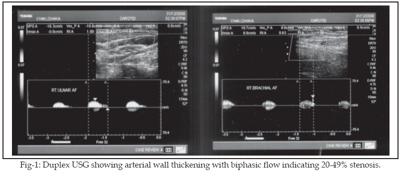
st hour and C reactive protein was 12 mg/L. TPHA was non-reactive and CPK was 45 U/L. Serological study for ANA, c-ANCA and p-ANCA were all negative. Her serum bilirubin, alanine amino transferase, routine microscopic examination of urine, serum creatinine, albumin and lipids profile were normal. Mantoux test was positive and chest X-ray posterior/anterior view was normal.USG of whole abdomen showed normal study and echocardiography reported normal cardiac chambers with normal valvular morphologies. Duplex vascular Ultrasonogram (USG) revealed significant narrowing of both subclavian arteries with descending abdominal aorta (Fig-1).
Histopathology of left axillary lymph node showed caseating tubercles suggestive of granulamatous tuberculous lymphadenitis. After confirmation of diagnoses we started standard anti-tubercular therapy along with steroid and aspirin. She was discharged with advice for periodic follow up.
DISCUSSION
TakayasuÆs arteritis was first described by Dr. Mikito Takayasu in 1905. The disease has been reported in all parts of the world. TakayasuÆs disease is one of the first vasculitides to be associated with a specific infectious agent. Despite the association with tuberculosis, and the similarity between granulomatous lesions in TA and tuberculosis, the exact role of Mycobacterium tuberculosis in the pathogenesis of TA is still unknown. Most recent reports suggest that cross- reactivity between mycobacteria and a human heat shock protein (HSP) might have a key role.
5,6 This hypothesis is further supported by the increased expression of 65 kDa HSPs of the involved vessels, as well as the activation of l - d subpopulations of T cells which may cross-react with host HSPs.5,6 It has also been speculated that M. tuberculosis can be the triggering factor through its production of superantigens, the suggested role of which is thought to be via the stimulation of autoreactive T cells that induce vascular damage.In our patient the tuberculin test was positive and caseating granulomatous lesions were found in auxiliary lymph node. These findings are suggestive of the association between the tuberculosis and the disease process. Almohammadi AA reported two cases wherein a tuberculous process is documented prior to / concomitant with TA and both of them responded well to prednisolone and anti tuberculosis therapy.
7 In our patient, we at first treated with only standard anti tuberculous treatment. Initially she showed some degree of improvement. It might be a result of removing the triggering infectious agent or reducing the triggering antigen load. Later on, rapid response was observed after addition of Prednisolone.Takayasu
,s arteritis is a systemic vasculopathy that can progress to cause vital organ ischaemia. Therefore, long term follow up is recommended. We advised our patient for regular follow up. We planned for ESR, CRP and assessment of arterial wall thickness using duplex vascular USG in each time follow up.In the light of this report, we suggest that tuberculosis should be kept in mind during exploration of aetio pathology of TakayasuÆs arteritis and we found the use of anti tubercular drug rational during treatment of this disease.
Authorship: First two authors revised the article critically for improvement of intellectual content and gave final approval of the version to be published. Last three authors were involved in acquisition of data, drafting the article and also had substantial contributions to conception and design. There is no potential conflict of interest.
REFERENCES
1. Hall S, Buchbinder R. TakayasuÆs arteritis. Rheum Dis Clin North Am 1990;16:411.
2. Moraes MF, Ordway D, Oliveira L. Cellular immune responses to Mycobacterium tuberculosis in a patient with TakayasuÆs arteritis. Rev Port Cardiol 1999;18:359-67.
3. Soto ME, Vargas-Alarc¾n G, Cicero-Sabido R. Comparison distribution of HLA- B alleles in mexican patients with takayasu arteritis and tuberculosis. Hum Immunol 2007;68:449.
4. Modi G, Modi M. Cold agglutinins and cryoglobulins in a patient with acute aortoarteritis (TakayasuÆs disease) and tuberculous lymphadenitis. Rheumatology (Oxford) 2000;39:337-8.
5. Aggarwal A, Chag M, Sinha N, Naik S. TakayasuÆs arteritis: role of Mycobacterium tuberculosis and its 65-kDA heat shock protein. Int J Cardiol 1996;55:49-55.
6. Seko Y, Minota S, Kawasaki A. Perforin-secreting killer cell infiltration and expression of a 65 kDa heat shock protein in aortic tissue of patients with TakayasuÆs arteritis. J Clin Invest 1994;93:750-8.
7. Almohammadi AA, Consunji-Araneta R. Two Cases of TakayasuÆs Arteritis and Tuberculosis. Chest 2007;134(4):675S-676S.
HOME | SEARCH | CURRENT ISSUE | PAST ISSUES
Professional
Medical Publications
Room No. 522, 5th Floor, Panorama Centre
Building No. 2, P.O. Box 8766, Saddar, Karachi - Pakistan.
Phones : 5688791, 5689285 Fax : 5689860
pjms@pjms.com.pk