|
|
||||
|
Published by : PROFESSIONAL MEDICAL PUBLICATIONS |
||||
|
ISSN 1681-715X |
||||
|
||||
|
- |
||||
|
ORIGINAL ARTICLE |
||||
|
- |
||||
|
Volume 22 |
October - December 2006 |
Number 4 |
||
|
|
||||
|
||||
|
|
||||
|
Published by : PROFESSIONAL MEDICAL PUBLICATIONS |
||||
|
ISSN 1681-715X |
||||
|
||||
|
- |
||||
|
ORIGINAL ARTICLE |
||||
|
- |
||||
|
Volume 22 |
October - December 2006 |
Number 4 |
||
|
|
||||
|
||||
Peadiatric Visceral Leishmanisis in the
South West Part of Iran:
A study of 215 Cases
Ahmad Shamsizadeh1, Roya Nikfar2, Sharif Maraghi3 , Nahid Zaker4
ABSTRACT
Objective: To study the epidemiological, clinical charactristics and laboratory findings of visceral leishmaniasis (VL) in children.
Design: It is a retrospective study, hospital records of all children with diagnosis of VL were reviewed from 1991 through 2003.
Setting: Hospitals affiliated to Ahvaz Jondi-Shapour University of Medical Sciences in the southwestern part of Iran.
Subjects: Two hundreds and fifteen patients (153 males and 62 females) were enrolled in the study.
Results: The mean age of the patients was 31 ± 22 months. Fever and splenomegaly were present in >95% of the patients and hepatomegaly and lymphadenopathy in 76% and 3.7% of cases, respectively. Common laboratory findings were anemia, leukopenia and thrombocytopenia.All patients were treated with meglumine antimoniate.Relapse were observed in 4% of the patients and fatality rate was 5%.
Conclusions: Visceral leishmaniasis is endemic in the southwestern part of Iran. The clinical profile of the disease is typical of the Mediterranean infantile form.
KEY WORDS: Visceral leishmaniasis, Children, Iran.
Pak J Med Sci October - December 2006 Vol. 22 No. 4 461-464
1. Ahmad Shamsizadeh
Assistant Professor of Pediatric Infectious Diseases
2. Roya Nikfar
Assistant Professor of Pediatric Infectious Disease
3. Sharif Maraghi
Professor of Parasitology
Department of Mycoparasitology,
School of Medicine,
4. Nahid Zaker
Assistant Professor of Pediatric Infectious Diseases
1-4: Infectious and Tropical Diseases Research Center,
Ahvaz Jondi-Shapour University of Medical Sciences,
Ahvaz, Iran.
Correspondence:
Dr. Ahmad Shamsizadeh
Email: shamsizadeh@ajums.ac.ir
* Received for Publication: September 30, 2005
* Accepted: May 15, 2006
INTRODUCTION
Visceral leishmaniasis (VL) or Kala- azar, is most commonly caused by leishmania donovani, L. infantum and L. chagasi, but on occasion other leishmania sPP. such as L. amazonensis is isolated from patients with typical VL.1 A viscerotropic syndrome caused by L. tropica was identified among a small number of American military personel who served in the Middle East.2 The first case of VL in Iran was reported from north of the country in 1949. Therefore, increasing number of cases were diagnosed from other parts of the country.3
Presently, the major foci of the disease are located in Meshkin–Shahr in the northwest and Shiraz in the south of Iran. The epidemmiological form of diseases in Iran is Mediterranean type, with a canine (dog, fox, Jackal) reservoir, and the etiologic agent is L. infantum which is transmitted by the bite of sand flies.4
Khuzestan Province (site of study) is located in the southwest of Iran and consists of two geographic regions (a plain and a mountainous). The population of this region is actually estimated to be 4,586,178 persons, and those younger than age 15 years comprise 30 %. We performed a retrospective study of epidemiological, clinical, therapeutic features and laboratory findings of VL in children.
PATIENTS and METHODS
Medical records of all children younger than 15 years with final diagnosis of VL who were admitted to two hospitals affiliated to Ahvaz Jondi-Shapour University of Medical Scienses (center of the province) were reviewed from 1991-2003. Inclusion criteria were the presense of lieshmania in the bone marrow samples and/or a positive serologic test result (indirect immunofluorescence antibody [IFA] test >1/128). All patients’ records were reviewed for age, sex, place of abode, clinical manifstations, Laboratory findings and clinical course.
RESULTS
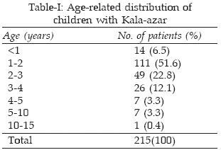
Two hundred fifteen patients were enrolled in the study, of which 153 were males (71%). The ages of the patients ranged from 4 months to 11 years (mean = 32 months, SD 22). The disease was most common in those younger than 2 years of age (Table-I).
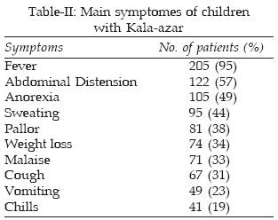
The majority of the children (80%) were from rural areas and nomads. The disease was commonly observed during March-May. The most common symptom was fever. Table-II demonstrates the frequency destribution of symptoms. Fifty seven percent of the patients had abdominal distension and 49% anorexia.
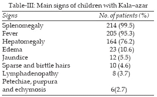
Fever and splenomegaly were present in >95% of the patients (Table-III). Hepatomegaly were seen in 76% and edema in 10% of the cases, but Lymphadenopathy was found in only 3.7% of the children. Table-IV shows the hematolagic features of the children.

Serum asparatate aminotransferase (AST) was increased in 45.5% of the cases and alanine aminotransferase (ALT) in 19%. Hypoalbuminemia was observed in 26% and hypergammaglobulinemia in 29%. The diagnosis was confirmed in 80% of the patients by demonstration of leishmania amastigotes in aspirate from bone marrow and 11% of the patients were diagnosed by a positive IFA test (>1/128). Both tests were positive in 90% of the patients.
All children were treated with meglumine antimoniate. The mean dose was 16-mg antimony /kg/day (SD 5) and the mean duration of treatment was 19 days (SD 4). Relapse was observed in 9(4%) of the patients. These patients received a second course of meglumine antimoniate plus allapurinol or amphotricin B. Eleven patients (5%) expired due to infection or bleeding.
DISCUSSION
The results of the present study indicated that VL is a relatively common disease in the southwestern part of Iran. The disease was more common in younger children. Several studies in countries where infantile form of VL is common showed similar results.5-8 About 70% of the patients were males. Most of the patients were from rural areas and nomads. With considering the reservoirs of the disease, are canine, this is expected.4,9 The clinical manifestations of VL exhibited by the children in this sample were similar to those in published description.9-13
Fever and splenomegaly were noted in more than 95% of the patients but patients were less likely to have lymphadenopathy.6,14
Anemia, leukopenia and thrombocytopenia were found with greater frequency among VL patients.9,15,16 Most of our patients were also cytopenic. Detection of parasites bymicroscopic examination of the bone marrow confirms the diagnosis of VL, but it may be negative in some samples. Because of such negative results, serologic tests like immunofluorescence test are sometimes necessary. In this study, 11% of the patients were diagnosed by IFA test.
Meglumine antimoniate was the first-choice treatment and the side effects were very low. Clinical response was rapid, however the optimal treatment duration was shorter and the mean dose of drug was lower than the world Health Organization recommendation.17 In the present study, 4% of the patients who were treated with meglumine antimoniate suffered a relapse. All of these patients responded well to the second course of meglumine antimoniate plus alluporinol or amphotericin B. Maltezou et al in a study of childhood VL in southern Greece found relapse rate of 6% in their patients.9 There were 11 deaths due to associated complications (5% mortality). The mortality rate was similar to those in published literature.10,18
In summary, VL is endemic in southwestern Iran and the disease is more common in young children. The patients respond well to meglumin antimoniate with shorter duration and lower doses than recommend.
ACKNOWLEDGMENT
We thank nursing staff of Abouzar and Golestan hospitals for taking care of the patients and Mrs Shohreh Nabi-davoodi for preparation of the manuscript.
REFRENCES
1. Jeromino SMB, Sousa Aq, Pearson RD. Leishmania Species: Visceral (Kala-Azar), Cutaneous and Mucocutaneous leishmaniasis. in: principles and Practices of Infectious Diseases, (sixth edition), Mandel GL, Bennett JE & Dolin R (Eds). Saunders, Philadelphia, USA 2005; Pp 3145-56.
2. Magil AJ, Grogl M, Gasser RA, Sun W, Oster CN. Visceral infection caused by leishmania tropica in veterans of Operation Desert Storm. N Eng J Med 1993; 328: 1383-7.
3. Edrissian GH, Nadim A, Alborzi A, Ardehali S. Visceral leishmaniasis; the Iranian experience. Arch Irn Med 1998; 1(1): 22-6.
4. Mohebali M, HamzaviY, Edrissian GH, Forouzani A. Seroepidemiologecal study of visceral leishmaniasis among human and animal reservoirs in Bushehr Province, Islamic Republic of Iran. East Mediterr Health J 2001; 7(6): 912-7.
5. Minodier P, Piarroux R, Garnier JM, Unal D, Perrimond H, Dumon H. Pediatric visceral leishmaniasis in southern France. Pediatr Infect Dis J 1998; 17(8): 701-4.
6. Rathor MH, Buksh D, Hassan M. Visceral leishmaniasis in Pakistani children. South Med J 1992; 89(5): 491-3.
7. Soleimanizadeh G, Edrissiam GH, Movahhed Danesh AM, Nadim A. Epidemiological aspects of Kala-azar in Meshkin-shahr, Iran: human infection. Bull World Health Organ 1993; 71(6): 759-62.
8. Totan M, Dagdemir A, Muslu A, Albayrak D. Visceral childhood Leimaniasis in Turkey. Act Paediatr 2002; 91(1): 62-4.
9. Maltezou C, Siafas C, Mavrikoh M, Spyridis P, Stavinadis C, Karpathios TH, et al. Visceral leishmaniasis during childhood in southern Greece.Clin Infect Dis 2000; 31(5): 1139-4.
10. Elnour IB, Akinbami FO, Shakeel A, Venugopalan P. Visceral leishmaniasis in Omani children: A review. Ann Trop Paediatr 2001; 21(2): 159-63.
11. Jeronimo SM, Oliveira RM, Mackay S, Costa RM, Sweet J, Nascimento ET. An urban outbreak of visceral leishmaniasis in Natal , Brazil. Trans R Soc Trop Med Hyg 1994; 88(4); 386-8.
12. Rahim F, Rehman F, Ahmad S, Zadab. Visceral leishmaniasis in District Dir, NWFP. J Pak Med Assoc 1998; 48(6): 161-2.
13. Lita G, Pavachi F, Sulcebe G, Bregu H, Basha M. Padiatric visceral leishmaniasis in Albania. Int J Infect Dis 2002; 6(1): 66-80.
14. Al-juraygan NA, al Ayed IH, al- Nasser MN, al– Mugeiren MM, Boohene AG, Al Herbish AS. Visceral leishmaniasis in infancy and childhood, epidemiology and clinicopathological study of 63 cases in Al- Baha province, Saudi Arabia. J Trop Pediatr 1992; 38(1); 12-6.
15. Cascio A, Colomba C, Antinori S, Orobello M, Paterson D, Titone L. Pediatric visceral leishmaniasis in western Sicily, Italy; a retrospective analysis of 111 cases. Eur J Clin Microbiol Infect Dis 2002; 21(4): 277-82.
16. Queiroz MJA, Alves JGB, Correila JB. Visceral leishmaniasis; chinical and epidemiological features of children in an endamic area. J Pediatr (Rio J) 2004; 80(2): 141-6.
17. Berman KD. Chemothrapy for lishmaniasis: biochemical mechanisms, clinical effiacacy, and future strategies. Rev Infect Dis 1998; 10:560-86.
18. Ben Salah AB, Ben Ismail R, Amri F, Chlif S, Ben Rzig F, Karrat H, et al. Investigation of the spread of human visceral leishmaniasis in central Tunisia. Trans R Soc Trop Med Hyg 2000; 94(4): 382-6.
HOME | SEARCH | CURRENT ISSUE | PAST ISSUES
Professional
Medical Publications
Room No. 522, 5th Floor, Panorama Centre
Building No. 2, P.O. Box 8766, Saddar, Karachi - Pakistan.
Phones : 5688791, 5689285 Fax : 5689860
pjms@pjms.com.pk