|
|
||||
|
Published by : PROFESSIONAL MEDICAL PUBLICATIONS |
||||
|
ISSN 1681-715X |
||||
|
||||
|
- |
||||
|
ORIGINAL ARTICLE |
||||
|
- |
||||
|
Volume 24 |
October - December 2008 (Part-I) |
Number 5 |
||
|
|
||||
|
||||
|
|
||||
|
Published by : PROFESSIONAL MEDICAL PUBLICATIONS |
||||
|
ISSN 1681-715X |
||||
|
||||
|
- |
||||
|
ORIGINAL ARTICLE |
||||
|
- |
||||
|
Volume 24 |
October - December 2008 (Part-I) |
Number 5 |
||
|
|
||||
|
||||
Hypotonia in infants and young children:
An etiological analysisAmirsalari S1, Kavehmanesh Z2, Khalili Matinzadeh Z3, Afsharpayman S4,
Torkaman M5, Javadipour M6, Kakoie Shourkaie J7, Ghazavi Y8ABSTRACT
Objective: To determine the relative frequency of specific disorders that present with hypotonia in Iran.
Methodology: It is a retrospective, cross–sectional study in which 107 children with hypotonia, aged one month to three years, were evaluated in Baqyiatallah Hospital between June 2003 and June 2006. Children were categorized into groups of central and peripheral hypotonia, and specific diagnosis of each of the two groups was made by clinical findings, neuro imaging, metabolic and genetic tests, muscular enzymes, EMG-NCV (Electromyography-Nerve conduction velocity) and thyroid function tests.
Results: Of the 107 infants, one hundred one (94.4%) children had central hypotonia, four (3.7%) had peripheral hypotonia and in two (1.9%) the hypotonia had other causes. The most common cause of central hypotonia was idiopathic central hypotonia thirty four (31.8%), followed by cerebral palsy in twenty two (20.6%), brain structural abnormality in nineteen (17.8%), inborn errors of metabolism fourteen (13.1%), genetic disorders seven (6.5%) and the TORCH(Toxoplasma, Rubella, Cytomegalovirus, Herpes simplex) syndrome three (2.9%).
Conclusion: In our study "central hypotonia" was most prevalent etiology of hypotonia. The most common cause of central hypotonia was idiopathic central hypotonia followed by cerebral palsy, brain structural abnormality, inborn errors of metabolism, genetic disorders and the TORCH syndrome.
KEYWORDS: Hypotonia, Central hypotonia, Peripheral hypotonia.
Pak J Med Sci October - December 2008 (Part-I) Vol. 24 No. 5 744-747
How to cite this article:
Amirsalari S, Kavehmanesh Z, Khalili Matinzadeh Z, Afsharpayman S, Torkaman M, Javadipour M, et al. Hypotonia in infants and young children: An etiological analysis. Pak J Med Sci 2008;24(5):744-47.
1. Amirsalari S, MD,
Assistant Professor,
2. Kavehmanesh Z, MD,
Associate Professor,
Department of Pediatrics,
Faculty of Medicine,
3. Khalili Matinzadeh Z, MD,
4. Afsharpayman S, MD,
5. Torkaman M, MD,
3-5: Assistant Professor,
Department of Pediatrics,
Faculty of Medicine,
6. Javadipour M, MD,
7. Kakoie Shourkaie J, MD,
General Physician,
8. Ghazavi Y,
Student
1-8: Baqyiatallah University of Medical Sciences,
Tehran – Iran.Correspondence
Susan Amirsalari,
Child Neurologist, Dept. of Pediatrics,
Faculty of Medicine,
Baqyiatallah University of Medical Sciences,
Molla-Sadra St,
Tehran, Iran.
E Mail: susanamirsalari@yahoo.com
* Received for Publication: February 11, 2008
* Accepted: July 26, 2008
INTRODUCTION
Hypotonia is a decreased resistance to passive movement. Hyperextensibility, an abnormaly increased range of joint movement, usually accompanies hypotonia.
1 The central and peripheral nervous system modify tone but intrinsic physical characteristics of tendons, joints and muscles also contribute significantly to tone.2 There is an extensive list of conditions associated with the diagnosis of hypotonia, including hundreds of rare diseases.3 Several longitudinal studies support the opinion that majority of hypotonic infants are floppy because of central nervous system dysfunction; some hypotonic infants have genetic disorders and metabolic disturbances, whereas the minority of hypotonic infants have neuromuscular and connective tissue disease.4-7However controversy exists over which of the causes, play a greater role in the frequency of hypotonia.
2,8 Literature available today shows an extensive list for disorders which may present with hypotonia, and the diagnostic profiles are often complex. As knowledge of the relative frequency of particular disorders that present with hypotonia might help in delineation of appropriate investigations, prevention and treatment, our aim was to gain an insight into the causes of hypotonia in Iran.METHODOLOGY
A cross- sectional study was performed in the Baqyiatallah Hospital, a tertiary care center and educational hospital, through a search of outpatient records. Children aged between one month and three years, whose predominant problem was hypotonia and had been referred between June 2003 and June 2006 to the pediatric neurology clinic of Baqyiatallah Hospital were enrolled in the study. Hypotonia has been clinically confirmed by the attending pediatric neurologist. Criteria used by the pediatric neurologist to define hypotonia were that the hypotonic child looked floppy, felt floppy, or had hyperextensible joints. Hypotonia is classified as central or peripheral according to the clinical findings (quality of antigravity limb movements, deep tendon reflexes, the child’s psychosocial responses) and the result of investigations. If the patients had normal or increased DTR (deep tendon reflexes) and other clinical signs of central nervous system involvement like seizures and mental retardation, they were included in the central group and cranioneuroimaging (CT scan or MRI) was carried out, whereas if they had abnormal facies or dysmorphology, genetic tests were done. Muscle enzymes and EMG-NCV (Electromyography-Nerve conduction velocity) were done for the remaining patients with hypoactive or absent DTR in order to confirm peripheral hypotonia. Thyroid function tests and metabolic work up were done for all patients.
Eventually 138 infants were included, with 31 of these being excluded from the final analysis because of insufficient information. We also obtained demographic data, prenatal and perinatal factors, physical examination findings (DTR) and paraclinic findings (neuroimaging, EMG-NCV, genetic and metabolic tests, muscular enzymes, thyroid function tests) from the clinical charts. To analyze the findings, we used SPSS software (version 11.5).
RESULTS
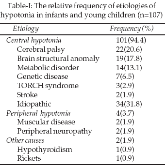
Forty nine (45.8%) infants were male and fifty eight (54.2%) were female the mean age of infants was 18.2 ± 9.76 months. Of 107 infants, one hundred one (94.4%) had central hypotonia, four (3.7%) had peripheral hypotonia and two (1.9%) had other causes of hypotoina. The relative frequencies of etiologies are presented in Table-I.
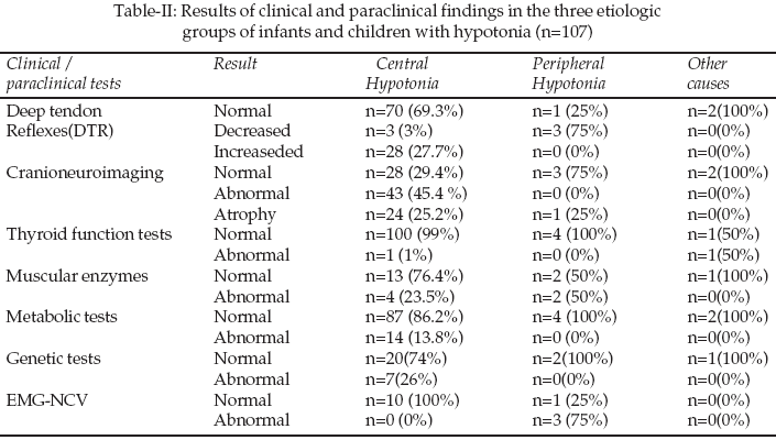
In central hypotonia, the most common causes were idiopathic central hypotonia (31.8%) followed by cerebral palsy (20.6%) and brain structural anomaly (17.8%) respectively. In the central hypotonia group thirty one (30.2%) cases had been delivered by normal vaginal delivery (NVD) and seventy (69.3%) cases by cesarean section. Among children twenty four (23.8%) had a positive history of birth asphyxia, all of them being from the central hypotonia group. Neither of the two remaining groups had a history of birth asphyxia. There were fifty three (52.5%) cases with neonatal jaundice in the central hypotonia group and three (75%) in the peripheral hypotonia group. There was no history of neonatal hospitalization due to causes other than jaundice in the peripheral hypotonia group, while there were 48 (47.5%) cases with such histories in the central hypotonia group. List of clinical and paraclinical findings in the three groups are summarized in Table-II.
DISCUSSION
Results of this study showed central hypotonia in 101 patients (94.4%) and peripheral in four patients (3.7%). In the Paro-Panjan D et al, study, they found that 88% of cases had central causes of hypotonia, 9% had peripheral causes and 3% remained undiagnosed.
9The study conducted by Eng GD that included children aged under three years, similar to our study, in this aspect reported that 85% of cases had central causes and 15% had peripheral causes of muscle tone disturbance.10 Richer et al studied a group of neonate admitted to intensive care unit and found that 66% had central hypotonia and 34% had peripheral causes.6In the majority of studies, cerebral palsy is the most common cause of central hypotonia, while in our study the frequency of idiopathic central hypotonia was higher than cerebral palsy. This may be due to various reasons; in our country, specific genetic tests like FISH (fluroscent insitu hybridization) method and molecular genetic testing are not available. In cases with abnormal facies and dysmorphology, gentic study has been limited to karyotype, as a result of which it is possible to mistakenly classify some of these cases in the idiopathic group. Again in our study, serum aminoacid chromatogrophy was performed using the qualitative method, and we could not measure serum and urine organic acids, and some cases of inborn errors of metabolism may remain undiagnosed and classified in the idiopathic group.
In patients examined after three months of age, TORCH study (Toxoplasma, Rubella, Cytomegalovirus, Herpes simplex) was not reliable, because it could not differentiate between congenital and acquired infections; again some of these patients may have been classified in the idiopathic group. Birdi K et al, could not find a clear etiology for hypotonia in 31% of their cases, indicating difficulty in determing the etiology in hypotonic infants and neonates who present with floppiness and hypomobility.
11 Some profile have been proposed for determination of the causes of infantile hypotonia but, to the best of our knowledge, none of them had good diagnostic value and were widely agreed upon.12-16 It is necessary to prepare an algorithm with high accuracy in order to facilitate determination of the etiology of hypotonia in infants and young children. It is also necessary in our country to constitute new diagnostic methods including specific genetic and metabolic tests.CONCLUSIONS
In this study "central hypotonia" was most prevalent etiology of hypotonia. The most common cause of central hypotonia was idiopathic central hypotonia followed by cerebral palsy, brain structural abnormality, inborn errors of metabolism, genetic disorders and the TORCH syndrome.
REFERENCES
1. Behrman RE, Kliegman RM, Jenson HB. Nelson Textbook of Pediatrics. 17th edition; Philadelphia, Saunders, 2004; PP: 1986-1987;2059-64.
2. Swaiman KF. Pediatric Neurology, Principles and Practice, 3rd ed, Mosby 1999;54-59,1189-90.
3. Crawford TO. Clinical evaluation of the floppy infant. Pediatr Ann 1992;21(6):348-54.
4. Paine RS. The future of "floppy infant": A follow – up study of 133 patients. Dev Med Child Neurol 1963;5:115-24.
5. Carboni P, Pisani F, Crescenzi A, Villani C. Congenital hypotonia with favorable out comes. Pediatr Neurol 2002;26:383-6.
6. Richer LP, Shevell MI, Miller SP. Diagnostic profile of neonatal hypotonia: An 11- year study. Pediatr Neurol 2001;25:32-7.
7. Dua T, Das M, Kabra M. Spectrum of floppy children in Indian scenario. Indian Pediatr 2001;38:1236-43.
8. Menkes JH, Sarnat HB. Child Neurology, 6th ed, williams and wilkins, 2000;1032-1040,1136-8.
9. Paro-Panjan D, Neubauer D. Congenital hypotonia: is there an algorithm? J Child Neurol 2004;19:439-42.
10. Eng GD.Neurormuscular disease, in Avery GB, Fletcher MA, Macdonald MG (eds). Neonatology: Pathophysiology and Management, 4th ed. Philadelphia, JB Lippincot Company 1994;1174-6.
11. Birdi K, Prasad AN, Prasad C, Chodivker B, Chudlog. The floppy infant: retrospective analysis of clinical experience (1990-2000) in a tertiary care facility; J Child Neurol 2005;20(10):803-8.
12. Dubowitz V. Hypotonia in infancy. Acta Univ Carol Med Monogr 1976;75:13-8.
13. Dubowitz V. The floppy infant, in Clinics in Developmental Medicine, Vol .76, 2nd ed. London, Spastics International Medical Publications, William Heinemann Medical Book, 1980;133-8.
14. Darras BT. Neuromuscular disorders in the newborn. Clin Peri -Natal 1997;24:827-44.
15. Bergen BJ. Evaluation of the hypotonic or floppy infant. Minn Med 1985;68:341-7.
16. Prasad AN, Prasad C. The floppy infant: contribution of genetic and metabolic disorders. Brain Dev 2003;25:457-76.
HOME | SEARCH | CURRENT ISSUE | PAST ISSUES